Reading...
![]()
Play button
![]()
Play button
![]()
Use LEFT and RIGHT arrow keys to navigate between flashcards;
Use UP and DOWN arrow keys to flip the card;
H to show hint;
A reads text to speech;
103 Cards in this Set
- Front
- Back
|
1) What surrounds the CNS?
2) A cluster of cell bodies in the CNS is called? 3) A cluster of cell bodies in the PNS is called? |
1) Meninges
2) Nucleus 3) Ganglion |
|
|
1) What is the NS composed of?
2) What 2 types of cells compose the nervous system? 3) Which of these is are non-conducting cells? |
1) Nervous tissue; connective tissue, and blood vessels
2) Neurons & supporting cells 3) Supporting cells |
|
|
1) Why are neurons amitotic?
2) What are Nissl bodies/Chromatophilic substances? 3) What are neurofilaments? |
1) They don't have centrioles
2) Well-developed rough-ER that functions to perform protein synthesis 3) Intermediate filaments throughout the cell body |
|
|
1) What is the biosynthetic region of a neuron?
2) What are the receptive regions of a neuron? 3) What is the conductive region of a neuron? |
1) Cell body
2) Dendrite 3) Axon |
|
|
1) How many axons can a neuron have?
2) What are the branches and bulbs of an axon termed? |
1) 1
2) Branches=telodendria; Bulbs=synaptic knobs=boutons |
|
|
Name & describe the three types of neurons
|
1) Multipolar: at least 3 processes/poles
2) Bipolar: has 2 processes 3) Pseudounipolar: one short process that branches out into two |
|
|
1) What is the most abundant type of neuron in the body?
2) What type of neuron is: a. motor neuron b. sensory neuron c. interneuron 3) What type of neuron is only found in the eye and ear? |
1) Multipolar
2) a. multipolar; b. unipolar c. multipolar 3) Bipolar |
|
|
1) 99% of neurons in the body are _________________
2) What are bundles of axons called in the CNS/PNS |
1) Association neurons/interneurons
2) CNS=tract; PNS=nerve |
|
|
1) What wraps:
a. axon b. fascicle c. bundle of fascicle 2) What is another name for supporting cells? |
1) a. endoneurium; b. perineurium; c. epineurium
2) Neuroglia |
|
|
1) What are the 4 supporting cells in the CNS?
2) What are the 2 supporting cells in the PNS? |
1) Astrocytes; microglia; oligodendrocytes; ependymal cells
2) Schwann cells; satellite cells |
|
|
What are:
a. Astrocytes b. Microglia c. Ependymal cells d. Oligodendrocytes |
a. BBB, regulate chemical environment of brain, regulate brain fctn
b. primary immune response - act as macrophages c. Ciliated columnar cells that circulate CSF; line ventricles in brain d. myelinate up to 50 axons in the CNS |
|
|
1) What is the most abundant supporting cell?
2) What are: a. Schwann cells b. Satellite cells 3) What is another name for Schwann cells? 4) What are 2 components of a schwann cell? |
1) Astrocytes
2) a. myelinate axons in the PNS; b. like astrocytes of CNS 3) neurolemmocytes 4) Myelin sheath and the neurilemma(contains cytoplasm and the nucleus) |
|
|
1) The axons in the CNS are myelinated by ______a_____ from the oligodendrocytes hence, ______b_______ is absent
2) What are the 3 functions of the myelin sheath? |
1) a. extensions; b. neurilemma
2) Protection, electrical insulation, speed |
|
|
1) Can axons in the PNS regenerate? Why or why not?
2) Can axons in the CNS regenerate? Why or why not? |
1) Yes b/c immune cells clean up the damaged area of cell debris (through debridement) and the neurilemma of the schwann cells forms a regeneration tube to guide regeneration of the axon
2) No b/c microglia function poorly to clean up damage area. Debridement isn't complete. No neurilemma to form regeneration tube. Growth-inhibiting proteins inhibit regeneration |
|
|
Describe depol, repol, & hyperpol phases
|
Depol=Na+ influx, mp less negative, AP occurs once threshold is reached/ Repol=Na+ channels close, K+ channels open - K+ efflux toward neg pot./Hyperpol=more K+ leaves and pot. goes below RMP/RMP restored by Na+/K+ pump
|
|
|
1) What is a propagated or transmitted AP called?
2) What is the difference b/t a strong and weak stimulus? 3) What are 2 characteristics of an AP? |
1) Impulse
2) A stronger stimulus causes a higher frequency 3) All-or-nothing; Self-propagating |
|
|
Describe the 2 refractory periods of an AP
|
1) Absolute refractory period: depol phase, another AP cannot be generated
2) Relative refractory period: Repol phase, another AP can be generated |
|
|
1) What is the difference between continuous conduction and saltatory conduction?
2) What are two factors effecting conduction velocity? |
1) Continuous cond. is through an unmyelinated axon. The lack of myelin sheath allows for ion leakage and no saltatory conduction - it's slower
2) a. Diameter of the axon (thicker hose=less resistance); b. Degree of myelination (saltatory conduction) |
|
|
What are 3 types of nerve fibers (based on diameter & myelination)? Give ex. of each
|
a. Group A Fibers: Largest diameter, heavy myelination - Ex. Motor neurons
b. Group B Fibers: Intermediate diameter, lightly myelinated - Ex. Preganglionic autonomic fibers c. Group C Fibers: Smallest diameters, unmyelinated - Ex. Postganglionic autonomic fibers |
|
|
What is Multiple Sclerosis (MS)?
|
Autoimmune disease that results in demyelinations of axons in the CNS
-impulse travels slowly and interferes w/brain communication to body |
|
|
What are the 4 protective structures of the brain?
|
1) Cranium
2) Meninges 3) Cerebrospinal Fluid 4) BBB |
|
|
What are the 3 connective membranes of the meninges and describe them?
|
1) Dura matter - double layered; above =outer periosteal layer/under=inner meningeal layer; under=subdural space
2) Arachnoid matter - under=subarachnoid space; web-like extensions 3) Pia matter - clings to surface of brain |
|
|
1) What is the CSF?
2) What produces CSF? 3) What is the BBB? |
1) Filtered from the blood; located in ventricles & subarachnoid space; acts as a liquid cushion, provides bouyancy, nutrients, removes metabolic wastes
2) Choroid plexus 3) Barrier that prevents harmful substances in blood from crossing to brain |
|
|
1) What is a ventricle?
2) How many are there? Name them |
1) A space in the brain containing CSF
2) 4. 2 lateral ventricles and 3rd and 4th ventricles |
|
|
1) What membrane separates the 2 lateral ventricles?
2) Name the channel that connects the 2 lateral ventricles to the ventricles below it 3) What connects the 3rd and 4th ventricle? |
1) Septum Pellucidum
2) Interventricular foramen 3) Cerebral aqueduct |
|
|
What are the four major regions of the brain?
|
Cerebrum; diencephalon; brain stem; cerebellum
|
|
|
1) Describe the cerebrum
2) What is a gyrus, fissure, and sulcus? 3) What is the corpus callosum? |
1) Accounts for 80% of brain mass; 2 cerebral hemispheres (both divided into 5 lobes); divided by longitudinal fissure;
2) Gyrus - elevated ridge/Fissure - deeper groove/Sulcus - shallow groove 3) Holds together the 2 cerebral hemispheres |
|
|
1) What are the 5 lobes of the cerebral hemispheres?
2) What are the 3 sulci of the brain? |
1) Frontal, occipital, parietal, temporal, insula
2) Central, lateral, parieto-occipital |
|
|
1) What are the ridges before and after the central sulcus termed?
2) What are the 3 regions in each cerebral hemisphere? Describe them |
1) Precentral and Poatcentral gyri
2) a. Cerebral cortex: gray matter (cell bodies, dendrites); 40% of brain mass; location of our conscious mind/ b. Cerebral white matter: deep to cerebral cortex; only myelinated axons present(bundled into tracts)/ c. Basal nuclei: clusters of neurons cell bodies within the white matter |
|
|
What are the 3 types of tracts within the cerebral white matter? Describe them
|
1) Commissural tracts: connects the 2 cerebral hemispheres (ex. corpus callosum)
2) Projection tracts (2 types): connect cerebrum to lower brain areas and the spinal cord -Descending - brings info from cerebral cortex to lower brain reigons, like spinal cord (ex. pyrimidal and corticospinal tracts) -Ascending - transmits sensory info to the thalamus, then to the cerebral cortex 3) Association tracts - connects areas w/in the same cerebral hemisphere (ex. arcuate fasciculate) |
|
|
What are the 3 functional areas in the cerebral cortex?
|
1) Motor areas: control voluntary movements
2) Sensory areas: for the conscious awareness of sensation 3) Association areas: integrates and interprets sensory input from sensory area so each primary sensory area has an association area |
|
|
Describe the 4 functional motor areas of the cerebral cortex.
|
1) Primary motor cortex: controls voluntary mvmt of skeletal muscles
2) Broca's area (motor speech area): Controls the muscles involved in speech production. Found only in the LEFT cerebral hemisphere 3) Frontal eye field: controls voluntary movements of the skeletal muscles that position the eye 4) Premotor cortex: controls learned/repetitive motor skills like typing. If this area is damaged you will need to relearn skills. |
|
|
1) Where are pyramidal cells located?
2) What is decussation of the pyramids? 3) What is hemiplegia? |
1) In the precentral gyrus of the frontal lobe
2) The crossing over of pyramidal tracts or corticospinal tracts 3) Damage to the left precentral gyrus resulting in paralysis of the right side of body. |
|
|
1) What do axons of pyramidal cells bundle to form?
2) Damage to the left side of the frontal lobe will have what affects? |
1) Pyramidal tract or corticospinal tract
2) Right side paralysis & loss of speech |
|
|
__________a________ connects the Broca's area to the _______b_______ area located in the left temporal lobe to produce _______c________
|
a. Arcuate Fasciculus
b. Wernicke's Area c. Language |
|
|
1) What is aphasia?
2) What is the result of damage Wernicke's area? 3) What is the result of damage to the frontal eye field? |
1) Loss of speech
2) "Word salad" speech 3) Eyes become fixed; inability to move eyeballs |
|
|
What are the 5 sensory areas in the cerebral cortex?
|
1) Somatosensory cortex in the parietal lobe
2) Primary visual cortex in the occipital lobe 3) Primary auditory cortex in the temporal lobe 4) primary olfactory cortex in the frontal and temporal lobes 5) Primary gustatory in the insula lobe |
|
|
1) What are basal nuclei?
2) What are 3 major basal nuclei? |
1) Islands of gray matter in the cerebral white matter that start & stop mvmnts; they inhibit unnecessary mvmnts
2) a. Caudate nucleus b. Putamen c. Globus Pallidus |
|
|
1) The putamen + globus pallidus = ?
2) The lentiform nucleus + caudate = ? 3) Where is the 3rd ventricle located? |
1) Lentiform nucleus
2) Corpus stratum 3) Thalamus |
|
|
Name and describe the 3 paired structures of the diencephalon
|
1) Thalamus: the relay station for sensory input to cerebral cortex
2) Hypothalamus: Controls endocrine function (produces 9 hormones); activities of ANS; Emotional response, sleep-wake cycle, etc. 3) Epithalamus: Contains pineal gland that secretes melatonin (sleep-inducing hormone); forms roof of 3rd ventricle |
|
|
1) What is the term for the visual relay center in the thalamus?
2) What is the term for the auditory relay center in the thalamus? |
1) lateral geniculate nucleus (LGN)
2) medial geniculate nucleus (MGN) |
|
|
Name and describe the 3 regions of the brain stem
|
1) Midbrain: Contains the: cerebral aqueduct, corpora quadrigemina (4 colliculi); red nuclei & substantia nigra
2) Pons: contains respiratory centers; are b/t the midbrain & medulla oblongata; connect motor cortex & cerebellum 3) Medulla oblongata: decussation of the pyramids on its ventricle surface; most inferior region; contains autonomic reflex & cardiovascular centers, etc. |
|
|
1) What is the substantia nigra?
2) What does degeneration of the dopaminergic neurons from the substantia nigra cause? 3) What is the function of red nuclei? 4) In the midbrain, what acts as the visual reflex center? the auditory reflex center? |
1) Contains dopamine-releasing neurons that project and modulate activities of the basal nuclei
2) Parkinson's disease 3) Controls flexion of limbs 4) a. 2 superior colliculi; b. 2 inferior colliculi |
|
|
1) What makes up 11% of the total brain mass?
2) What separates the cerebellum and the cerebrum? 3) What holds together the 2 cerebellar hemispheres? |
1) Cerebellum
2) Transverse fissure 3) Vermis |
|
|
1) Name the 3 cerebellar lobes
2) What is the term for cerebellar white matter? 3) What is the function of the cerebellum? |
1) a. Anterior lobe; b. Posterior lobe; c. Flocculomodular lobe
2) Arbor vitae 3) Processes info from cerebral motor areas, visual & equilibrium inputs; voluntary skel. muscle mvmnts; maintenance of balance |
|
|
1) What common drink can affect the function of the cerebellum?
2) The spinal cord extends from the _____________ to the second ______________? 3) What is the name of the cone-shaped structure at the end of the spinal cord? |
1) Alcohol
2) medulla oblongata; 2nd lumbar vertebra 3) Conus medullaris |
|
|
1) What is the fibrous extension from the conus medullaris?
2) What is its function? 3) What laterally anchors the spinal cord? |
1) Filum terminale
2) Anchors the spinal cord vertically to the coccyx 3) Denticulate ligaments |
|
|
1) What type of matter forms the denticulate ligaments?
2) What type of matters composes the spinal cord? |
1) Pia matter
2) Outer white matter and inner gray matter |
|
|
Name and describe the 3 protective structures of the spinal cord
|
1) Vertebral column: bony structure of cord
2) Meninges: -Dura mater=spinal dural sheath, single layer -Arachnoid mater=separated from spinal dural sheath by the subdural space -Pia mater=innermost meninx 3) Cerebrospinal Fluid: in central canal in core of spinal cord and inside subarachnoid space |
|
|
1) What is the space b/t the spinal dural sheath and the internal surface of the vertebral column?
2) What 2 things is this space made of? 3) What is the space b/t the arachnoid space and the pia mater? |
1) Epidural space
2) Veins & fat 3) Subarachnoid space |
|
|
1) What forms the denticulate ligaments and the filum terminale?
2) What are 3 functions of the CSF? |
1) Pia mater
2) a. acts as a liquid cushion b. provides nutrients c. removes metabolic wastes |
|
|
1) What is spinal bifida?
2) What tracts compose the outer white matter of the spinal cord? |
1) The laminae and spinous processes of the vertebrae are missing
2) a. Ascending tracts = sensory inputs b. Descending tracts = motor outputs c. Transverse tracts = commissural fibers |
|
|
Name and describe the horns that compose the inner gray matter of the spinal cord
|
1) Dorsal (posterior) horns: house cell bodies of afferent fibers
2) Ventral (anterior) horns: house mainly cell bodies of somatic neurons 3) Lateral horns: in thoracic & upper lumbar parts of spinal cord and has cell bodies of sympathetic neurons |
|
|
1) In what part of the spine are ventral horns largest?
2) What does the amount of gray mater present at a certain level of the spinal cord signify? 3) How many pairs of spinal nerves come out of the spinal cord? |
1) The cervical and lumbar enlargements that innervate the upper and lower limbs
2) The amt of gray mater reflects the extent at which it innervates the skeletal muscle served by those fibers 3) 31 |
|
|
1) What is the term for the collection of nerve roots below the spinal cord?
2) What collection of neuronal cell bodies in the spinal gray matter is largest in the cervical and lumbar enlargements? |
1) Cauda equina
2) Ventral horns (innervate the upper & lower limbs) |
|
|
Name and describe the 2 spinal enlargements
|
1) Cervical enlargement - nerves that innervate and control voluntary nerve function & sensation in the upper limbs
2) Lumbar enlargement - nerves that innervate and control voluntary motor function & sensation in the lower limbs |
|
|
1) What is the result of damage at/above the cervical enlargement?
2) What is the result of damage at/above the lumbar region? |
1) Quadriplegia = paralysis & paresthesia of all 4 limbs
2) Paraplegia = paralysis & paresthesia of lower limbs |
|
|
1) What are endocrine glands?
2) What is the difference b/t hormones & NTs? 3) Hormones are released in response to homeostatic imbalance called _______________. |
1) Ductless glands that release hormones directly into the ECF (interstitial fluid & blood)
2) Hormones are released into the ECF and NTs are released into synaptic cleft 3) Stimuli |
|
|
1) Hormones work via _________________ to maintain homeostasis
2) Name and describe the 3 types of general stimuli for the release of hormones |
1) Negative feedback
2) a. Humoral stimuli: changes in the levels of chemicals in the body's humors b. Neural stimuli: activation of the NS c. Hormonal stimuli: released hormones |
|
|
What 3 things are hormones named based on?
|
1) Organ of origin - the endo gland releasing the hormone (ex. PTH)
2) Function (ex. follicle stimulating hormone) 3) Chemical structure (ex. T3 -triiodothyronine) |
|
|
1) Name and describe the 3 chemical structures of hormones
2) Of these 3, which one is non-polar? |
1) a. Biogenic amine hormones-hormones derived from the a.a. tyrosine/ b. Peptide, protein, glycoproteins hormones-hormones composed of a sequence of a.a.'s; protein w/a carb attached/ c. Steroid hormones-hormones derived from cholesterol
2) Steroid hormones |
|
|
1) Can a target cell be bound to more than one hormone simultaneously?
2) Target cells/tissues express __________________ & __________________ receptors that the hormone binds to |
1) Yes
2) Accessible and functional |
|
|
1) What the two respiratory control centers? What are their functions?
|
1) Apneustic center: controls rate of breathing & pneumotaxic center: controls depth of breath
|
|
|
1) Describe the three sensory receptors?
2) What sensory receptors are used in hearing? 3) What sensory receptors are used in olfaction? 4) What sensory receptors are used in gustation? |
1) Photoreceptors - sensory receptors that respond to light energy; in the retina; used in vision
Mechanoreceptors: Respond to touch Chemoreceptors: Respond to chemical changes 2) Mechanoreceptors - hair cells 3) Chemoreceptors - olfactory cells 4) Chemoreceptors - gustatory cells |
|
|
1) Name the 3 major parts of the eye
2) Name and describe the 3 layers of the wall of the eye. |
1) Eye wall, humors, lens
2) Fibrous layer(outer layer;dense CT; 2 regions: sclera&cornea) Vascular layer-Uvea(middle layer; highly vascularized; 3 regions: choroid, middle ciliary body, anterior iris) Sensory Layer(Innermost layer;confined to posterior wall;2 layers: pigmented & neural) |
|
|
Fibrous layer - What is the:
1) Sclera 2) Cornea |
1) Maintains shape, protects, and provides site of attachment for extrinsic muscles of the eye - sclera forms white of the eye
2) allows light to enter the eye because it's avascular and transparent |
|
|
Vascular layer - What is the:
1) Choroid 2) Ciliary processes 3) Ciliary body 4) Iris |
1) Provides nutrients to the sclera and the sensory layer
2) The folds at the end of the ciliary body; contain capillaries that secrete aqueous humor 3) ends in folds called ciliary processes & has string-like structures extending from those processes (ciliary zonule/suspensory ligaments) that hold the lens upright 4) Colored part of eye; central opening is the pupil; has 2 types of smooth muscle: circular & radial smooth muscles that control size of pupil |
|
|
1) What causes pupillary dilation?
2) What causes pupillary constriction? |
1) SNS causes radial smooth muscles to contract when viewing distant objects
2) PNS causes circular smooth muscle to contract when viewing close objects |
|
|
Sensory layer - What is the:
1) Pigmented layer 2) Neural layer/retina |
1) Single layer of cells that provide nutrients to the neural layer; contain melanin & VitA
2) Extends anteriorly from pigmented layer; 3 layers: photoreceptors, bipolar neurons, ganglion cells |
|
|
1) What does melanin do in the pigmented layer of the eye?
2) What is the function of VitA? |
1) Absorbs light & prevents it from scattering
2) Required for the synthesis of the light-absorbing pigment - Retinal |
|
|
Retina - What is/are:
1) Photoreceptors? 2) the two types of photoreceptors? 3) the trichromatic theory of vision? |
1) Border pigmented layer; respond to light and generate electrical signals
2) Rods(more numerous, more sensitive to light, used in dim-light & peripheral vision, provide images in shades of gray) & Cones(function in bright light, provide color vision, 3 types: blue, green, & red) 3) Depending on which/how many cones are activated, several colors are seen |
|
|
What are:
1) Bipolar neurons 2) Ganglion cells 3) What are the only neurons in the retina that can generate & transmit APs? From where is the AP generated? |
1) Receive electrical signals from photoreceptors & conduct signals to ganglion cells
2) Neurons that receive electrical signals from bipolar neuron 3) Ganglion cells. Axons |
|
|
1) Describe the pathway of light
2) What do bundles of axons of the ganglion cells form? 3) Where is this structure (#2) located? |
1) Cornea-->aqueous humor-->pupil-->lens-->vitreous humor-->ganglion cells-->bipolar neurons-->photoreceptors
2) Optic nerve 3) The optic disc is on the posterior wall where the optic nerve exits the eye |
|
|
1) What is another name for optic disc? Why?
2) Name & describe the structure that is lateral to the optic disc? |
1) Blind spot b/c it lacks photoreceptors
2) Macula lutea - contains mostly cones; at its center is the fovea centralis that contains only cones and is used for hard focus |
|
|
1) What are humors?
2) Describe the aqueous humor 3) Describe the vitreous humor |
1) Fluids in the body
2) Filtered blood from capillaries flows into ciliary processes to the anterior chamber of the eye; supplies nutrients 3) gel-like fluid in posterior segment beind lens; supports posterior surface of lens; pushes neural layer against pigmented layer; maintains intraocular pressure |
|
|
1) What is glaucoma?
2) How is aqueous humor drained? 3) Is vitreous humor replenished at points throughout our lives? 4) What is retinal detachment? |
1) rate of synthesis of aq. humor>rate of drainage, intraocular pressure rises causing damage to the retina & optic nerve
2) By the canal of Schlemm - aq. humor is formed & drained continuously 3) No. It is formed in the embryo & lasts a lifetime 4) Retina detaches from pigmented layer & vitreous humor seeps into space. W/out the pigmented layer's nutrient source, the photoreceptors die leading to blindness |
|
|
1) Describe the structure of the lens
2) Function of the lens 3) lens is composed of transparent proteins called? |
1) Avascular, transparent, biconvex and flexible
Held in upright position behind the pupil and the iris by the ciliary zonule 2) Focuses light on retina; can change shape to focus light on retina (known as accomodation) 3) Crystallins |
|
|
1) Is accomodation required for distant vision?
2) What is the far point of vision? 3) What is close vision? Is accomodation required? 4) What is the near point of vision? |
1) No, the eyes are adapted to distant vision
2) Distance beyond which accomodation is not needed = 6m = 20ft (20/20 vision) 3) less than 20ft;involves accomodation of lens - lens bulges and pupils contract in close vision 4) The distance at which the lens can bulge maximally to focus the object on the retina = 10cm = 4in |
|
|
1) What is Myopia?
2) How is #1 corrected? 3) What is hyperopia? 4) How is #3 corrected? |
1) Nearsightedness - occurs when distant objects are focused in front of the retina; eyeball too long.
2) Wear concave lenses to diverge light before it enters the eye or flatten cornea using LASIK 3) Farsightedness – occurs when light from close objects are focused behind the retina; eyeball too short 4) Wear convex lenses to converge the light onto the retina |
|
|
1) What are cataracts? How are they formed?
2) Describe the pathway of electrical signal transmission |
1) Clouding of the lens from thickening and hardening of the crystallins
2) Light hits the photoreceptors and they generate electrical signals--> Bipolar neurons-->Ganglion cells |
|
|
Describe the pathway of impulse transmission
|
Axons of ganglion cells form the optic nerves; generate and transmit impulses via the optic nerves; medial fibers of optic nerves cross over to opposite sides at the OPTIC CHIASMA and continue on as the OPTIC TRACTS.
|
|
|
Name and describe the 3 major parts of the ear
|
External ear: Pinna + external auditory canal
Middle ear: Contains the three ossicles (malleus,incus,stapes) and the tympanic membrane. Internal ear: (Labyrinth) Bony labyrinth and Membranous labyrinth; separated from the middle ear by a bony wall with an oval window and round window; the stapes sits atop the oval window |
|
|
What is the tympanic membrane?
|
Cone-shaped membrane that separates the external ear from the middle ear; the broad base faces the external auditory canal and the apex abuts the malleus
|
|
|
1) What is the Perilymph?
2) What is the Endolymph? 3) What is the bony labyrinth composed of? 4) What is the membranous labyrinth composed of? |
1) CSF-like fluid that fills the bony labyrinth
2) Fluid found inside the structures of the membranous labyrinth 3) The vestibule, semicircular canals, cochlea. 4) Of interconnecting sacs and ducts located inside the structures of the bony labyrinth. |
|
|
1) Name in order (from the tympanic membrane) the 3 auditory ossicles.
2) What are the utricle and saccule? 3) Hair cells are located in between what two membranes? |
1) 1) malleus 2) Incus 3) Stapes
2) Membranous sacs located inside the vestibule; contain the equilibrium receptors called MACULAE that respond to the pull of gravity and head position 3) The inferior membrane (basilar membrane) & superior gel-like membrane (tectorial membrane). |
|
|
1) What are the semicircular ducts?
2) What is the cochlear duct? |
1) Membranous ducts located in the semicircular canals; expanded ends called AMPULLAE house equilibrium receptors called CRISTAE AMPULLARES that respond to the rotational movements of the head
2) Membranous duct located in the cochlea |
|
|
1) Name the ossicle in direct contact with the a) tympanic membrane b) oval window.
2) Name the 3 regions of the bony labyrinth. |
1) a) The malleus b) The stapes
2) 1: Vestibule: the central cavity of the bony labyrinth of the ear 2: Semicircular canals: three tiny, fluid-filled tubes in your inner ear that help you keep your balance. 3: cochlea: the auditory portion of the inner ear. It is a spiral-shaped cavity |
|
|
Give the location of the following: Saccule, utricle, maculae, cristae ampullares,
perilymph, endolymph, organ of Corti, hair cells, auditory cortex. |
Saccule, utricle, and maculae: all found in the vestibule
Cristae ampullares: in the semi-circular canals Perilymph: within the bony labyrinth Endolymph: within the membranous labyrinth Organ of Corti: located in the cochlear duct and contains endolymph. It rests on the basilar membrane Hair cells: In the organ of corti trapped in the tectorial membrane Primary auditory cortex: in the temporal lobes |
|
|
1) What is stereocilia?
2) What receptor inside the semicircular canals responds to rotational movements of the head? 3) What receptor inside the vestibule responds to the pull of gravity and head position? 4) Name the equilibrium receptors for rotational movements |
1) Microvilli stiffened by actin filaments. These are attached to the apical surfaces of the hair cells and trap the hair into the gel-like tectorial membrane.
2) Maculae 3) Maculae 4) Cristae ampullares |
|
|
1) Give the location of the (a) Basilar membrane (b) Tectorial membrane
2) Name the mechanoreceptors in the ear. |
1) a) within the cochlea of the inner ear is a stiff structural element that separates two liquid-filled tubes that run along the coil of the cochlea, the scala media and the scala tympani
b) located above the sulcus spiralis internus and the spiral organ of Corti and extends along the longitudinal length of the cochlea parallel to the basilar membrane. 2) Hair cells |
|
|
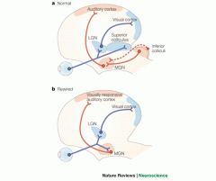
1) Give the location and function of a) medial geniculate nucleus b) inferior colliculi.
2) The organ of corti is composed of two type of cells: 3) What nerve wraps around the bases of the hair cells? |
1) a) In the thalamus it is the auditory relay center
b) In the midbrain it is the auditory reflex center 2) Supporting cells and hair cells. 3) The afferent fibers of the cochlear nerve (a division CN VIII). |
|
|
Go through the route of sound wave entry through the ear/impulse transmission.
|
Pinna -> External Auditory canalà Tympanic membrane
vibrates -> Malleus -> Incus -> Stapes-> Perilymph + Endolymph move -> Basilar Membrane oscillates-> Hair cells move -> stereocilia bend -> electrical signals develop -> transferred to the cochlear nerve -> cochlear nerve generates and transmits action potentials via the vestibulocochlear nerve (CNVIII) -> the auditory reflex centers in the midbrain called the INFERIOR COLLICULI -> the auditory relay center in the thalamus called MEDIAL GENICULATE NUCLEUS (MGN) -> primary auditory cortex in the Temporal lobes |
|
|
1) What are gustatory cells?
2) How often are basal cells replaced? How? 3) What are gustatory hairs? |
1) Receptors for taste - specialized cells w/microvilli bathed in saliva and subjected to tremendous friction
2) Every 7 days by the differentiation of basal cells into new gustatory cells 3) Long microvilli that extend to the surface of the tongue through taste pores |
|
|
1) Where are taste buds located?
2) What are the four types of papillae? 3) What two types of taste buds make up the taste buds? 4) _____________ ____________ coil around the gustatory cells |
1) In peg-like projections of the tongue called papillae
2) fungiform, foliate, vallate and filiform papillae ( the filiform papillae lack taste buds) 3) Basal cells & gustatory cells (chemoreceptors) 4) Afferent fibers |
|
|
What are 3 types of cranial nerves involved in the gustatory pathway to the brain?
|
-Chorda tympani, a branch of the facial nerve (CN VII)
-Glossopharyngeal nerve( CN IX) -Vagus nerve ( CN X) |
|
|
1) What must happen to the tastant before the gustatory cells are activated?
2) Describe what happens after activation |
1) It must dissolve in saliva
2) Dissolved chemical binds to gustatory hairs resulting in depol. The impulse is transferred to the afferent fibers. Impulses from the 3 cranial nerves (facial, glossopharyngeal, & vagus nerves) are sent to the solitary nucleus in the medulla oblongata, then to the gustatory relay center in the thalamus (VENTRAL POSTEROMEDIAL NUCLEUS), impulse is then relayed to the primary gustatory cortex in the insula |
|
|
1) chorda tympani of the _________________ generates and transmits action potentials from gustatory cells in the anterior two-thirds of the tongue
2) ________________ generates and transmits action potentials from the posterior third of the tongue and the superior part of the pharynx 3) ________________ generates and transmits action potentials from the inferior part of the pharynx |
1) Facial nerve (CN VII)
2) Glossopharyngeal nerve (CN IX) 3) Vagus nerve (CN X) |
|
|
1) Taste is 80% _____________ because ______________ .
2) How come food doesn't taste good when I have a cold? |
1) Taste b/c the same chemicals activate both types of chemoreceptors = olfactory & gustatory cells
2) Because most of what we call "taste" is in fact smell, triggered by odor molecules from our food and drink. Some molecules we smell in the air, others vaporize as we chew, then rise into the nasal passages at the back of the mouth. |
|
|
1) What are Olfactory receptors cells?
2) These bipolar neurons, the olfactory cells, are unique in humans - explain why they are unique? 3) What are the two criteria a chemical must meet for olfactory processing to occur? |
1) Bipolar neurons with ciliated dendritic end.
2) They need replacement! 3) a) Must be volatile: in the air b) Must be soluble in the thin coat of mucus covering the olfactory hairs (cilia). |
|
|
1) Bundle of axons of which cells form (a) olfactory nerve (b) olfactory tract?
2) Name the two types of chemoreceptors: 3) Describe olfactory cells: |
1) a) olfactory bipolar cells
b) Bundle of axons of the Mitral cells 2) Olfactory cells & Gustatory cells. 3) These process the sense of smell (olfaction). They are BIPOLAR neurons located in the olfactory epithelium. |
|
|
1) Describe the olfactory epithelium. What cells does it consist of?
2) Which type of neuron die and are replaced every 60 days? 3) Axons of the olfactory bipolar cells bundle to form? |
1) Its a yellowish patch in the roof of nasal cavity.
Olfactory cells, supporting cells, & basal cells. 2) Olfactory cells are replaced by differentiation of basal cells as the olfactory cells are damaged by odorants. They do not exhibit longevity. 3) Olfactory nerve (CN I) |
|
|
1) The olfactory nerve projects to second order neurons in the olfactory bulb called?
2) True or false: axons of mitral cells form the olfactory tract. |
1) Mitral cells. Many olfactory nerves bundle to contact one mitral cell.
2) False |
|
|
Describe how we smell? The pathway?
|
Dissolved chemicals attach to the cilia resulting in depolarization which eventually causes the axons of the olfactory nerve to generate and transmit action potentials ( the impulse)
Impulse is transmitted to a second type of neurons called MITRAL cells in the olfactory bulb. Bundle of axons of the mitral cells form the OLFACTORY TRACT Impulse is transmitted from the olfactory tract to the olfactory relay center in the thalamus called the medial dorsal nucleusà relayed to the olfactory cortex in the frontal lobes and the temporal lobes Impulses are also transmitted to the mammillary bodies -> limbic system including the amygdala, responsible for the emotional and autonomic responses to odors. |