![]()
![]()
![]()
Use LEFT and RIGHT arrow keys to navigate between flashcards;
Use UP and DOWN arrow keys to flip the card;
H to show hint;
A reads text to speech;
31 Cards in this Set
- Front
- Back
|
What do we typically use for the subcuticular stitch? What is the stitch's typical half life?
|
Monocryl. This is a synthetic absorbable suture that is monofilament, which makes it less reactive to the tissue. It comes in both dyed and non-dyed and it is usually used for closing the skin. It has a half life of 7-14days.
|
|
|
When closing small robotic 8mm ports or a 5mm port site, what suture technique does Dan McRackan use that makes it look much more asthetically pleasing?
|
He instead of attempting a subcuticular stitch, uses a simple horizontal mattress suture, that really looks much better.
|
|
|
What is Vicryl and what is its typical half life?
|
icryl (polyglactin 910) is an absorbable, synthetic, braided suture, manufactured by Ethicon Inc., a subsidiary of Johnson and Johnson. [1] It is indicated for soft tissue approximation and ligation. The suture holds its tensile strength for approximately three to four weeks in tissue, and is completely absorbed by hydrolysis within 70 days. Vicryl and other polyglycolic acid sutures may also be treated for more rapid breakdown in rapidly healing tissues such as mucous membrane, or impregnated with triclosan to provide antimicrobial protection of the suture line.
Although the name "Vicryl" is a trademark of Ethicon, the term "vicryl" has become something of a generalized trademark referring to any synthetic absorbable suture made primarily of polyglycolic acid. Other brands of polyglycolic acid suture include Surgicryl, Biovek, Visorb, Polysorb and Dexon, all of which are manufactured by different companies We use it for the ureteral - bowel anastamoses, use it for closing the bladder sometimes,like chromic for the inside stitch and vicryl for the outside stitch in a two layer closure. it is one of our go to stitches. The half life is about 3-4weeks |
|
|
When mobilizing the omentum off of the colon, what does Dr. Calvo state is your mantra?
|
There are no big vessels here, the blood vessels all come from the stomach via the gastroepiploics. There is no embryological origin in common to the two structures. The technique that Dr. Calvo also states is that you should cheat towards the colon cause you want as much omentum as possible. He also states that you should have one person holding up on the omentum and the other person putting traction on teh colon and you just go through layer by layer and this will prevent you from inadvertently going through the colonic mesentary posteriorly as it will dive down.
|
|
|
Why is it important not to pull to hard on the omentum when working on the left lateral side?
|
You pull to hard and Dr. Calvo states that you can rip the capsule of the spleen whether it's cause the omentum gets stuck to it or what, but he says if you do that you get all kinds of bleeding and you get to do a splenectomy!
|
|
|
When doing our ileostomy do we go lateral to the rectus musculature or in it?
|
We don't go lateral we do a rectus splitting position and the reason for that I think is to try and reduce the ocurrence of hernias.
|
|
|
When we had finished sowing up our rectal injury repair on Mr. Leech how did we test it?
|
Dr. Wallen suggested a way they used to do it with teh syringe we use in the OR and blowing air in the rectum, Dr. Calvo used a rigid anoscope and blew air in it that way and this caused air bubbles to blow up into some water that we had poured into the pelvis and that showed us that we had a leak and we had to put in more sutures.
|
|
|
Dr. Calvo made a comment about how much serosa and mucosa to get when doing a closure as well, what was that?
|
He said get a lot of serosa and some mucosa, he said just a little mucosa is all that you need.
|
|
|
Is there a way to change the spacing between the eyes on the Da Vinci system?
|
Yes, which is valuable I think for me, should play with it next time I am on the console. according to the module it is on the underneath side of the viewer area and will change the spacing like with a pair of binoculars.
|
|
|
When docking the robot and you have each arm docked you might look at it and think that the arms are going to clash, what is a way to test to see if that is true?
|
One way to test is to just hit the clutch on the arm and see if they clash because the only movements that the arms will make when in an operation are those at that joint.
|
|
|
Should you use the clutch when docking the arms?
|
No, you should avoid using the clutch if you can.
|
|
|
Have to ask Dr. Pruthi and Dr. Wallen about how far apart the robotic ports should be, what is the difference between what we say and what the site says?
|
10cm versus 8cm.
|
|
|
How do you decide how far away the camera port is for the surgery?
|
The site says that you want it 10-20cm away from the surgical site in question, so the prostate for example then we make a line 20cm up and that is our camera port.
|
|
|
What is the so called sweet spot for the Da Vinci system?
|
10cm spacing between the center column and the first arm of the camera port.
|
|
|
What is a good estimate of 10cm distance apart that the Da Vinci arms should be from the camera port?
|
About a hand's width.
|
|
|
If someone wants to remove an endowrist instrument to change it out for something else and I am at the console, what should I do?
|
You have to straighten out the instruemnt and open the jaws because otherwise the instrument shouldn't be easily removed as a saftey mechanism of the system.
|
|
|
When doing robotic prostatectomy what does Dr. Pruthi say about speed when tying down your knots?
|
*He says not to get in a hurry, don't try to be fast. He would rather you go slow and be in the right planes for tying down your knot.
|
|
|
How do you hold the needle when throwing the first throw on the DVC stitch?
|
*You hold it on top of the needle with the convexity toward the pubic bone and the concavity towards the prostate. Dr. Pruthi kind of angles it down a little bit when loading it.
|
|
|
How do you throw the DVC stitch?
|
* You again hold it with the instrument hand on top with it angled down a little with the convexity towards the pubic bone. When you find the spot that you want to go in you then push as if you are going towards the contralateral knee. You then will use your left hand to push against the prostate when you find where your needle is coming out to give you some tension to push against. You can use your left hand initially as well to hold over on the prostate towards the left when going to place the DVC stitch.
*Once you get the throw through you will pull your suture all the way through until you have a small tail. then take your left hand and grab both tails to pull up and tight and throw your DVC stitch again. Once you have pushed it through on the right side you can let go of the tails with your left hand and use your left hand to do the same maneuver as in the first throw to find your needle. *You then pull up through and if you need to at that point you can pull your loop through a little bit as well to shorten your right sided tail to sow to. *So then you will pull up towards the pubic bone with both sides to sinch it down. |
|
|
What do you have to do when tightening down your first knot of the DVC stitch?
|
* you have to pull straigth up, then pull down and out, then regrab closer to the knot and repeat that move to make sure that your knot is tight.
|
|
|
When cutting through the endopelvic fascia with the robot what is the move that Dr. Pruthi likes to use?
|
* He wants you to use the heat to buzz in and then migrate towards the feet or the apex of the prostate. He doesn't want you to use heat towards the base of the prostate as that is where the nerves are going to be entering in.
|
|
|
What is the move at the apex of the prostate when you are trying to better define your DVC?
|
*When taking down the endopelvic fascia more anteriorly as well as releasing the puboprostatic ligaments the move is to buzz superficially and then when you feel like you have a hole there slide your instrument in and tease away laterally and either more apically or laterally and more towards the base.
*The move not to make is to put your instrument in and spread or to dig to deep in a hole that you can't see then you will get into the DVC. |
|
|
What if after you have incised through the endopelvic fascia you are trying to tease the levator muscles off laterally and they won't seperate?
|
*If you really feel like you need the area for throwing or getting out the DVC stitch then you can grab the bundle with the bipolar and bipolar it
*Once you have bipolared it then you cut cold through it. |
|
|
What is the move when trying to clear the superficial DVC and middle of the prostate over the DVC?
|
*Basically the move is sweeping in and under with one of the jaws of your maryland and bipolaring it, if you can't get everything in your jaws then you can push over with your scissors and load your bipolar up.
*If you really feel that you have a big vein in there or the superficial DVC then you should bipolar it twice once closer to the pubic bone and once closer to the prostate so as not to just cut it and allow it to retract up under the pubic bone and bleed. |
|
|
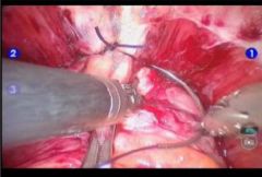
How do you hold the needle with the robot when preparing to throw the DVC stitch?
|

|
|
|
When dropping the bladder and entering the retropubic space, how can you tell if you are too close to the bladder?
|
*Dr. Pruthi was stating that when you start to see more bleeding and don't get into that layer of thing flimsy stuff then you are probably too close to the bladder.
|
|
|
When entering the endopelvic fascia lateral to the prostate where do we tend to enter and why?
|
*Towards the base but somewhere around the middle of the prostate. When you put good traction and counter traction on the prostate you really can see where the endopelvic fascia and the levators come together. We buzz into the endopelvic fasica instead of just cold cutting into the layer.
*Once in the layer we put one tine of the scissors in and cut towards the base and then towards the apex buzz superficially and then mobilize the space between curving towards the puboprostatics. |
|
|
What is chromic suture?
|
Absorbable biological suture material. Chromic is an absorbable suture made by twisting together strands of purified collagen taken from bovine intestines. Due to undergoing a ribbon stage chromicisation (treatment with chromic acid salts), the chromic offers roughly twice the stitch-holding time of plain catgut. The natural chromic thread is precision ground in order to achieve a monofilament character and treated with a glycerol containing solution. Chromic is absorbed by enzymatic degradation. Note – catgut is no longer used in the UK for human surgery.
Proteolytic enzymatic digestion complete in 70 days. Absorption by enzymatic digestion and starts losing tensile strength on implantation from 18–21 days of catgut chromic |
|
|
What type of suture would you use for an orchiopexy on a torsion patient?
|
A fine nonreactive nonabsorbable suture such as an ethibond or a prolene, like 4-0.
|
|
|
How much bupivicaine can you give a patient?
|
The toxic dose is at 2.5mg/kg. But an easy way to get really close is that bupivicaine starts with a B, and that is the second letter of the alphabet.
|
|
|
What is a pringle maneuver?
|
In 1908, Pringle first described a technique to minimize blood loss during hepatic surgery by clamping the vascular pedicle.[1] The inflow of blood to the liver is via the hepatic artery and portal vein. Surgeons must be able to isolate and control these sources of blood flow not only to control bleeding in traumatic injuries to the liver, but also in elective hepatic resections. Clamping of the hepatic pedicle, also known as the Pringle maneuver, allows surgeons to evaluate traumatic liver injury. It does so by allowing one to determine if the hemorrhage is from branches of the hepatic artery or portal vein. When a clamp is applied to the pedicle, hemorrhage ceases if from either of these sources. If hemorrhage continues then the other likely sources of bleeding include the retrohepatic vena cava or hepatic veins.
|

