Reading...
![]()
Play button
![]()
Play button
![]()
Use LEFT and RIGHT arrow keys to navigate between flashcards;
Use UP and DOWN arrow keys to flip the card;
H to show hint;
A reads text to speech;
70 Cards in this Set
- Front
- Back
|
solitary fibrous tumor definition...
a____of_____, occuring mostly in ____. |
benign proliferation of spindle cells, occuring in adults
|
|
|
what is this?
a benign proliferation of spindle cells, occuring in adults |
solitary fibrous tumor
|
|
|
Histological description of a solitary fibrous tumor...
|
haphazard arrangement of spindle cells
positive for cd34 |
|
|
haphazard arrangment of spindle cells, that is positive for CD34 is the histological description of...
|
solitary fibrous tumor
|
|
|
are solitary fibrous tumors benign?
|
yes, but 10-15 are agressive
|
|
|
SOsoft tissue myxoma occurs oten on the ____
|
palate
|
|
|
this benign neoplasm has NO age/sex predilection, is gelatinous material with myxoid appearence
|
soft tissue myxoma
|
|
|
What does this describe - non encapsulated, may show infiltration, stelatespindle cells in a myxoid stroma
|
soft tissue myxoma
|
|
|
soft tissue myxoma histology
peripheral is - shape of cells - cells arrange in - |
non-encapsulated/infiltrating
stellate spindle cells in myxoid sroma |
|
|
nasopharyngeal angiofibroma occurs in _____, and is 4 things to decribe
|
10-20 yr old boys
benign slow growing invasive unencapsulated |
|
|
what is this?
10-20 yr old boys benign slow growing invasive unencapsulated |
nasopharyngeal angiofibroma
|
|
|
symptom triad of epistaxis, nasal obstruction, mass effect in Nasopharyngeal is characterstic of---
|
nasopharyngeal angiofibroma
|
|
|
what type of neoplasm is often seen as a palatal expansion, which is blue
|
nasopharyngeal angiofibroma
|
|
|
what histology is this?
well collagenized cleft like vascular channels |
nasopharyngeal angiofibroma
|
|
|
describe nasopharyngeal angiofibroma hisologically
|
well collagenized
cleft-like vascular channels |
|
|
WHen treating nasopharyngeal angiofibroma, treat with....and is there a risk of recurrence?
|
surgical excision
recurrence high as 50%, due to incomplete surgery, difficult location or aggresive nature |
|
|
what is this?
after treating with surgery, this neoplasm has a recurrence rate high as 50% due to incomplete removal, difficult location or aggresive nature |
nasopharyngeal angiofibroma
|
|
|
Giant cell angiofibroma was first described in the ____, and has since been described in 3 places
|
orbit
oral cavity submandibular area posterior mediastinum |
|
|
giant cell angiofibroma
is it rare or common is it benign or malignant what is it covered by metastasize or no |
rare
benign covered by mucosa no metastasize |
|
|
this neoplasm was first described in the orbit, and is also since been found in the oral cavity, the submandibular area, or the posterior mediastinum.
|
giant cell fibroma
|
|
|
hisology
paternless growth of cells, that range from round to spindle, arrange in a stroma of collagen or myxoid. Has pseudovascular spaces. |
giant cell fibroma
|
|
|
histology of giant cell fibroma
|
paternless growth of round to spindle cells
arranged in stroma of collagen to myxoid pseudovascular spaces |
|
|
where are giant cells in a giant cell fibroma
|
lining pseudovascular spaces, interspersed among spindle cells
|
|
|
Spindle giant cells of giant cell fibroma identification
|
positive for cd34
positive for vimentin |
|
|
what cells are these?
postiive for cd34 positive for vimentin |
giant cell fibroma
|
|
|
what type of lesion occurs deep in the dermis or submucosa, with rapid growth?
|
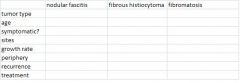
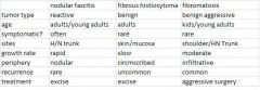
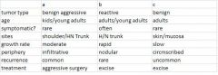
nodular fascitis
|
|
|
nodular fascitis occurs in teh _____ or ____ and grows____. It occurs in (m/f) and agewise____
|
dermis
submucosa rapid growth both m/f young adults/adults |
|
|
the most common location of nodular fascitis is...
|
trunk/extremities
|
|
|
when nodular fascitis doesn't occur in its most common lcoation, it occurs in ____, specifically the ____about 10% of the time. Or if it occurs in te _____, its in the _____
|
the head/neck, specifically the face or parotid sheath
oral cavity - buccal mucosa |
|
|
This neoplasm occurs in the trunk/extremities, or 10% of the time in head/neck, specifically the face/parotid sheath or the buccal mucosa
|
nodular fascitis
|
|
|
for the histology of nodular fascitis, where do the MN giant cells come from?
|
neadby muscle or from fusion with macrophages
|
|
|
what does this describe histologically
-mitotic firgures present that are normal - MN giant cells RBCs present inflammatory cells present |
nodular fascitis
|
|
|
name 4 things you would find in the histology of nodular fascitis
|
Normal mitotic figures
mN giant cells RBCs inflamm cells |
|
|
nodular fascitis immunochemistry that make it identifiable in a DD?
|
smooth muscle actin
NO desmin |
|
|
|
|
|
a = fibromatosis
b - nodular fascitis c - fibrous histiocytoma |
|
|
2 types of myofibroblastic tumors, what is thjeir main difference?
|
myofibromatosis - in infants, multifocal
myofibroma - wide age range, solitary |
|
|
what type of benign neoplasm occurs in infants, and may be fatal?
|
myofibromatosis
|
|
|
myofibroblastic tumors (a,b) tend to occur where?
|
myofibromatosis and myofibroma tend to occur in head/neck
|
|
|
what is this the histology of...
"pushing", well demarcated borders, with a hemagioperycotoma appearance |
myofibromatosis or myofibroma (myofibroblastic tumors)
|
|
|
what is a "hemangioperycytoma appearance" and where can it be seen?
|
= alternating zones of Cellular, and pauicellular(sparce cells) in a hyaline or collagen stroma
occurs in myofibroblastic tumors |
|
|
myofibroblastic tumors are positive for what, and negative for what?
|
SMA +
s100, cd34, desmin negative |
|
|
how can a myofibroblastic tumor be differentiated from leiomyoma and leiomyosarcoma?
|
it is desmin negative
|
|
|
what type of neoplasm is this...
has 2 ypes, superficial and deep(desmoid) and can be part of familia adematous polyposis(FAP) |
fibromatosis
|
|
|
What type of neoplasm is seen in multiple numbers in Familial adenomatousis polyposis?
|
fibromatosis
|
|
|
3 types of deep fibromatoses, what are they? frequency?
|
extra-abdominal - 60%
abdominal wall - 25% intrabdominal - 15% |
|
|
fibromatosis occur more in m/f, what age? where? symptoms? recurrence? borders?
|
F 2:1 M
kids/young adults shoulder/trunk recurrence rate significant (20-60%) infiltrative borders asymptomatic |
|
|
what's this
F 2:1 M kids/young adults shoulder/trunk recurrence rate significant (20-60%) infiltrative borders asymptomatic |
fibromatosis
|
|
|
extra-abdominal - 60%
abdominal wall - 25% intrabdominal - 15% what are these examples of? |
deep fibromatosis
|
|
|
multiple fibromatosis lesions are seen in what disorder?
|
familial adenomatosis polyposis
|
|
|
Histo of fibromatosis
borders overall look immunochem (3 things) |
unencapsulated - infiltrative
Bland look, of fibroblasts and collagen neg for s100, desmin, actin |
|
|
unencapsulated - infiltrative
Bland look, of fibroblasts and collagen neg for s100, desmin, actin |
fibromatosis
|
|
|
What neoplasm has a HERRINGBONE/interlacing pattern?
|
fibrosarcoma
|
|
|
fibrosarcoma histology pattern
|
herringbone
|
|
|
histo appearence of fibrosarcoma
overall ct markers type of cell borders immunochem (5 things) |
overall - herringbone appearance
no ct markers fibroblasts malignant - spindle cells ill defined borders no s100, actin, desmin, keratin, epith membrane antigen |
|
|
survival rate 5 yrs for fibrosarcoma?
|
30-50%
or 55-70% |
|
|
overall - herringbone appearance
no ct markers fibroblasts malignant - spindle cells ill defined borders no s100, actin, desmin, keratin, epith membrane antigen what is this? |
fibrosarcoma
|
|
|
fibrohistiocytic tumor has 2 types...
|
benign fibrous histiocytoma
malignant fibrous hjistiocytoma |
|
|
benign fibrous histiocytoma occurs in ____(patients) often seen in (2 words), not usually seen in (1 thing).
|
40 y/o adults
trunk/extremities not usually seen in oral |
|
|
40 y/o adults
trunk/extremities not usually seen in oral what's this? |
benign fibrous histiocytoma
|
|
|
histo - well demarcated
has storiform of cartwheel/mat-like appearance of fibroblasts infreq/normal mitotic figures, no cell atypia, tumor giant cells |
benign fibrous histocytoma
|
|
|
benign fibrous histiocytoma histology
borders? overall appearance? mitotic figures? cell atypia? other cell? |
well demarcated borders
storiform/carwheel/mat-like apperance of fibroblasts infrequent, but normal mitotic figures no cell atypia may see tumor giant cells |
|
|
malignant fibrous histiocytoma has 5 subtypes, which is the prototype?
|
pleomorphic storiform
|
|
|
pleomorphic storiform
myxoid giant cell inflammatory angiomatoid 5 subtypes of what? |
malignant fibrous histiocytoma
|
|
|
what is the msot common soft tissue sarcoma diagnosis, where does it occur most often?
|
malignant fibrous histiocytoma
leg |
|
|
radio appearnce of a malignant fibrous histiocytoma?
|
lucent, ill defined borders
|
|
|
histo
pleomorphic spindle/fibroblast morphology abnormal mitotic figures present necrosis, cell atypia seen |
malignant fibrous histiocytoma
|
|
|
malignant fibrous histiocytoma histology
cellular arrangemment? mitotic figures? cells? |
pleomorphic arrangement of spindle/fibroblasts morphology
abnormal mit figures present cells atypia, necrosis |
|
|
tx of malignant fibrous histiocytoma
survival 5 yr recur % |
wide surgery, radio/chemo
20-60% survival 40% recur |
|
|
tx = wide surgery, radio/chemo
20-60% survival 5 yr 40% recur whats this? |
malignant fibrous histiocytoma
|