Reading...
![]()
Play button
![]()
Play button
![]()
Use LEFT and RIGHT arrow keys to navigate between flashcards;
Use UP and DOWN arrow keys to flip the card;
H to show hint;
A reads text to speech;
57 Cards in this Set
- Front
- Back
|
Question:
What is the definition of joints? |
Answer:
The regions of skeleton where bones articulate, sometimes allows movement between bones and facilitate growth of bones |
|
|
Question:
What is the definition of a synarthrosis joint? |
Answer:
A type of joint characterized by limited or no movement between opposing bones |
|
|
Question:
What are the 3 types of synarthrosis joints? |
Answer:
1. Synostosis 2. Synchondrosis 3. Syndesmosis |
|
|
Question:
What is a synostosis joint? |
Answer:
- bones joined with bone - does not allow movement to take place - stablizes parts of skeleton after cessation of bone |
|
|
Question:
What is an example of a synostosis joint? |
Answer:
Cranial bones |
|
|
Question:
What is a synchondrosis joint? |
Answer:
- bones connected by hyaline cartilage - allow little movement - pertmits bone growth and expansion by endochondral ossification |
|
|
Question:
What are 2 examples of a synchondrosis joint? |
Answer:
1. epiphyseal plates 2. union of first rib at sternum |
|
|
Question:
What is a syndesmosis joint? |
Answer:
- bones that are far apart united by a ligament composed of dense CT - allows limited movement - functions as shock absorbers and sites of appositional bone growth |
|
|
Question:
What is an example of syndesmosis joints? |
Answer:
The union between the distal tibia and fibula |
|
|
Question:
What is an amphiarthrosis joint? |
Answer:
- a type of joint found where fibrocartilage is the interosseous medium - capable of slight movement |
|
|
Question:
What are 2 examples of amphiarthrosis joints? |
Answer:
1. intervertebral joints 2. pubic symphysis |
|
|
Question:
What is a diarthrosis joint (synovial joint)? |
Answer:
- a type of joint usually found between long bones - permits wide range of movements |
|
|
Question:
The diarthroidal joint is organized into 3 articular structures. What are they? |
Answer:
1. Articular capsule 2. Articular cavity 3. Articular cartilage |
|
|
Question:
The bones of a diarthroidal joint are linked by ligaments and an outer capsule that are composed of 2 layers. What are they? |
Answer:
1. Fibrous Layer 2. Synovial membrane (synovium) |
|
|
Question:
Describe the Fibrous Layer of a diarthroidal joint. |
Answer:
- an outer layer made of dense CT continuous with periosteum - contains blood vessels and nerves |
|
|
Question:
Describe the synovial membrane (synovium) of the diarthroidal joint. |
Answer:
- an inner layer lined by 1-3 layers of synovial cells (synovocytes) - function is to produce and resorb synovial fluid and to remove material shed into the articular cavity |
|
|
Question:
Describe the structure of synovial membrane (synovium) of the diarthroidal joint. |
Answer:
- a loose sheet of vascularized and innervated CT facing the articular cavity and arranged in fold and villi - does not line the articular cartilage |
|
|
Question:
Describe the cells type of the synovial membrane (synovium) of the diarthroidal joint. |
Answer:
1. Type A (macrophage-like cells) - light staining phagocytic cells 2. Type B (fibroblast-like) cells - dark staining cells that secrete hyaluronic acid and glycoprotein lubricant in the synovial fluid |
|
|
Question:
What are the bones of a diarthroidal joint surfaced with? |
Answer:
articular cartilage |
|
|
Question:
The articular cartilage of the diarthroidal joint is what type of cartilage? |
Answer:
hyaline cartilage |
|
|
Question:
The articular cartilage of the diarthroidal joint is characterized in part by the absence of what? |
Answer:
Perichondrium |
|
|
Question:
Describe how the collagen fibers of the diarthroidal joint are arranged. |
Answer:
- the collagen fibers of the articular cartilage form wide arches - the collagen fibers are perpendicular to the surface of the bone - they are parallel to the articular surface when near the articular surface |
|
|
Question:
Desrcibe what is stacked near the osseous surface of the articular cartilage of the diarthroidal joint. |
Answer:
chondrocytes - found near the articular surface and are solitary and flat in shape |
|
|
Question:
What makes the articular cartilage of the diarthroidal joint so resilient? |
Answer:
The cartilage is an efficient absorber of mechanical pressures |
|
|
Question:
What is the articular cartilage of the diarthroidal joint consist of? |
Answer:
- type II collagen - proteoglycans - 8o% water |
|
|
Question:
What do the proteoglycans of the articular cartilage of the diarthroidal joint contain and what does this enable them to do? |
Answer:
- these molecules contain large amounts of water, enabling them to function as a biomechanical spring |
|
|
Question:
Describe how the proteoglycan molecules of the articular cartilage works as a spring. |
Answer:
- when pressure is applied, water is forced out of the matrix into the synovial fluid - reciprocal elctrostatic repulsion of negatively charged carboxyl and sulfate groups in the GAG molecule occurs - this separates the GAG branches and creates spaces to be occupied by water - when pressure is released, water is attracted back into these spaces. |
|
|
Question:
Water movements brought about by use of the diarthroidal joint is responsible for what? |
Answer:
This is responsible for the resiliency of the cartilage and for its nutrition |
|
|
Question:
What are the 3 zones in the articular cartilage? |
Answer:
1. superficial zone 2. middle (transitional) zone 3. deep zone |
|
|
Question:
Describe the superficial zone of the articular cartilage. |
Answer:
- chondrocytes are small and flattened - collagen fibers arranged parallel to the surface |
|
|
Question:
Describe the middle (transitional) zone of the articular cartilage. |
Answer:
- chondrocytes arranged in columns - collagen fibers begin to change their orientation from horizontal to vertical |
|
|
Question:
Describe the deep zone of the articular cartilage. |
Answer:
- contains calcified cartilage with small chondrocytes - located adjacent to the subchondral bone - collagen fibers run perpindicular to the surface |
|
|
Question:
Describe the intra-articular menisci (discs). |
Answer:
- cushions of fibrocartilage or dense CT - may be free or transverse the joint |
|
|
Question:
Describe the 2 classifications of the articular cartilage. |
Answer:
- simple: two articulating surfaces (ex. knee joint) - compound: three or more articulating surfaces (ex. TMJ) |
|
|
Question:
The TMJ is a type of what kind of joint? |
Answer:
This is a type of gliding-hinge synovial joint found in mammals |
|
|
Question:
Describe the articulation of the TMJ |
Answer:
This is the articulation of the mandibular condyle with the temporal bone, by way of the glenoid fossa |
|
|
Question:
Describe the mandibular condyles of the TMJ. |
Answer:
These are ovoid structures with long axes oriented medially |
|
|
Question:
Identify the 5 layers of the TMJ. |
Answer:
The layers of this structure are 1. dense fibrous CT 2. proliferative zone 3. fibrocartilage (hyaline during growth period) 4. calcified cartilage 5. sub-articular bone |
|
|
Question:
Describe the proliferative zone of the TMJ. |
Answer:
this zone consists of a layer of cells acting as a replicating pool for underlying cartilage |
|
|
Question:
Describe the layers of the articular eminence of the TMJ from superficial to deep. |
Answer:
1. dense CT (fibrous layer) 2. proliferative layer 3. fibrocartilage 4. bone |
|
|
Question:
What is the glenoid fossa? |
Answer:
- This is the concave depression in the squamous portion of the temporal bone. - Covered by thin film fibrous layer (periosteum) |
|
|
Question:
What is the capsule of the TMJ? |
Answer:
This is the fibrous membrane that surrounds the joint and incorporates the articular eminence. |
|
|
Question:
What is the capsule of the TMJ composed of? |
Answer:
This is composed of dense fibrous CT. |
|
|
Question:
Describe the attachments of the TMJ capsule. |
Answer:
1. the upper half forms a loose envelope with a posterior attachment at the squamotympatic fissure 2. an anterior attachment at the articular eminence 3. a lateral attachment at the glenoid fossa 4. attached medially, laterally, and posteriorly at the articular disk 5. firmly attached to the neck of the mandibular condyle |
|
|
Question:
What is the function of the articular disk of the TMJ? |
Answer:
This structure functions as an articular surface and divides the TMJ into 2 distinct compartments. |
|
|
Question:
Describe the composition, innervation, and vascularization of the TMJ articular disk. |
Answer:
This structure is: 1. composed of fibrous connective tissue 2. it is avascular and aneural centrally 3. it is vascular and innervated at its periphery |
|
|
Question:
What can be said about the attachment of the bioconcavity structure in the TMJ articular disk? |
Answer:
This structure is attached to the medial and lateral poles of the condyle. |
|
|
Question:
Describe the upper lamella of the TMJ anterior articular disk. |
Answer:
This region runs forward and fuses with the capsule and periosteum of the anterio slope of the articular eminence. |
|
|
Question:
Describe the lower lamella of the TMJ anterior articular disk. |
Answer:
This region is attached to the anterior surface of the condylar neck. |
|
|
Question:
What inserts between the lamellae of the TMJ articular disk? |
Answer:
The superior head of the lateral ptyerygoid is found here. |
|
|
Question:
Describe the upper lamella of the TMJ posterior articular disk. |
Answer:
This region is composed of fibrous and elastic CT and inserts into the squamotympatic fissure (as part of the capsule) |
|
|
Question:
Describe the lower lamella of the TMJ posterior articular disk. |
Answer:
This region merges with the periosteum of the condylar neck |
|
|
Question:
What 2 things are found in the zone between the posterior lamallae? |
Answer:
1. retrodiscal tissue 2. comprised of loose vascular CT |
|
|
Question:
Name and describe the lateral portion of the TMJ capsule. |
Answer:
This portion is thickened and fanshaped and is known as the temporomandibular ligament. |
|
|
Question:
Describe how the temporomadibular ligament is placed within the TMJ. |
Answer:
This structure runs in an oblique course posteriorly and inferiorly from the later aspect of the articular eminence to the posterior part of the condylar neck. |
|
|
Question:
Identify the muscle of the TMJ. |
Answer:
The muscle of this structure is called the lateral pterygoid. |
|
|
A - mandibular condyle
B - glenoid fossa D/C - articular disc E - articular eminence H - region of squamotympanic fissure *** There's more! Check your notes! *** |
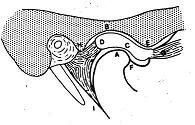
Image Question:
Can you name all the structures in the TMJ? |