![]()
![]()
![]()
Use LEFT and RIGHT arrow keys to navigate between flashcards;
Use UP and DOWN arrow keys to flip the card;
H to show hint;
A reads text to speech;
367 Cards in this Set
- Front
- Back
|
frequency |
the interval from a given point on one sound wave to the equivalent point on the next sound wave
inverse of wavelengthcy |
|
|
hertz |
cycles per second |
|
|
azumith |
horizontal plane |
|
|
elevation |
vertical plane |
|
|
receptive field |
the region of a sensory surface that when stimulated changes the membrane potential of a neuron |
|
|
tonotopic map |
the organization of the auditory strucutre based on characteristic of frequency |
|
|
hair cell |
auditory cell that transduces sound into change in membrane potential can be a vestibular cell that transduces head movements into a changei n membrane potential |
|
|
mechanoreceptor |
any sensory receptor selective or mechanical stimuli, hair cells in inner ear, receptors on skin etc.
sensitive to pressure or distortion of membrane |
|
|
pinna |
funnel shaped outer ear consisting of cartileage covered by skin |
|
|
organ of corti |
an auditory receptor organ that contains hair cells, rods of Corti and supporting cells |
|
|
dorsal and ventral cochlear nucleus |
a nucleus in the medulla that receives afferents from the spiral ganglion in the cochlea of the inner ear |
|
|
phototransduction |
process by which light is converted into electrical signals in photoreceptor cells |
|
|
cochlea |
spiral bony structure in the inner ear that contains hair cells that transduce sound |
|
|
inferior colliculus |
a nucleus in the midbrain from which all ascending auditory signals project to the medial geniculate nucleus |
|
|
basilar membrane |
a membrane separating the scala tympani and scala media in the cochlea in the inner ear |
|
|
rod |
a photoreceptor in the retina containing rhodospin and specialized for low light levens |
|
|
cone |
a photoreceptor in the retina containing one of three photopigments that are sensitive to different wavelengths of light
concentrated in the fovea and specialized for daytime vision
responsible for color
|
|
|
spiral ganglion |
a collection of neurons in the modiolus of the cochlea that receives input from hair cells and sends output to the cochlear nuclei in the medulla via the auditory nerve
fires action potentials |
|
|
middle ear |
tympanic membrane plus the ossicles |
|
|
hindbrain |
region of brain derived from the caudal primary embryonic brain vesicle
structures are cerebellum, pons and medulla |
|
|
diencephalon |
a region of the brain stem derived from prosencephalon(forebrain). Diencephalic structures are thalamus and hypothalamus |
|
|
lemniscal pathway |
d |
|
|
topographic map |
d |
|
|
motor neuron |
a neuron that synapses on a muscle cell and causes muscle contraction |
|
|
medulla |
the part of the hindbrain caudal to the pons and cerebellum |
|
|
thalamus |
dorsal part of the diencephalon in forebrain highly interconnected with cerebral neocortex |
|
|
spinothalamic pathway |
an ascending somatic sensory pathway traveling from spinal cord to thalamus via lateral spinothalamic columns, mediates information about pain temp and some forms of touch |
|
|
dorsal root ganglio |
a collection of cell bodies the sensory neurons that are part of the somatic PNS. There is one dorsal root ganglion for each spinal nerve |
|
|
grey matter |
a generic term for a collection of neurnal cell bodies in the CNS. grey because it is unmyelinated. |
|
|
pons |
the part of the rostral hindbrain that lies ventral to the cerebellum and fourth ventricle |
|
|
ventricles |
CSF filled spaces inside the brain lateral, third, cerebral aqueduct and fourth ventricle |
|
|
dorsal root |
a bundle of sensory neuron axons that emerges from a spinal nerve and attaches to the dorsal side of the spinal cord. Dorsal root axons bring information to spinal cord |
|
|
white matter |
collection of CNS axons |
|
|
midbrain |
region of brain derived from the middle primary embryonic brain vesicle. Mesencephalon.
strucutres tectum and tegmentum |
|
|
forebrain |
region of brain derived from the rostral primary embryonic vesicle. Prosencephalon.
Structures telencephalon and diencephalon |
|
|
ventral root |
a bundle of motor neuron axons that emerges from the ventral spinal cord and joins sensory fibers to form a spinal nerve. Carry info away from spinal cord |
|
|
tract |
collection of CNS axons with a common site of origin and destination |
|
|
somatotopic map |
orginization of somatic senosry pathways in which neighboring receptors in the skin feed information to neighboring cells in target strucutre |
|
|
telencephalon |
a region of the brain derived from the prosencephalon. Telencephalic structures are cerebral hemispheres that have cerebral cortex and basal telencephalon |
|
|
roger sperry |
expirement with frogs that cut optic nerve rotated and let ot regrow to optic tectum caused frog to have oppositemovment
must be optic mechanism to make connection |
|
|
Normal connections of retina |
Nasal retina to posterior tectum temporal retina to anterior tectum |
|
|
neuromuscular junction synaptioc competition |
refinement is activity dependent |
|
|
What are the submodalities of the somatosensory system? |
Light touch, joint pain, deep visceral pain, pressure, vibration |
|
|
What is a novel aspect of the submodalities of touch? |
it can be transduced by different pathways |
|
|
proprioception |
the sense of where your joints are |
|
|
what are the types of skin? |
hairy and glabrous which is the surface of the hands and feet where there is no hair but are fingerprints |
|
|
free nerve endings |
reach epidermis no specialization at ends have high threshold sense cuts, tears |
|
|
pacinian corpuscle |
large receptive field elastic, pressure on membrane that is gelatinous will ADAPT to pressure. |
|
|
ruffini ending |
can sense sustained pressure no adapting |
|
|
meissner's corpuscle |
small receptive field |
|
|
merkel disk |
senses vibration very good at adaptive response |
|
|
What varies among the receptors in the somatosensory system? |
different receptors can carry different types of information and have different types of ion channels
receptors vary in strength required for response |
|
|
What types of receptive fields can somatosensory neurons have? |
inhibitory and excitatory |
|
|
What are the three key findings about receptive fields? |
1. different neurons have different receptive fields LOCATIONS 2. different neurons have different receptive field SIZES 3. different neurons require different stimulus intensities to cause action potentials |
|
|
transient receptor potential |
TRP resonds to deflection of cell membrane responds to chemical and temperature stimuli ion channels that respond to stimuli listed above |
|
|
What is the relationship between neighboring neurons and receptive fields? |
they will have neighboring areas in the nervous system |
|
|
What are the four segments of the dermatomes? |
cervical, thoracic, lumbar and sacral |
|
|
What is a dermatome? |
area of skin innervated by a spinal segment |
|
|
how many dermatomes are there? |
33 |
|
|
What is the best way to survey the plan of the nervous system? |
from the perspective of the body as a quadriped |
|
|
How many pathways from the skin to the brain? |
Two main |
|
|
Dorsal column lemiscal pathway |
axons rise in region called dorsal column and make way to medial lemiscus in brain
dorsal root axons make first synapse in dorsal column nuceli, then axons cross the midline in the medullas and rise through the medial lemniscus. The next synapse is in the thalamus where their projections are then sent to the primary somatocortex |
|
|
What is the job of the thalamus? |
Almost all sensory information sent through thalamus to reach cortices |
|
|
What will injury to the dorsal column in spinal cord cause? |
Numbness on the ipsilateral side |
|
|
Which neurons are the longest? |
lemniscal primary affrents |
|
|
What is the pattern of white/gray matter in the spinal cord? |
From caudal to rostral there is more white matter as you approach the cervial region because all information has to be passed through the region to reach the brain |
|
|
What is the pattern noticed in the cortex/body plan? |
there is systematic change as you move across the cortex |
|
|
What is the relationship between the use of a sense and the representation in the cortex? |
The more the sense is used the larger the representation in the cortex.
Semi-plastic especially when limbs are removed |
|
|
How are sensory fibers distinguised? |
diameter and velocity of signal |
|
|
delta A fiber |
can take dorsal/lemiscus pathway myelinated, conduct quickly used for rapid withdrawl movements |
|
|
c fibers |
unmyelinated, slow, small diameter secondary pain |
|
|
What pathway dose the C fiber take? |
spinothalamic pathway
|
|
|
c fiber pathway |
cell body is still located in dorsal root ganglion but axon synapses in dorsal horn of spinal cord. Post synaptic neurons send axons across midline and up white matter all the way up to the brain. |
|
|
What does damage to the spinothalamic pathway affect? |
temperature, pain not light touch |
|
|
spinothalamic pathway |
first synape ipsilateral to receptive field, dessicates at midline in spinal cord and moves up tract, passes medulla and synapses on thalamus, information is processed in thalamus and then sent to primary somatosensory cortex |
|
|
What type of receptive field does the spinothalamic path have? |
contralateral |
|
|
What are the differences in touch pathways? |
medial lemniscal pathway dessicates at medulla where spinothalamic does in lower spinal cord near dorsal horn.
medial lemniscal pathway synapses in medulla and then onto thalamus where spinothalamic synapses onto thalamus |
|
|
How does the cortical map change? |
We dont know, we just noticed a relationship between use and representation` |
|
|
What side of the brain sees the left visual world? |
The right |
|
|
Where do laminae project?
|
laminae project into seperate zones of visual cortex interdigitating areas one eye or the either |
|
|
Where is visual information converged ? |
in the layers before the IV cortex, information converges |
|
|
What do visual receptive fields require? |
constrast withedges and partciular orientation |
|
|
What is unique about some of the cells in the visual system? |
concentric cells can synapse onto one cell establishing a type of cell heirarchy so a ray of light that is orientated over all three cells causes greatest rate of fire |
|
|
What is the relationship between the spatial arrangement of cortex and light orientation? |
preferred light orientation changes systematically as you move across layer of cortex, maps across the width of the cortex |
|
|
hypercolumn |
chunk of cortex where receptive field properties go through a cycle, vertical to vertical and alternating eyes |
|
|
What do hypercolumns cause? |
The ability to process visual world in any orientation or field, units that could process each part of the world
areas are continous |
|
|
How much of the cortex of a primate is devoted to processing visual information? |
50% |
|
|
Where is most visual information sent in the cortex? |
V1 and then is distributed to other areas |
|
|
What is V4 responsble for? MST? |
shapes, color motion information |
|
|
What are the similarites between visual and somato world? |
multiple submodalities cortical maps that are systematic of the contralateral world feature detection |
|
|
cochlea |
organ of corti fluid filled, coiled scala tympani, scala media, scala vestibuli basilar membrane tectorial membrane
|
|
|
Why is it considered more difficult to be deaf than blind? |
Social interaction |
|
|
external ear structures |
ear canal tympanic membrane-where external ear and middle ear meet pinna |
|
|
pinna |
collects sound waves |
|
|
hair cells |
transduce sound into electrical signals |
|
|
sound |
waves of pressure that eminate from sound source |
|
|
amplitude |
determines intensity of sound |
|
|
What happens with an ear infection? |
bacterial infection cause fluid to fill air space and prevents ossicles from vibrating |
|
|
what drives the movement of the ossicles? |
tympanic membrane sounds counds vibrations in tympanic membrane |
|
|
What drives the movement of the oval window? |
stapes window has constant volume |
|
|
what causes the basal membrane to vibrate? |
pressure on the oval window |
|
|
basilar membrane |
tapered runner band, narrow at base of cochlea and wider at apex |
|
|
What regions of the basilar membrane vibrate to which frequencies? |
lower frequency at more flexible base region higher frequency at less flexible apex region |
|
|
organ of corti |
specilized sensory cells that have hair cells embedded in them
has inner and outer hair cells. |
|
|
tectorial membrane |
move up and down with the traveling wave, the hinge mechanism causes the tectorial membrane to move laterally over the hair cells. This lateral shearing motion bends the cilia atop the hair cells, pulls on the fine tip links, and opens the trap-door channels |
|
|
What is a special ability of the basilar membrane? |
Can segregate different sound frequencies simultaneously |
|
|
Hair cells |
have stereocilia imbedded in tectorial membrane three sets of outer hair cells and one set of inner hair cell
|
|
|
which hair cells allow you to hear? |
do not regenerate the 1 row of inner hair cell 3500 |
|
|
what protein forms links in stereocilia? |
cadherin 23 |
|
|
what role does cadherin 23 play? |
allows tension change in stereocilia and opens ion channels that are crucial for transduction process |
|
|
What will light in surround cause in an ON cell? |
inhibition of firing rate |
|
|
What do we know about how signals lead to our concious perception of contrast? |
nothing |
|
|
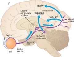
What are the four targets of the retinal ganglion axons and what is the function of each? |
superior colliculus-orienting movements pretectum-pupillary light reflex suprachiasmatic nucelus-sets circadian rythms lateral geniculate nucelus-concious vision |
|
|
where is the lateral geniculate nucleus and what does it look like? |
a nucleus within the thalamus that is responsible for concious vision that processes information of contralateral side of vision 6 layers look like bent knees |
|
|
superior colliculus |
very fast transmittance important for generating orientation movements located in midbrain |
|
|
What does the superior colliculis contol? |
whole movements of body head movements in body eye movements in head |
|
|
pretectum |
controls pupillary light reflex rostral to superior colliculus |
|
|
suprachiasmatic nucleus |
sets phase of circadian rythms according to day light cycle, jet lag etc located in hypothalamus, ventral portion of diencephalon |
|
|
Where do visual ganglion axons synapse? |
lateral geniculate nucleus and then send axons up to visual cortex |
|
|
How many lateral geniculate nuceli are there? |
2 |
|
|
retinotopic organization |
neighboring cells have similar receptive fields, l receptive field locations move systematically through space in the cortex |
|
|
where are optic nerves located? |
straight back from the lens at the optic disk |
|
|
Where do axons that pass through optic chiasm synapse? |
some stay ipsilateral and some diverge contralateral but all pass through white matter on way to brain |
|
|
Where does the left portion of visual field appear in our perception? |
right portion of each eye axons coming from nasal of right eye and left eye converge onto optic tract |
|
|
how do tracts find their correct position? |
chemical signals |
|
|
How are the four target regions positioned? |
close to each other |
|
|
What are the differences in the geniculate layers? |
Layers 1,4,6 receive information from contralateral eye and 2, 3, 5 recive from ipsilateral eye |
|
|
geniculate nucleus |
sheet of tissue that has a map of contralateral visual hemifield.
If located in left side of the brain it has a map of right visual hemifield |
|
|
Where does the LGN send axons? |
Primary visual cortex in the occiptal lobe |
|
|
Names for primary visual cortex |
Area 17 of Brodmann V1 Striate cortex-stria of gennari
|
|
|
Primary visual cortex structure |
layered and each layer has differnt functions heavily myelinated layer of axons |
|
|
V1 |
2D retinotopic systematic orginization of receptive field position representation of fovea is magnified similar to somatosensory cortex |
|
|
What expirement led to the discovery of alternating layers in the eyes? |
inject radioactive labeled prolene was transported to LGN in particular layers depending on whcih eye was injected. Showed pattern of activity in cortex. Looked like zebra |
|
|
What higher level function is the merging of information crucial for? |
depth perception |
|
|
What are the common themes through sensory systems? |
submodalities, receptive fields and patterns of activity across brain |
|
|
structures of the eye |
cornea, iris, pupil, lens, viterous humor, blood vessels, retina(extension of nervous system) |
|
|
what layers of the retina must light pass through to reach photoreceptors? |
all layers |
|
|
fovea |
specialization of retina, tissue is thinned where we have our highest resolution higher density of photoreceptors all cones
|
|
|
What is the function of the specilization of the fovea? |
Less tissue allows for less degredation of image if all tissue were thinned the image would be too degraded and delayed |
|
|
lens |
projects and focuses image of outside world into retina |
|
|
What are the layers of the retina? |
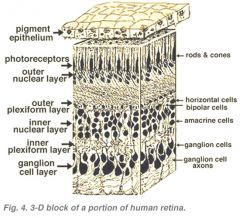
ganglion, inner plexiform, inner nuclear, outer plexiform, outer nuclear, photoreceptor |
|
|
Which layer contains the photoreceptors? |
outer nuclear layer |
|
|
What are the types of neuronal retina cells? |
photoreceptors horizontal cells bipolar amacrine ganglion |
|
|
What order is information sent from photoreceptor to retinal surface? |
photoreceptor to bipolar cell to ganglion cell which sends axons to back of eye |
|
|
What are the classes of photoreceptors |
rods and cones |
|
|
rods |
surrounded by plasma membrane have membrane disks stacked next to each other specialized for darkness and low light
|
|
|
What purpose does disk stacking in rod serve? |
increases surface area |
|
|
cones |
have disks that are folds of the membrane but they are part of the membrane insensitive to darkness attuned to bright light and color 3 types |
|
|
Cone wavelengths |
blue-short 430 green-530 red-560 |
|
|
optic disk |
ganglion cells send axons through back of eye to the brain and pierce through the retina, no photoreceptors, cant see at all |
|
|
What important process does the optic disk control? |
photo to electrical transduction |
|
|
What is in the cell during the resting state in dark of a rod or cone? |
cGMP in high levels Resting potential -40mV no action potentials sodium channels open high concentration of neurotransmitter |
|
|
Effect of light absorbtion in rods |
cascade of enzymatic reactions: cGMP drops, closes ion channels which causes cell to hyperpolarize to -70mV Adaptation can occur |
|
|
Properties of disk membrane |
contains proteins opsin in rods and rhodopsin in cone
each cone has different opsin protein |
|
|
What occurs in photoreceptor when photon absorbed? |
protein changes shape whcih causes conformation change and triggers cascade which leads to reduction in cGMP which hyperpolarizes the cell |
|
|
What is the photo effect cascade similar to? |
Metabotrophic effect but has photon instead of ligand binding |
|
|
Light reaction in rod(transduction) |
photon causes opsin to bind to transducin, activates g protein that has GTP which in turn activates phoshodiesterase which degrades cGMP. Gives rise to reduction in cGMP and closing of channels |
|
|
Why does the complex system of absorbing protons thrive? |
Amplification |
|
|
Facts of amplification |
one rhodopsin absorbs one photon 1:1 transducin to phosphodiesterase one enzyme can degrade 10^5 cGMP 250 channels close and blocks 1-^-6 ions per second depolarizes membrane by 1mV |
|
|
What is the direction of the flow of current in a rod? |
inward through outer segment and out through inner segment
ion pumps working continously in dark |
|
|
What keeps cells at resting potential? |
potassium sodium pumps |
|
|
What is a characteristic of visual response to light? |
Its graded small amount of light causes small hyperpolarization |
|
|
What is the sign of inward current? |
negative |
|
|
What happens to wavelengths that are not absorbed? |
They pass through the eye |
|
|
What do nocturnal animals use to increase their sensitivity to light? |
Tempidum reflects light so it allows a second chance to absorb |
|
|
What is the relationship between photoreceptors and bipolar cells? |
Many photoreceptors can syanpse onto one bipolar or horizantal cell |
|
|
What is the receptive field of a photoreceptor? |
a spot the size of the photoreceptor |
|
|
Bipolar receptive field |
center is supplied by small group of receptors, or surround is in relation with indirect pathway and horizontal cells |
|
|
What cells follow bipolar cells? |
ganglion cells |
|
|
Which cells fire action potentials in the visual system? |
Only ganglion cells, respond to contrast between surround and center |
|
|
Off center ganglion |
responds to contrast between center and surround
light in center causes no response but dark in center and light on surround causes response. dark in center only causes greatest response |
|
|
How many types of bipolar and ganglion cells are there? |
two on/off |
|
|
Effect of hyperpolarization of bipolar cell |
reduces neurotransmitter glutamate responds when light is on the center causes ganglion on cells to fire direct |
|
|
OFF cell types |
AMPA recetors that depolarize post synaptic cell. Light in center hyperpolarizes. Indirect. |
|
|
Relationship between bipolar and ganglion cell |
sign is conserved if one depolarizes the other will too |
|
|
dorsal horn activities |
afferent sensory |
|
|
ventral horn activities |
efferent motor |
|
|
midsagittal plane |
directly down the midline |
|
|
parasagittal |
parallel to midsagittal but off midline |
|
|
coronal plane |
through the ears vertical |
|
|
sagittal fissure |
fissure that divides the two hemispheres |
|
|
cerebrum |
largest part of brain four parts: parietal, temporal, frontal and occipital responsible for higher level function forebrain |
|
|
cerebellum |
hindbrain, brainstem two hemispheres motor control |
|
|
brain stem |
unconcious processes breathing, heart rate etc |
|
|
telencephalic vesicles |
most rostral part of the forebrain develop into the cerebral hemispheres
|
|
|
diencephalon structures |
hypothalamus posterior lobe thalamus third ventricle forms in center |
|
|
telencephalon structures |
cerebral cortex hippocampus basal ganglia |
|
|
diencephalon location |
dorsal to the telencephalon in forebrain |
|
|
optic vesciles |
located on either side of the diencephalon form the optic nerve and retinas |
|
|
caudal hindbrain |
develops into medulla, fourth ventricle and medullary pyramids |
|
|
rostral hindbrain |
develops into pons and cerebellum |
|
|
midbrain |
contains tectum, superior and inferior colliculus and central aqueduct |
|
|
superior colliculus function |
vision/eye movement |
|
|
inferior colliculus |
hearing |
|
|
pons |
serves as a relay for the cerebrum and cerebellum |
|
|
forebrain structures |
diencephalon, telencephalon, corpus collosum, thalamus, hypothalamus, cerebral cortex, basal telencephalon, lateral and third ventricles, internal capsule, cortical white matter |
|
|
dura mater |
hard layer for protection |
|
|
arachnoid membrane |
cushions the CNS |
|
|
pia mater |
adheres to surface of the brain encasing it |
|
|
Cerebral Spinal Fluid |
Brain floats in it Moves from choroid plexus in ventricles where it is produced, from caudal to rostral Ventricles are filled with it. |
|
|
choroid plexus |
tissue in all ventricles that produce CSF |
|
|
occipital lobe |
visual |
|
|
parietal lobe |
sensory, association |
|
|
temporal lobe |
hearing high level vision |
|
|
frontal lobe |
motor, planning, high function |
|
|
coupling |
transformation of stimulus energy of external world into a physical change of the sensory dendrite. |
|
|
transduction |
transformation of the physical change of dendrite into a trans-membrane current and voltage change
hair cells taste cell photoreceptors |
|
|
encoding |
transformation of stimulus into a train of action potentials that travel into the brain |
|
|
What is the problem with encoding? |
Max 500Hz min 0Hz |
|
|
Solutions to encoding limitations |
nonlinear encoding range fractionation detection of change in stimulus sensory adaptation rate vs temporal code |
|
|
nonlinear encoding |
can be sigmoid, log etc must most common is sigmoid curce |
|
|
range fractionation |
divide encoding up with multiple cells to cover larger range. If stimulus crosses one to get to the other both fire |
|
|
detection of change in stimulus |
tonic always fires while stimulus is present phasic fires when there is a change in the stimulus phasic tonic is a combination of the two |
|
|
lateral inhibition |
neurons can inhibit the firing of neighboring cells |
|
|
adaptation to stimulus |
neuron responds the same to a stimulus even when it is increased
sensitivity shifts with long exposure |
|
|
rate and temporal code |
rate code affected by frequency temporal code is the timing of hte stimulus to cause neuron to fire ex song |
|
|
nociceptor |
responds to damage to tissue(pain) |
|
|
proprioceptors |
give information about limbs in space |
|
|
What are the five types of taste? |
sweet, salty, umami, sour, acidic |
|
|
How is salt taste transduced |
direct depolarization by |
|
|
How is acidic transduced? |
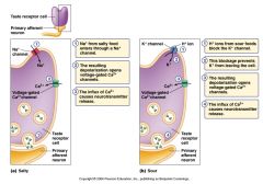
direct depolarization by protons and closes K channels |
|
|
How are umami, bitter and salty transduced? |
|
|
|
Taste receptor classes |
Bitter: T2Rs Sweet: T1R2 + T1R3 Umami: T1R1 + T1R3
|
|
|
What are the pathways for taste to get to brain? |
the tongue, then to left gustatory nucleus in the medulla where they synapse then synapses in ventral medial nucleus in thalamus, then to brainstem, then to thalamus, then to cortex via 3 different cranial nerves X, IX, VII
|
|
|
population coding |
large number of broadly tuned neurons, gathers info and sends to brain where it is processed. |
|
|
Where are smells coupled? |
nasal epithelium |
|
|
olfactory transduction process |
Odorant + R -> Golf(protein) -> AdenylCyclase -> cAMP -> inward cation current -> outward Cl- current -> depolarization
|
|
|
Where does the graded potential occur in olfactory system? |
in the cilia does not fire action potentials |
|
|
where are action potentials fired in the olfactory system? |
olfactory receptor cell |
|
|
properties of receptor cells in olfactory system |
Each receptor cell expresses one receptor gene Each cell has a different response profile to different odorants
|
|
|
Where do olfactory cells project? |
directly into olfactory bulb |
|
|
Where do olfactory receptors send their information? |
Glomeruli, many send to just one golmerulis many receptors with same protein project to same glomerulus |
|
|
Where do olfactory bulbs project? |
Some to thalamus in the medial dorsal nucleus, some straight to olfactory cortex in temporal lobe and some to orbitofrontal cortex |
|
|
metabotrophic receptor |
A metabotropic receptor influences the activity of a cell indirectly by first initiating a metabolic change in the cell. This metabolic change may ultimately affect the opening or closing of an ion channel or may alter some other activity of the cell such as protein transcription. |
|
|
Where is the primary visual cortex located? |
in the occipital lobe in the V1 layer of cortex |
|
|
What side of the brain perceives the left visual field? |
the right hemisphere |
|
|
parallel processing |
same sensory inputs routed through brain in different pathways, to different locations. * information about the same sensory stimulus is used for different purposes |
|
|
What are coupling steps prior to transduction in auditory system? |
Outer ear (pinna) funnels sound pressure waves. Sound waves hit tympanic membrane and passes into middle ear where the malleus is attached to the tympanic membrane and vibrates when the tympanic membrane vibrates the malleus in turn moves the incus and finally the stapes that creates waves in the fluid of the inner ear |
|
|
How does transduction in the ear work and where does it happen? |
Fluid causes cells of specific frequencies on the basilar membrane to vibrate which in turn pulls on the hair cells causing the mechanoreceptors to depolarize and cause action potentials.
Louder cells cause hair cells to more hair cells to move |
|
|
What does the direction of the stereocilia deflection cause? |
towards kinocilium depolarize, away hyperpolarize |
|
|
What are transduction channels in auditory system permeable to when open and what is it that moves to open them? |
cations, tip links toward kinocilium |
|
|
What ions are found in endolymph? |
potassium and calcium that cause neurotransmitter release |
|
|
How many ganglion cells do the inner hair cells connect to? |
Many |
|
|
What type of orginization is found mapped in the cortex? |
frequency based organization each hair cell has specific frequency and each ganglion cell as well as nerve fiber on basilar membrane has specific frequency |
|
|
interaural intensity difference |
sound will be louder on one side of the head or the other
high frequency |
|
|
interaural frequency difference |
low frequency
the timing between the sound and the ears allows us to calculate where sound will be |
|
|
What is phase locking? |
auditory nerve fires action potentials are specific part of sound wave.
used for low frequency sounds, lower than 2kHz
|
|
|
What are delay lines and where do they occur? |
Sound hits one ear sends input into superior olive while a small time later sound hits other ear and input is sent to superior olive. The inputs will reach a neuron in superior olive closer to the opposite side of the initial stimulus. |
|
|
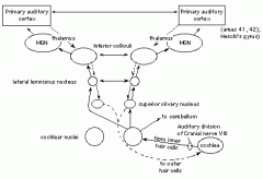
Describe the path of a sound to the brain |
starts in spiral ganglion that has received action potential and sends signals via the auditory nerve that is synapsed in the ventral cochlear nucleus in the hindbrain axons are then sent to the superior olive in the pons which sends information via the lateral lemniscus nerve to the inferior colliculus in the midbrain to the medial geniculate nucleus in the thalamus and finally to the auditory cortex |
|
|
where is the auditory cortex located? |
in the temporal lobe A1 |
|
|
What is the function of the visual ganglion cells? |
fire action potentials that are sent to the forebrain |
|
|
what is the function of visual bipolar cells? |
to directly or indirectly send information from photoreceptors to ganglion cells
communicate with GRADED POTENTIAL |
|
|
what is the function of visual horizontal cells? |
lateral inhibition(inhibit other cells to fire action potentials) usually inhibitory helps in edge detection |
|
|
what is the optic disk and what is its function? |
point at which the image in a visual field
Blind spot because it is so dense with neurons to send information |
|
|
What is the receptive field of a photoreceptor? |
The spot of light in the visual field that corresponds to the visual range of that receptor |
|
|
What is the receptive field of a bipolar cell? |
No action potentials fired just hyper or de polarization the center or surround depending on the type of cell |
|
|
What is the receptive field of a ganglion cell? |
The same as bipolar but responds by firing or not firing action potetnials |
|
|
What is the direct pathway of a light processing |
from photoreceptor to the bipolar cell |
|
|
What is the indirect pathway of light processing |
from photoreceptor to horizontal to bipolar cells |
|
|
What pattern of orgnization of visual information the the visual cortex is seen? |
Alternative layers by eyes in the IV layer of cortex |
|
|
Where and how is information from the two eyes merged? |
In layers past the IVc layer where the representation of the eyes overlap creating binocular vision |
|
|
How are neurons stimulated in the V1 corte |
specific orientation of bars of light |
|
|
WHat is the physical pattern of mapping in the V1 cortex? |
Orientations of light are arranged systematically through cortex |
|
|
What is the function of a cortical hypercolumn? |
Processes specific orientations of light for both eyes in V1 cortex |
|
|
Where is the gustatory nucleus located and what is its function? |
Information from potestrior tongue, anterior tongue and epiglottis is sent to it in the brainstem |
|
|
Where is the ventral posterior medial nucleus found and what is its function? |
In the thalamus, receives gustatory information from the gustatory nucleus in the brainstem and sends to primary gustatory cortex |
|
|
Where is the primary gustatory cortex located? |
Frontal lobe |
|
|
Where do receptor cells of the oflactory system send their axons? |
to oflactory bulb |
|
|
How many genes are expressed in an olfactory cell? |
JUST ONE |
|
|
What are the two main classes of mechanism that give rise to formation of maps in the brain? |
Chemical and activity dependence |
|
|
Visual pathway |
|
|
|
auditory pathway |
|
|
|
What is the function of the vitreous humor? |
Involved in coupling, light passes through after hitting the lens |
|
|
Which ion channels play a role in the rythmic firing of a single neuron? |
Calcium dependent potassium channels |
|
|
What does the topographic map of the visual region represent? |
Magnification of the fovea |
|
|
What is the neurotransmitter released by photoreceptors? |
Glutamate |
|
|
Where is the somatosensory homunculus found? |
parietal lobe |
|
|
Where does information from the left retinal ganglion cell send axons to? |
The right and left lateralgeniculate nucleus |
|
|
What is the stimulus coding scheme in which features are encoded by prescise timing? |
temporal code |
|
|
What will happen to the region of cortex associated with an amputated appendage? |
it will die |
|
|
Why does light touch stimulation occur faster than temperature change? |
larger axonal diameter |
|
|
What sensory modality does not have relay station in thalamus? |
hearing |
|
|
When a sound increases in intensity which change is most likely to occur in hair cells? |
higher amplitude movement of stereocilia |
|
|
evidence of parallel processing |
vision through both eyes in frog |
|
|
critical perod |
deprivation of animals visual or auditory systems in this stage caused cause abnormal development in cortical areas of brain
Based on the use of neurons, if not used will not stay |
|
|
what are spiral ganglia neurons and what is their function? |
neurons that have dendrites that synapse with root of auditory hair cells and have axons that project into cns as part of auditory nerve |
|
|
function of pinna |
allows ability to locate sounds opn Y axis |
|
|
What feature of natural stimili and fundamental limitation of all spiking neurons make encoding by single neuron challening? |
Action potentials can only fire at rate of 500Hz due to refractory period which is not a large range of stimuli |
|
|
What does sound intensity affect in the hair cell? |
amplitude of membrane potential oscillations in single neuron |
|
|
What does sound waveform phase affect in haircell |
phase of membrane potential oscillations |
|
|
What does sound intesity affect in neurons of medial superior olive? |
firing rate of the neuron |
|
|
What dies the interaural time difference affect in medial superior olive? |
location where firing rate is highest |
|
|
What does interaural intensity difference affect in medial superior olive? |
nothing |
|
|
What does the overall light intensity of a rod photoreceptor affecT? |
membrane potential(hyper/depolarize) |
|
|
What does the size of the spot affect in rod receptor? |
nothin |
|
|
what does the color of light affect in rod receptor? |
nothing |
|
|
What does the orientation of the light affect in a rod receptor? |
not applicable |
|
|
What does the orientation of light in a single ganglion cell affect? |
not in the structure |
|
|
What does the intesnity of light affect in the V1 cortex? |
nothing not represented |
|
|
what does the orientation of light affect in the v1 cortex? |
rate of firing |
|
|
How are the cranial nerves involved in the gustatory pathway? |
Information sent from taste buds to any of the 3 cranial nerves attached to tounge then to the gustatory nucleus in the brainstem then to thalamus then to primary gustatory cortex in the frontal lobe. |
|
|
What are the three different cranial nerves involed in taste? |
VII Facial IX Glossopharyngeal X: Vagus |
|
|
What respsonse do cilia in nasal epithileum have to odorants? |
Graded change of membrane potential they do not fire action potentials. |
|
|
Olfactory receptor cells function |
receive graded response from cilia and encode PSP into action potential |
|
|
Layers of LGN |
1,4,6 attached to contralateral eye 2,3,5 attached to ipsilateral eye |
|
|
Are reflexes complicated? |
They can be |
|
|
What makes up a behavior |
all systems in the body nervous, sensory, muscle, skeletal etc |
|
|
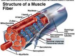
Myofibril |
organelle within muscle cell chain of sarcomeres
each is wrapped in sarcoplasmic reticulum |
|
|
Muscle cell fiber |
many muscle cell fibers make one bundle of muscle fibers
Can only be innervated by 1 motor neuron |
|
|
sacromere |
cell that contracts or expands due to response from stimuli
z lines
smallest functional unit of muscle
includes the tick and thin filaments |
|
|
What is the thick filament protein? |
Myosin |
|
|
What is the thin filament protein |
actin |
|
|
What protein covers the filament head? |
troponin |
|
|
Structure of muscle |
|
|
|
electro mechanical coupling in muscle contraction |
ACh binding causes action potential which stimulates release of Ca in the sarcoplasmic reticulum. Ca binds to tropinin which allows micro and thick filament to bind and cause contaction |
|
|
How is a contraction ended? |
AChE removes ACh from cleft and Ca pump pumps Ca out of cell. |
|
|
T tubule |
channels in the myofibril that allow for movement of neurotransmitters and ions |
|
|
Tetrad |
4 calcium channels formed together |
|
|
How does calcium enter the cytosol in a muscle fiber cell? |
Tetrads and calcium release channels |
|
|
Main type of neuron associated with contraction of muscle |
alpha motor neuron, thick |
|
|
where are alpha motor neurons found? |
in the ventral horn ` |
|
|
What do motor neurons innervate? |
Several muscle cells in one muscle |
|
|
motor unit |
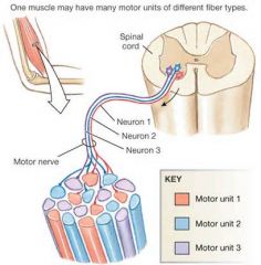
One motor neuron! and all the muscle cells it! innervates
|
|
|
motor pool |
all motor units of a given muscle |
|
|
How is muscle for controlled? |
by frequency regulation
temporal summation
dont want to fire an action potential and get all the way back down to resting, high energy cost |
|
|
Which types of motor neurons innervate more cells? |
Large cells innervate more than small cells |
|
|
Which cells are recruited first ? |
Small cells(fine motor control) than onto larger AKA size principle |
|
|
What type of motor pool does a fine control neuron have? |
Muscles that are used for fine control have a large motor pool, |
|
|
Where do alpha motor neurons receive information? |
input from spinal interneurons sensory input from spinal signals input from upper motor neurons in brain |
|
|
What is a muscle spindles function? |
to measure length |
|
|
What type of neurons are found in a muscle spindale? |
Ia |
|
|
What is the function of the myotatic reflex? |
To resist change in muscle
weight added causes increase in length causes high frequency action potential firing causes alpha motor neuron to fire and contract |
|
|
extrafusal muscle fiber |
"normal" the ones on the outside of the muscle cell |
|
|
intrafusal fiber |
attached to spindle organs, where gamma neurons synapse ` |
|
|
gamma motor neuron |
attached to intrafusal fibers, activated by contraction of muscle fiber
involved in reflexes
adjust tension on muscle spindles |
|
|
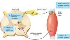
What is the function of Golgi tendon? |
to encode the muscle tension |
|
|
What do alpha motor neurons encode? |
length |
|
|
What do golgi tendon afferents encode? |
Muscle Tension |
|
|
What type of axons do Golgi tendons have? |
Ib |
|
|
What is the relationship between interneurons and alpha motor neurons? |
Some interneurons mediate reciprocal inhibition Other interneurons mediate excitation Other interneurons mediate reflexes! across limbs (e.g. crossed-extensor reflexes). |
|
|
reciprical inhibition |
one side of muscle contracts while other relaxes |
|
|
Type of muscles |
flexors and extensors |
|
|
What is the pathway of the golgi tendon responding to tension? |
|
|
|
What are the receptive fields of retinal ganglion? |
Center-surround areas ON/OFF exhibitors/inhibitors |
|
|
Temporal coding |
Stimulus features are encoded by the precise timing of action potentials |
|
|
How does the auditory nerve encode the frequency of a simple tome? |
By which axons are firing action potentials |
|
|
Why is the onset of a muscle twitch faster than offset? |
Calcium must. Be pumped back into the Sarcoplasmic reticulum |
|
|
What compounds are found in the olfactory transduction system&? |
Cyclic AMP adenylyl cyclase cyclic nucleotide gated channels(also found in visual system) |
|
|
What are the mechanisms by which motor neurons regulate strength of muscle contraction? |
Muscle recruitment |
|
|
Receptive field |
The region of space which when stimulated affects the firing of a neuron |
|
|
Where do eyes project their information'? |
To both sides of the brain |
|
|
What do the tip links of the cilia help with'? |
Stabilization of of the cilia |
|
|
What do owls different orientation of their eyes allow'? |
Sound detection on the y axis |
|
|
Do muscles fire action potentials&? |
Yes |
|
|
Visual information from the ventral stream |
Shape/what is the object |
|
|
Visual information from the dorsal stream |
Location |
|
|
Two acoustic cues humans use for horizantal sound location |
Interaural intensity and frequency |
|
|
Where are the cell bodies of alpha and gamma neurons |
Ventral horn |
|
|
Where does a gamma neuron synapse? |
Intrafusal fiber of muscle spindles |
|
|
Where does alpha motor neurons synapse |
Extra fussy fibers of muscle spindles |
|
|
Where are Ia cell bodies located? |
Dorsal root ganglion |
|
|
Where do Ia axons synapse? |
Directly on alpha motor neuron in ventral horn |
|
|
What kind of mechanism is involved in muscle contention? |
Electro mechanical coupling |
|
|
What do spindle organs encode? |
Length |
|
|
What types od axons are associated with Golgi tendon? |
Ib |
|
|
What process do Golgi tendons serve'? |
Measure muscle tension send information to spinal cord interneuron which inhibits alpha motor neuron and causes relaxation |
|
|
What can Interneurons do in muscle systems |
Inhibit |