Reading...
![]()
Play button
![]()
Play button
![]()
Use LEFT and RIGHT arrow keys to navigate between flashcards;
Use UP and DOWN arrow keys to flip the card;
H to show hint;
A reads text to speech;
14 Cards in this Set
- Front
- Back
|
What is an afferent neuron? efferent neuron?
|
Afferent neurons carry information from the periphery into the CNS. These are also known as sensory neurons
Efferent neurons carry information from the CNS to the structures in the periphery (either smooth muscle, skeletal muscle or blood vessels, cardiac muscle and glands). Also known as motor neurons |
|
|
What is a somatic neuron? visceral neuron? proprioreception neuron?
|
Somatic neurons carry information from external body surfaces (or structures deep to the body surface that are stimulated by changes in the external environment) or to striated muscle
Visceral neurons carry information to or away from structures internally within the animal (the viscera) Proprioreception neurons carry information about proprioreception. They are a type of afferent, or sensory neuron |
|
|
What is a general neuron? special neuron?
|
General neurons carry information to or from most of the structure of the body except those involved with the special senses or GI tract
Special neurons carry information about the special senses (all except touch: taste, vision, hearing, small) or neurons that develop in association with the GI tract |
|
|
What types of afferent neurons are there?
|
♦SOMATIC (S)
a) General somatic afferent (GSA) neurons carry information to the CNS about temperature, touch, noxious stimuli; this includes neurons in all spinal nerves, plus CNV b) Special somatic afferent (SSA) - neurons carry information about vision (CNII) or hearing (CNVIII) to the central nervous system ♦VISCERAL (V) a) General visceral afferent (GVA) neurons carry information about organ content, distention and chemicals via the splanchnic branches of the spinal nerves, plus cranial nerves VII, IX, and X ♦PROPRIORECEPTIVE neurons (P) a) General proprioreption (GP) neurons carry information about all muscle and joint movement to the CNS via spinal nerves + CNV; General proprioreception includes both conscious and unconscious proprioreception systems |
|
|
What types of efferent neurons are there?
|
♦SOMATIC
a) General Somatic Efferent (GSE) neurons carry information to the striated skeletal muscle -this includes neurons within all spinal nerves, as well as CNIII, VII, IX, X and XI ♦VISCERAL (V) a) General Visceral Efferent (GVE) neurons carry information to smooth and cardiac muscles and glands -this includes neurons within spinal nerves (parasympathetic) and neurons within CN III, VII, IX, X, and XI |
|
|
What is a pathway?
|
A pathway describes the series of neurons involved in communicating a particular set of information from one part of the nervous system to another e.g. papillary light response (PLR) pathway describes the series of neurons (and their locations) that take part in changing pupil size in response to light
-the name of the pathway expleains where the information is coming from (the first part of the name) and going to (the second part of the name) e.g. Corticospinal pathway contains axons taking information from the cerebral cortex to the spinal cord |
|
|
What is a tract?
|
A tract describes a group of axons in white matter all of which are taking information from the same origin to the same destination e.g. corticospinal tract in the spinal cord is located in the ventral white matter and contains only axons of neurons involved in the corticospinal pathway
|
|
|
What is a reflex? What is a response?
|
A reflex is a hard-wired pathway of neurons (ie. present at birth) that produces a sterotypic response to a given stimulus e.g. patellar reflex involves a predictable contraction of the quadriceps muscle in response to stretching of the patellar tendon
A response is a LEARNED pathway of neurons that produces a sterotypic response to a given stimulus e.g. the menace response, which produces a blink of the eye when it is approached by a fast moving stimulus, only develops in dogs at 14-16 weeks of age |
|
|
What is a motor neuron? Upper motor neuron? Lower motor neuron?
|
Motor neurons carry information from the brain to striated muscle for voluntary contraction of muscle
-they are divided into upper motor neurons (UMNs), which have their dendrities, cell body, and axons all within the CNS, and lower motor neurons (LMNs), which have their cell body and axon within the CNS but whose axon continues into the PNS and ultimately terminates on a neuromuscular junction |
|
|
What are the 4 groups of sensory modalities?
|
Sensory modalities are sensations that the cerebral hemisphere is concious of - does not include unconcious proprioreception
♦Touch, pressure, proprioreception: The receptor endings of these modalities are low threshold mechanoreceptors, and they respond to purely mechanical stimulation -they are transmitted within the spinal cord along the fasciculus gracilus and fasiculus cuneatus ♦Pinprick pain, heat and cold -they are transmitted within the spinothalamic tract in the spinal cord in man, and probably in domestic animals (although this is unproven) ♦True pain -this is transmitted within the ascending reticular formation in the spinal cord, in man -again not sure in animals but likely to be the sam ♦Vision, hearing, balance, taste & olfaction -these are the special senses, and travel within the brain rather than the spinal cord |
|
|
What is a reflex composed of? What's an example? What's the only reflex that doesn't involve an interneuron?
|
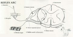
-a reflex is a response to an involuntary stimulus
-most reflex arcs involve a sensory neuron, an interneuron (internuncial neuron), and a motor neuron, making them multisynaptic (the patellar tap reflex lacks an interneuron [monosynaptic]) -specialized receptors at the end of sensory neurons receive a stimulus (e.g. a pinprick), resulting in an impulse (nerve action potential) -the impulse travels through the dendrite, body, and axon of the sensory neuron into the spinal cord -in the spinal cord, the sensory neuron synapses on an interneuron which then carries the impulse to a synapse on a motor neuron -the motor neuron stimulates a receptor organ (muscle or gland) to complete the reflex e.g. withdrawal from a stimulus 1. Stimulus - a change in the environment (e.g. pin-prick/noxious stimulus) 2. Receptor - the sensory neuron's dendritic zone (e.g. pain detector in finger) that responds to change (stimulus) by generating an impulse 3. Sensory (afferent) neuron - carries a sensory impulse to the CNS 4. Interneuron or association neuron -located in the spinal cord, connects a sensory neuron with a motor neuron -it can also connect with other neurons and send information up to the brain (so when the reflex is over, it can be perceived and commented on "ouch" 5. Motor (efferent) neuron or lower motor neuron -carries an impulse to an effector organ 6. Efferent (target) organ - the muscle or gland innervated by a motor neuron which reacts to the stimulus (e.g. pulling away from the stove) EXAMPLE e.g withdrawal reflex - fibers are activated by a noxious stimulus, travel to the spinal cord and activate multiple interneurons in the grey matter -the interneurons facilitate LMNs to flexor and extensor muscles on both sides of the spinal cord -contractions of flexor muscles on the side of the stimulus facilitate removal of a limb from the painful stimulus, and contractions of extensors in opposite limb provide postural support during limb withdrawal |
|
|
What are the 2 types of somatic efferent neurons?
|
There are 2 types of somatic efferent neurons in the ventral horn grey matter of the spinal cord:
♦the alpha motor neuron innervates extrafusal muscle fibers, is large in diameter and fast ♦the gamma motor neuron or fusimotor neuron innervates intrafusal muscle fibers within muscle spindles, is thinner and slower conducting -there are 2 types of intrafusal fibers: nuclear bag or neuclear chain fibers -stretching of the spindle stimulates class Ia afferent fibers which innervate the central region of the spindle and this in turn synapses on the cell body of the alpha motor neuron, which causes contraction of the extrafusal fiber -collaterals from the Ia afferent fibers travel to the cerebral hemispheres and cerebellum to transmit information to the brain about proprioception as well as being involved in reflex arcs -innervation of alpha and gamma neurons happens concurrently during stimulation of muscle contraction - this is co-activaton -in this way, when extrafusal fibers contract the intrafusual fibers also shorten to maintain the length of the mid-region of the muscle spindle just below the threshold for activation |
|
|
What are the 3 ways the intrafusal fiber can be stimulated to contract?
|
♦directly by upper motor neurons
-in quadripeds relatively little muscle contraction is the result of this ♦The gamma motor neuron can be stimulated by upper motor neurons descending from the brain and travelling within the spinal cord -this triggers contraction of the intrafusal fibers which stimulates the Ia fiber to stimulate alpha motor neurons -this accounts for the greater part of muscle activation ♦Stretching of the intrafusal fiber past the setpoint can occur if the bones of the limb are moved apart, causing firing of the Ia neuron which then synapses on the alpha motor neuron and causes contraction of the extrafusal fibers -this last method is the way in which reflexes work -collaterals from the Ia afferent fibers travel to the cerebral hemispheres and cerebellum to transmit information to the brain about proprioreception, as well as being involved in reflex arcs |
|
|
Describe muscle spindles. What are golgi tendon organs?
|
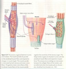
-the terminal branches of the sensory nerve fibers in the stretch reflex arcs are twisted around the central region of each of several thin, modified muscle cells in sensory organs called muscle spindles
-a single muscle may contain many muscle spindles -the specialized muscle cells within the spindles are called intrafusal muscle fibers -the central regions of the intrafusal muscle fibers are noncontractile -the remaining muscle fibers which are responsible for contraction are called extrafusal muscle fibers -the sensory nerve endings twisted around the intrafusal muscle fibers change shape when the muscle is stretched -this leads to depolarization of the nerve endings and increased frequency of nerve impulses in sensory nerve fibers -in the spinal cord these nerve fibers form excitatory synapses directly on motor neurons that extend axons to the same muscle -the subsequent reflexive muscle contraction counteracts stretching of the muscle and tends to restore muscle length -in the gray matter the sensory nerve fibers from the muscle spindles in a particular muscle branch and form synapses on interneurons that inhibit motor neurons innervating the anagonistic muscle -therefore, when a stretched muscle is stimulated by the stretch reflex, the antagonistic muscle will be simultaneously inhibited, causing the stretched muscle to return to its original length -if a muscle contracts and maintains a shorter length, the muscle spindles would slacken and lose their ability to respond to further stretching -the ends of the intrafusal muscle fibers are contractile and are innervated by special motor nerve fibers -these are called gamma motor nerve fibers -when the muscle is shortened, the impulse frequency of the gamma motor nerve fibers is increased, causing the ends of the intrafusal muscle fiber to contract -in this way, the central regions of these fibers are tautened, and the sensitivity of muscle spindles is restored GOLGI TENDON ORGANS -located at the muscle tendon junction -the sensory neurons have branched nerve endings interwoven between the collagen fibers of the tendon an the nerve endings are deformed when the muscle contracts and pulls on the tendon -this leads to depolarization of the nerve endings and increased frequency of nerve impulses conducted along sensory nerve fibers -arranged in series with extrafusal muscle fibers -response of these organs correlates with muscle tension and is independent of muscle length |