Reading...
![]()
Play button
![]()
Play button
![]()
Use LEFT and RIGHT arrow keys to navigate between flashcards;
Use UP and DOWN arrow keys to flip the card;
H to show hint;
A reads text to speech;
30 Cards in this Set
- Front
- Back
|
What does white matter consist of?
|
Consists of dendrites, neuroglia and LOTS of axons
|
|
|
What does the grey matter consist of?
|
Neuronal cell bodes, neuroglia, lots of dendrites
|
|
|
Where is the grey matter found?
|
On the outside of the brain and interior of the spinal cord
|
|
|
Where is white matter found?
|
On the inside of the brain and outside of the spinal cord
|
|
|
What does the nervous system do?
|
It conveys sensory (afferent) and effector or motor (efferent) information around the body in order to respond to both external & internal sitimuli
|
|
|
What does the central nervous system consist of?
|
Brain & Spinal Cord
|
|
|
What does the peripheral nervous system consist of?
|
Nerves & Ganglia
-or specifically: the spinal nerves and their branches, the cranial nerves and their branches, and the nerves of the autonomic nervous system |
|
|
What does the autonomic nervous system do?
|
-conducts impulses to the smooth muscle of the viscera and blood vessels, cardiac muscle and glands
|
|
|
Give names for the 3 main regions of the brain
|
✤FOREBRAIN = prosencephalon = cerebrum + diencephalon
●Cerebrum = cerebral hemisphares = telencephalon ●Dienephalon = thalamus (includes interthalamic adhesion) + hypothalamus (includes tuber cinereum & mammillary bodies) + metathalamus (includes medial & lateral geniculate bodies) + epithalamus (includes pineal gland) + subthalamus ✤MIDBRAIN = mesencephalon = tectum (corpora quadrigemina/ rostal & caudal colliculi) + tegmentum + crus cerebri (cerebral peduncles/ pyramidal tracts) tectum - dorsal region of mesencephalon tementum = forms floor of midbrain →contains the motor nuclei of oculomotor and trochlear nerves, the red nucleus & reticular formation ✤HINDBRAIN = rhombencephalon = metencephalon (pons + cerebellum + left & right rostral, middle & caudal cerebellar peduncles) + myelencephalon (medulla oblongata) + trapezoid body + pyramids |
|
|
Where is the cerebellum?
|
-large mass behind the cerebrum
-located above the pons and medulla, and caudal to the transverse fissue |
|
|
Where is the cerebrum (cerebral hemispheres)?
|
Located underneath the skill
|
|
|
Where is the connective tissue associated with the nervous system?
|
Regular connective tissue types surround the components of the central nervous system, but they are not found within the tissue.
|
|
|
What is a ganglion?
|
Collections of neuronal cell bodies within the CNS are called nuclei, and outside the CNS they are called ganglia
|
|
|
How many pairs of cranial nerves are there?
|
12
|
|
|
What are the 3 major dural folds within the cranial vault?
|
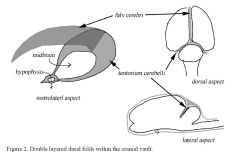
There are 3 major areas in which a double layered fold of dura arises from the internal surface of the skull
1) falx cerebri - the fold which lies in the longitudinal fissures between the 2 cerebral hemispheres -from ethmois to the osseous tentorium -contains the dorsal sagittal venous sinus draining the dorsal forebrain 2) tentorium cerebelli - is the fold which separates the caudal portion of the cerebral hemispheres from the cerebellum -runs from the petrosal crest to the osseous tentorium bilaterally -inverted "U" in shape and the midbrain passes through the notch -separates the cerebral hemisphere from the cerebellum - divides the cranial vault into the: ●rostral fossa which lies rostral to the tentorium and contains the forebrain ●caudal fossa which lies caudal to the tentorium and contains the cerebellum and caudal brain stem. In cats, the tentorium cerebelli often ossifies making it difficult to remove the brain. Ossification probably occurs in the young adult 3) diapgragma sella - bridges from the dorsum sella to the caudal clinoid process of the sphenoid bones near the optic canal area -it can make removal of the entire brain with the pituitary intact difficult |
|
|
What kinds of herniation can occur in the brain?
|
-brain herniation is the movement of a portion of the brain under another structure
❤Caudal transtentorial herniation -portions of the temporal cortex under the tentorium cerebelli into the caudal fossa ❤Rostral transtentorial herniation -of the rostral cerebellum rostrally under the tentorium cerebelli into the rostral fossa ❤Cingulate gyrus herniation -of the medial portion of the cerebral hemisphere under the falx cerebri from one side to the other ❤Foramen magnum herniation -of portions of the caudal cerebellum under the foramen magnum |
|
|
What is a tract?
|
Tracts or Fasciculi - are nerve axon bundles of a common origin in the brain and/or spinal cord
-they are usually named for their origin and destination -sensory and motor tracts of the spinal cord are continuous with sensory and motor tracts of the brain |
|
|
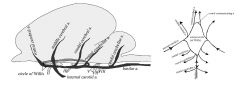
What is the arterial circle? What is its functional significance? What major arteries supply it in the dog?
|
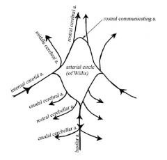
Circle of Willis
-maintains a constant blood pressure and provides collateral circulation -however, once the vessels leave the circle to enter the brain tissue, there are very limited connections with other vessels -therefore any disease which blocks one of these end vessels, a cerebrovascular accident or stroke, results in hypoxia and necrosis of the brain parenchyma -it is supplied by the internal carotids and basilar artery |
|
|
What is the rete mirabile and in which species is it found?
|
"rete mirable" is the name given to any blood vessel that breaks up into a plexus and reforms into a single blood vessel
-present in ruminants, pigs, and cats Function: possibly involved in thermoregulation by heat transfer from warmer arterial blood to the blood of the cavernous sinus blood which is draining the cooler nasal cavity -this may help protect the brain against rising body temperatures |
|
|
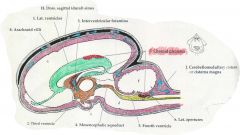
Which parts of the ventricular system are associated with each of the five divisions of the brain?
|

Lateral ventricles - largest of the ventricles. There is one located within each cerebral hemisphere each with a choroid plexus
Interventricular foramina - the channels connecting the two lateral ventricles with the third ventricle Third ventricle - located in the diencephalon, encircling the interthalamic adhesion -it has a choroid plexus in its roof Mesencephalic (midbrain) aqueduct - in midbrain -connects the third & fourth ventricles -it is the narrowest part of the ventricles Fourth Ventricle - associated with two brain divisions (metencephalon & myelencephalon) -located between the brainstem and the overlying cerebellum -lateral apertures (foramen of Luschke) which are openings between the 4th ventricle and the subarachnoid space at the level of CN VIII |
|
|
How are the lateral ventricles connected with the third ventricle?
|
Mesencephalic Aqueduct
|
|
|
How is the 4th ventricle connected with the subarachnoid space?
|
-it is connected through the lateral apertures (foramen of Luschke) which open at the level of CNVIII
|
|
|
Where are the choroid plexi found?
|

-in the dorsomedial aspect of the lateral ventricles
-in the dorsal aspect of the third ventricle or the roof -found in the fourth ventricle originating from the roof and protruding into the lateral apertures |
|
|
What is the histological structure of the choroid plexi?
|
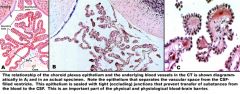
Each plexus consists of a tortuous loop or tuft of capillaries, overlain by a neatly cuboidal epithelial covering (ependymal cells). The cells covering the blood vessel have a sort of brush border consisting of irregular microvilli.The cells of this epithelium are sealed together by occluding junctions, to maintain the integrity of the blood-brain barrier.
|
|
|
What is the cerebrospinal fluid? Where is it produced?
|
It is an ultrafiltration of plasma blood. It is the clear straw colored or colorless fluid which surrounds the neuroaxis. It is produced by the choroid plexus.
Compared with the plasma: -it has less K+ and more calcium -more Cl-, Na+, and Mg+ -glucose - approx 80% of blood -protein - much less than blood, and it is mostly albumin -cells - virtually acellular, may contain a few WBC which are primarily mononuclear cells |
|
|
Describe the flow of the CNS and how it is absorbed.
|
Produced in the ventricle (lateral, third, & fourth) → flows caudally through the brain's ventricular system [Lateral ventricle → 3rd ventricle → Mesencephalic aqueduct →4th ventricle] → out the lateral apertures into the subarachnoid space → over the cerebral hemispheres and caudally along the spinal cord
-some also flows from the 4th ventricle along the central canal to the terminal end of the spinal cord |
|
|
What are 3 functions of the CSF?
|
1) Physical protection
-physical cushion -buffer against pressure changes within the CNS 2) chemical protection -more stable chemically than blood plasma -allows increased regulation of the neuronal environment -CSF pH directly influences the function of the medullary respiratory center 3) Nourishment -may transport nutrients between the blood and the brain -may transport neuroendocrine substances and neurotransmitters |
|
|
Describe the venous drainage of the brain
|
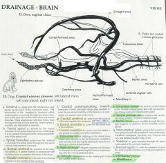
DORSAL SYSTEM:
-collects blood from the dorsal parts of the brain and the diploe of the bones of the cranial vault -it includes the dorsal sagittal sinus within the falx cerebri -the dorsal sagittal sinus receives numerous tributary beings directly from the cerebral hemispheres and is joined toward its caudal end by the straight sinus, which runs within the ventral part of the falx and collects blood from a major vein draining the deep parts of the brain -the dorsal sinus splits into bilateral transverse sinuses within the tentorium cerebelli → each later divides into a temproal and sigmoid sinus -coursing caudolateral to the petrosal part of the temporal bone, the temporal sinus extends to the retroarticular foramen where it emerges as the emissary veint of the retroarticular foramen and joins the maxillary vein -each signmoid sinus forms an S shaped curve -the ventral petrosal sinus joins the sigmoid sinus and from this anastamosis the vertebral and internal jugular veins arise and leave the typano-occipital fissue and course caudally -the basilar sinus is a branch of the signmoid sinus that continues caudally to the ventral internal vertebral venous plexus in the vertebral canal VENTRAL OR BASILAR SYSTEM: -drains the ventral part of the brain and also receives a major inflow from a vein that enters the cranial cavity from the orbit after draining much of the face, including the nasal cavity -emissary veins connect each cavernous sinus with the ophthalmic plexus of veins rostrally and with the maxillary vein laterally -these sinuses are continued caudally by the ventral petrosal sinus |
|
|
What are the 3 meningial coverings in the brain?
|
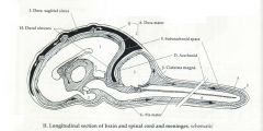
❤DURA MATER
-the outermost meninx made of strong connective tissue -the cranial dura mater has 2 layers where the spinal dura mater only has one -the two layers of the dura separate over the fissures of the brain; the inner layer extends into the fissues forming partitions and dural sinuses -the cranial dura mater is continuous with the spinal cord dura mater at the foamen magnum -because the cranial dura mater is fused with the periosteum of the bones of the cranium there is no epidural space surrounding the brain ❤ARACHNOID MATER -the delicate meninx pushed against the dura mater by the cerebrospinal fluid ❤SUBARACHNOID SPACE -the cavity between the arachnoid and pia mater where cerebrospinal fluid (CSF) circulates ❤PIA MATER -the innermost meninx closely investing the spinal cord and brain ●Dural sinuses -the venous spaces formed where 2 dural layers separate from each other -they are filed with venous blood returning to the general circulation ●Dorsal sigital sinus -a major part of the venous drainage system of the brain and cranial cavity, located within the split layers of the dura mater (falx cerebri) ●Cerebellomedullary cistern or cisterna magna -an expansion of the subarachnoid space located between the caudal surface of the cerebellum and the dorsal surface of he medulla, just inside the foramen magnum -this is a common site for a CSF "tap" (removal of cerebrospinal fluid) |
|
|
What are the arteries supplying the brain?
|
✤Arteries supplying the base of the brain:
-internal carotix -basilar (which is a direct continuation of the ventral spinal artery) ✤Cerebral brain supply: A) Rostral cerebral artery -arises lateral to the optic chiasm and forms the rostal portion of the arterial circle -supplies the rostromedial half of the the cerebral hemispheres B) Middle cerebral artery -largest vessel supplying the brain -arises just rostral to the hypophysis -supplies the lateral part of the cerebral hemispheres and rostral brain stem C) Caudal cerebral artery -arises near the occulomotor nerve -supplies the caudomedial part of the cerebral hemispheres ✤Cerebellar blood supply -rostral cerebellar artery arises from the arterial circle -the caudal cerebellar artery arises from the basilar artery ✤Brainstem blood supply -medullary and pontine branchs from the basilar artery supply the medullar and pons ✤Meningial arteries -3 major meningial branches: rostral, middle, and caudal meningial arteries |