Reading...
![]()
Play button
![]()
Play button
![]()
Use LEFT and RIGHT arrow keys to navigate between flashcards;
Use UP and DOWN arrow keys to flip the card;
H to show hint;
A reads text to speech;
13 Cards in this Set
- Front
- Back
|
What are some causes of cerebral edema?
|
Intracellular edema: hypoxia (dysfunction of Na/K ATPase pump) or hyponatremia causing an osmotic shift.
Extracellular edema (due to increased vessel permeability): acute inflammation (meningitis), metastasis, trauma, lead poisoning, respiratory acidosis, hypoxemia. |
|
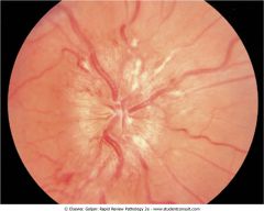
What other signs and symptoms may accompany this particular finding?
|
This is papilledema (swelling of the optic disk) that occurs when there is increased intracranial pressure. Other signs: headache, projectile vomiting without nausea, sinus bradycardia, HTN, herniation.
|
|
|
Cerebral herniation can be a complication of increased intracranial pressure. What are the three most common cerebral herniations?
|
1) Subfalcine herniation - cingulate gyrus herniates under the falx cerebri. Complication: compress ACA.
2) Uncal herniation: medial portion of temporal lobe herniates through tentorium cerebelli. Complications: compress midbrain, CN III, PCA. 3) Tonsillar herniation: cerebellar tonsils herniate into the foramen magnum. Complication: depressed respiratory center. |
|
|
Hydrocephalus is an increase in CSF volume causing enlargement of the ventricles. Describe the difference between communicating, noncommunicating, and hydrocephalus ex vacuo.
|
Communicating: increased CSF production or obstruction in reabsorption of CSF via arachnoid granulations.
Non: obstruction of CSF flow out of the ventricles. Ex vacuo: dilated appearance of the ventricles when the brain mass is decreased. |
|
|
1) What is the most common cause of hydrocephalus in a newborn?
2) What type of hydrocephalus would a choroid plexus papilloma cause? 3) What type of hydrocephalus would a medulloblastoma cause? 4) What is the clinical manifestation of hydrocephalus ex vacuo? |
1) Sticture of the aqueduct of Sylvius (btw 3rd and 4th).
2) Communicating. 3) Noncommunicating. 4) Dementia, gait disturbances, urinary incontinence. |
|
|
What is Arnold-Chiari malformation?
What is Dandy Walker malformation? |
AC: caudal extension of medulla and cerebellar vermis through foramen magnum, noncommunicating hydrocephalus, flattening of base of skull, meningomyelocele, syringomyelia., 50% have aqueduct stenosis.
DW: hypoplasia of the cerebellar vermis, cystic dilation of the 4th ventricle, noncommunicating hydrocephalus. |
|
|
Patient is brought in by some work mates who claim the patient put his hand on a hot metal plate, but didn't feel any burning sensation. Patient claims his hands have become progressively weak over the past couple months. MRI shows enlargement of the cervical cord. Diangosis?
|
Syringomyelia: degenerative disease of the spinal cord: cystic formation within cervical spinal cord. Cavity expands impeding: decussating spinothalamic tracts (decrease pain/temperature) and destruction of anterior horn (atrophy of intrinsic muscles of the hands).
|
|
|
Young child presents with seizures and mental retardation, as well as angiofibromas on the face. CT scan identifies 'candlestick drippings' in the ventricles. What test could be performed to identify other skin lesions?
|
Tuberous sclerosis (a neurocutaneous syndrome - disordered growth of ectodermal tissue) is an AD disorder characterized by the patients presentation. Hypopigmented skin lesions (ash leaf) is best identified with a Wood's lamp. Hamartomatous lesions create the astrocyte proliferations in subependyma as well as angiomyolipomas in the kidney. Rhabdomyoma in the heart also occur.
|
|
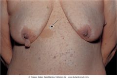
This patient also had a bp of 160/110. What is the inheritance of this disorder?
|
This is neurofibromatosis, an autosomal disorder, is characterized by cafe-au-lait macules, lisch nodules (hamartomas in the eye), kyphoscoliosis, and HTN due to pheochromocytoma. Other common findings: optic nerve glioma, meningioma, acoustic neuroma.
|
|
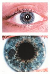
What is apparent in this eye?
|
Lisch nodules - hamartomas in the eye. Characteristic of neofibromatosis an autosomal disorder which is associated with pheochromocytoma, acoutic neuromas.
|
|
|
Define:
1) Porencephaly 2) Hydranencephaly 3) Schizencephaly 4) Holoprosencephaly |
1) Fluid filled cystic cavity
2) Fluid filled cystic cavity replacing the cerebral hemispheres. 3) Cerebral cortical cleft that communicates with the ventricles. Due to abnormal cell migration. 4) Prosencephalon fails to cleave down midline. |
|
|
Describe the role of amyloid precursor protein in the development of Alzheimer's.
|
APP is normally coded on chromosome 21 (trisomy 21 invariable develops Alzheimer's). Defects in degradation of APP by secretases causes an increase in beta-amyloid. Alpha-secretases cleave APP into fragments that cannot produce beta-amyloid. Beta and gamma secretases cleave APP into fragments that ARE converted to AB. AB deposits in neurons are neurotoxic.
|
|
|
What role do chromosome 14 and chromosome 19 play in the development of Alzheimer's?
|
14: mutations lead to abnormal tau protein which causes formation of neurofibrillary tangles.
19: apolipoprotein gene E has a high binding affinity for beta-amyloid. |