Reading...
![]()
Play button
![]()
Play button
![]()
Use LEFT and RIGHT arrow keys to navigate between flashcards;
Use UP and DOWN arrow keys to flip the card;
H to show hint;
A reads text to speech;
422 Cards in this Set
- Front
- Back
|
What are the two categories of PNS disease?
|
1)focal group -simple mononeuropathies and multiple mononeuropathies
2)non-focal group - polyneuropathies |
|
|
What 3 questions must be answered in the evaluation of PNS disease?
|
-are the sensorimotor signs symmetrical or asymmetrical
-are the sensorimotor signs and symptoms distal or both proximal and distal? -is the modality involved exclusively motor or sensory or mixed |
|
|
How many spinal nerves are there?
|
31
|
|
|
Describe the 2 components of the autonomic peripheral nervous system?
|
Sympathetic (thoracolumbar)
Parasympathetic (craniosacral) |
|
|
What are the 3 responses of the PNS to pathologic stimuli?
|
-myelinopathies (best prognosis)
-axonopathies (worse prognosis) -neuronopathies (worst prognosis) |
|
|
What are the 2 types of electrophysiologic testing used for PNS disorders?
|
-Nerve conduction studies (NCS)
-electromyography (EMG) |
|
|
List the causes of acute, emergent weakness and possible respiratory compromise
|
Autoimmune
-myelinopathies (GBS and CIDP) -myasthenia gravis Toxic -botulism -buckthorn -seafood -tick paralysis -metals (arsenic and thallium) Metabolic -dyskalemia -hypophosphatemia -hypermagnesemia -porphyria Infectious -polio -diptheria |
|
|
What are characteristic findings in GBS?
|
-symmetrical weakness
-usually worse distally -variable sensory findings |
|
|
What is Miller Fisher syndrome?
|
A rare form of GBS with ophthalmoplegia, ataxia and areflexia
|
|
|
What are common triggers form GBS?
|
-URTI
-gastrointestinal flora |
|
|
What are typical features of GBS?
|
-decreased DTR
-variable sensory findings -anal sphincter sparing -urinary retention |
|
|
Waht are common infectious organisms associated with GBS?
|
-campylobacter jejuni
-CMV -EBV -Mycoplasma pneumoniae |
|
|
What ancillary tests may be helpful in the diagnosis of GBS?
|
-csf analysis (shows increased protein and mild pleocytosis)
-MRI (selective enhancement of the anterior spinal nerves) -electrophysiologic testing |
|
|
What respiratory parameters are predictive of impending respiratory failure in GBS?
|
-FVC <20cc/kg
-NIF (MIP) <30cmH2O -increased pCO2 |
|
|
In patients with normal pulmonary fuction, how can impending ventilatory failure be predicted?
|
Assessing extensor neck strength
|
|
|
What are possible treatments for patients with GBS
|
-IVIG
-plasma exchange |
|
|
Should patients with GBS and elevated BP be treated?
|
No, it is typically transient and may be followed by hypotension
|
|
|
What type fo polyneuropathy is GBS?
|
It is a demyelinating polyneuropathy
|
|
|
What are the mostcommon distal symmetrical polyneuropathies (DSPN) encountered in the Ed?
|
-diabetic DSPN
-alcoholic DSPN -HIV neuropathies -toxic and metabolic neuropathies |
|
|
What is the 1st motor sign of diabetic DSPN?
|
-weakness of big toe dorsiflexion
|
|
|
what are possible medications used to treat neuropathic pain?
|
-gabapentin
-opioids -tramadol -TCA -pregabalin |
|
|
What is the most common cause of plexopathies?
|
-blunt trauma with young men
|
|
|
What are causes of plexopathy?
|
-trauma
-radiation -neoplastic -thoracic outlet |
|
|
What is teh presentation of brachial plexus injury?
|
Gradually progressive weakness and wasting fo the median and ulnar hand muscles
|
|
|
What are typical causes of mononeuropathies?
|
-trauma (blunt or penetrating)
-entrapment -compression (antecedent peripheral neuropathy is a risk factor) |
|
|
How do you determine whether radial mononeuropathy is axillary vs humeral in origin?
|
-test the triceps (triceps is involved if axillary compression and spared in the more common humearl form)
|
|
|
WHat is Saturday Night Palsy?
|
This is a radial mononeuropathy secondary to improper positioning of arm during sleep (it is also called bridegrooms palsy)
|
|
|
What are the findings in Saturday night Palsy?
|
-wrist drop
-finger drop -mild numbness over the first distal interosseous muscle (depending on the level, findings may be variable) |
|
|
What are the branches of the radial nerve and wehre does the branching occur?
|
-posterior interosseous (pure motor)
-superficial radial (pure sensory) This branching occurs at the antecubital fossa |
|
|
What is the management of patients waiting for spontaneous recovery of a radial mononeuropathy?
|
Splint at 60 degrees of dorsiflexion (referral form special splints to provide passive dorsiflexion of the fingers)
|
|
|
Which two branches of the ulnar nerve do not pass through Guyon's canal?
|
-dorsal cutaneous
-palmar cutaneous |
|
|
what is the function of the interossei muscles
|
finger abduction and adduction
|
|
|
What is the function of the lumbricals?
|
Flexion of the metacarpophalageal joints
|
|
|
In what locations of the elbow is the ulnar nurse at risk of being injured?
|
-ulnar condylar groove
-the cubital canal |
|
|
What is the most likely area for ulnar nerve injury
|
the elbow
|
|
|
How would you differentiate ulnar nerve damage at the elbow vs at the wrist?
|
-sensory abnormalities in an ulnar distribution in the hand and fingers strongly suggests a lesion in the elbow
-a lesion at the wrist would only affect sensation at the palmar surface of the 5th digit and ulnar 1/2 of the 4th digit (the dorsal surface of those digits would have normal sensation) |
|
|
What is the most common entrapment neuropathy?
|
Carpal tunnel syndrome
|
|
|
Which is affected first in carpal tunnel syndrome sensory or motor?
|
Sensory
|
|
|
When motor function is involved in CTS which muscles are involved?
|
LOAF
L-lumbricals thumb Opposition Abduction Flexion |
|
|
What is the most specific finding in CTS?
|
Splitting of sensation in the 4th digit
|
|
|
What is Tinel's sign?
|
percussion of the median nerve at the wrist
|
|
|
What is Phalen's sign?
|
Maximal palmar flexion at the wrist mimics symptoms
|
|
|
What conditions are associated with CTS?
|
Acromegaly
Amyloid DM Hypothyroid Obesity Pregnancy Renal Failure Rheumatoid Arthritis |
|
|
What are the branches of the sciatic nerve?
|
-the common peroneal
-the tibial nerve |
|
|
What are the most common causes of sciatic nerve lesions?
|
-Posterior hip dislocation
-Penetrating or blunt trauma that causes formation of a buttock hematoma |
|
|
What are the functions of the sciatic nerve?
|
Innervates the hamstrings
Provides all sensorimotor function distal to the knee |
|
|
What is the most common presentation of sciatic mononeuropathy?
|
-incomplete neuropathy
-usually involves the trunck which becomes the common peroneal |
|
|
What is the treatment for foot drop?
|
Posterior splint to maintain the ankle at 90 degrees
|
|
|
What is meralgia paresthetica?
|
Lateral femoral cutaneous mononeuropathy: a syndrom believed to be caused by injury to this pure sensory nerve as it passes through the inguinal ligament
|
|
|
What neuropathis are most commonly associated with HIV?
|
-Facial nerve neuropathy
-meralgia paresthetica |
|
|
Where is the common peroneal nerve most vulnerable to injury?
|
Where it winds around the fibular neck
|
|
|
Which muscles are weak in a peroneal mononeuropathy?
|
Foot dorsiflexors
Foot everters |
|
|
What is the most reliable clinical feature distinguishing sciatic from common peroneal mononeuropathy?
|
Foot inverters (innervated by the tibial nerve) are weak in sciatic and not affected in peroneal
|
|
|
What is mononeuropathy multiplex?
|
Asymmetrical sensory motor distal peripheral neuropathy - it is strongly associated with vasculitis
|
|
|
What are the PNS manifestations of early Lyme disease?
|
Facial nerve involvement (rarely other cranial nerves)
|
|
|
What are the PNS manifestations of late Lyme disease
|
Distal sensory polyneuropathy
|
|
|
What are the most useful diagnostic tests in patients with suspected Lyme disease?
|
ELISA
Western Blot LP |
|
|
What are the LP findings in Lyme disease
|
increased protein
normal glucose |
|
|
What is the treatment for facial nerve palsy in Lyme disease?
|
Doxycycline 100mg BID for 2 weeks
|
|
|
What is the treatment for all other neurologic abnormalities in Lyme disease?
|
Ceftriaxone 2g/day
|
|
|
What are UMN signs in ALS?
|
-Hyperreflexia (sustained clonus)
-Spasticity -positive Babinski |
|
|
What are LMN signs in ALS?
|
+fasciculations and cramps
- asymmetrical distal weakness, atrophy |
|
|
What are combined UMN + LMN signs in ALS?
|
-Dysarthria
-Dysphagia -Respiratory compromise |
|
|
What type of syndrome is ALS?
|
-Pure motor (it may affect UMN and LMN)
|
|
|
What is the clinical presentation of Ciguateral poisoning?
|
-history of ingestion of a large tropical fish
-nausea, vomiting, abdominal pain and diarrhea -painful paresthesia, altered hot/cold perception, ataxia, myalgia, bradycardia and hypotension |
|
|
List the peripheral causes of vertigo? (11)
|
FB in the ear canal
Cerumen or hair touching the TM AOM Meniere's Labyrinthitis BPV Vestibular neuronitis Perilymphatic fistula Trauma Motion sickness Acoustic neuroma |
|
|
List the central causes of vertigo? (10)
|
Infection (meningitis, encephalitis, brain abscess)
Vertebral basilar artery insufficiency Subclavian steal Cerebellar hemorrhage or infarction Vertebral basilar migraine Post-traumatic injury (temporal bone fracture) Post- concussive Temporal lobe epilepsy Multiple sclerosis Cervical spine muscle and ligamentous injury |
|
|
What is internuclear ophthalmoplegia?
|
When the eyes are in normal position on straight-ahead gaze, but on eye movement the adducting eye is weak or shows no movement while the abducting eye moves normally (possibly with a course nystagmens). This finding indicates an interruption of the MLF on the side of the cranial nerve 3 weakness
|
|
|
What are the differences between BPV, labyrinthitis and neuronitis?
|
BPV - short-lived, positional, fatiguable
Labyrinthitis - positional, often associated with an antecedent infection of the ear throat, meninges or nose. Accompanied by hearing loss. Vestibular neuronitis: presents as sudden onset of severe vertigo, increasing in intensity for hours, then gradually subsiding over several days. Auditory symptoms do not occur. |
|
|
Name on central cause of vertigo that can be provoked by position?
|
Subclavian steal syndrome
|
|
|
Describe the Epley maneuver?
|
Sequential movement of head into 4 positions.
1) lie patient down briskly and turn head 45 degrees to the symptomatic side - hold for 30 seconds 2) turn head 45 degrees to the other side - hold for 30 sec 3) turn body to asymptomatic side and keep head 45 degrees of midline - hold 30 seconds 4) sit up, keep head tilted downward for 1 min |
|
|
On history what are the classic properties of ictal events?
|
Abrupt onset - generalized seizures typically occur without an aura
Brief duration - seizures rarely last longer than 90-120sec, although bystanders typically overestimate duration Altered mental status - present by definition except for simple partial seizures Purposeless - automatisms and undirected tonic-clonic movement Unprovoked - with regard to emotional stimuli; fever in children and substance withdrawal are notable exceptions Postictal state - an acute confusional state that typically occurs with all seizures except simple partial and absence |
|
|
Where do most lacunar or small vessel strokes occur?
|
Basal ganglia
Pons Thalamus Internal Capsule These typically present with pure motor, pure sensory or ataxiac hemiparesis |
|
|
What is the clinical presentation of an occlusion of the anterior cerebral artery?
|
Altered mentation
impaired judgement and insight presence of primitive grasp and suck reflexes May have bowel and bladder incontinence Paralysis and hypesthesia of the lower limb opposite to the side of the lesion Leg weakness > arm weakness apraxia or clumsiness of the patients gait |
|
|
What is the clinical presentation of an occlusion of the middle cerebral artery?
|
Marked motor and sensory deficits contralateral to the side of the lesion
Usually worse in the arm and face > leg hemianopsia ipsilateral to the lesion agnosia (inability to recognize previously known subjects) aphasia (if the lesion is in the dominant hemisphere) Gaze preference towards the affected hemisphere |
|
|
What is the clinical presentation of an occlusion of the posterior cerebral artery?
|
May present with loss of consciousness
May have nausea and vomiting Impaired vision and thought processing (visual agnosia or alexia) Crossed deficits Vertigo Diplopia Ataxia Nystagmus Spasticity Weakness |
|
|
What are the NINDS recommended stroke evaluation targets for Potential thrombolytic candidates?
|
Door to doctor -> 10min
Door to CT -> 25min Door to CT scan reading -> 45 min Door to treatment -> 60min Access to neurologic expertise -> 15min Access to neurosurgical expertise -> 2 hours |
|
|
Describe your approach to Emergency Antihypertensive Therapy for Acute Ischemic Stroke?
|
Antihypertensives should be withheld unless planned fibrinolytic therapy or medical indication is present. Medical indications include: AMI, AD, True HTN encephalopathy, Severe LV failure
Withhold treatment unless SBP >220mmHg, DBP >120mmHg or MAP >130mmHg. Thrombolytics should be withheld if SBP >185mmHg, DBP >110mmHg Hypotension can also be detrimental and patients should be given fluid bolus to increase cerebral perfusion, if this is ineffective, the patient may require vasopressor therapy |
|
|
What are the inclusion and exclusion criterial for fibrinolytic therapy for acute ischemic stroke?
|
Inclusion
Age >18year Clinical diagnosis of ischemic stroke causing a measurable neurological deficit Time of onset well established to be less than 270min before treatment would begin Exclusion (13) Evidence of intracranial hemorrhage on a non contrast head CT Minor or rapidly resolving stroke symptoms High clinical suspicion of SAH even when normal CT findings Active internal bleeding within the last 21 days Known bleeding diathesis (platelets <100,000, patient has received heparin in the last 48 hours and PTT is greater than the upper limit of normal, recent use of warfarin with PT>15 sec) Within 3 months of intracranial surgery, serious head trauma or stroke Within 14 days of major surgery or serious trauma Recent arterial puncture at a non-compressible site Lumbar puncture in the last 7 days History of intracranial hemorrhage, AVM or aneurysm Witnessed seizure at stroke onset Recent AMI SBP >185mmHg, DBP >110mmHg on repeated measurements requiring aggressive treatment to lower BP |
|
|
How can seizures be categorized?
|
Generalized vs Partial(focal)
Primary (epileptic) vs Secondary/reactive |
|
|
What is the definition of status epileptics?
|
Seizures occurring serially without an intervening return to normal neurologic condition or seizure activity lasting longer than 30minutes. A working definition of >5min has been suggested
|
|
|
What is neurogenic pulmonary edema?
|
Neurogenic pulmonary edema is a relatively common, often subclinical complication of any structural CNS insult including seizure, trauma and hemorrhage. IT is probably caused by centrally mediated sympathetic discharge and generalized vasoconstriction, coupled with increased pulmonary capillary membrane permeability
|
|
|
Should anticonvulsant therapy be initiated after a first single seizure?
|
Seizures due to a structural lesion (stroke, tumor, or head injury) are likely to recur and may warrant anti-epileptics. In patients with a single unprovoked seizure, most authorities agree that anti epileptic therapy should not be initiated; rather the patient should be discharged with referral for neurologic consultation because the diagnosis may be incorrect, patients may not have a seizure recurrences (and recurrences do not affect long term prognosis), anti epileptics have side effects that may outweigh the benefit of treatment
|
|
|
What is the management of a tonic clonic seizure?
|
Assess the ABCs and a finger stick glucose
IV O2 monitor Place the patient in the left lateral decubitus position Attempt to place a bite block Administer BZD (IV/PR/IM) Dilantin load Phenobarbital Look for causes other than epilepsy |
|
|
Describe the aura of classic migraine
|
Focal neurologic symptoms that precede and herald the migraine attack
-scintillating scotoma (bright rim around an area of visual loss) -teichopsias (subjective visual image perceived with eyes open or closed) -fortification spectrums (zigzagged wall of fortress slowly drifting across the visual field) -photopsias (poorly formed flashes or sparks of light) -blurred vision -tingling or numbness -motor disturbances -cognitive or language disorders |
|
|
What is the clinical presentation and management of cluster headaches?
|
Sudden unilateral sharp stabbing pain in the eye. Patients present using a hand to protect the affected eye, rocking, rubbing the head and pacing.
-abortive therapy with SC sumatriptan -in the ED high flow oxygen at 7-10L/min -DHE 1mg IV or IM |
|
|
What is the clinical presentation of a giant cell arteritis? How is it diagnosed?
|
Headache is the most common initial manifestation (worse at night or on exposure to cold) - often 2-3months in duration
-sharp, throbbing, boring or aching and usually localized to the temporal region but may occur anywhere in the head -tenderness over the scalp in the area of the temporal artery -systemic signs include fever, anorexia or weight loss Diagnosis: abnormalities of the temporal arteries (tenderness, reduced pulsations, erythema, modularity) Elevation of ESR, mild anemia, elevated CRP. The diagnosis is confirmed by temporal artery biopsy |
|
|
What is the clinical presentation of a carotid dissection? How is it diagnosed?
|
Abrupt onset of pain in the neck or face
Neurologic findings usually occur within the first few hours Carotid -unilateral headache -ipsilateral Horner's syndrome -contralateral hemispheric findings (aphasia, neglect, visual disturbance, or hemiparesis) -acute severe retro-orbital pain in a previously healthy person with no history of cluster headaches in is particularly suggestive DIagnosis: initial imaging involves CT scan, further imaging including MRI, MRA or catheter angiography are required to confirm the diagnosis |
|
|
What are the risk factors for post lumbar puncture headache? What is the treatment?
|
Highest incidence in 18-30 year olds
Larger size or diameter of the spinal needle Orientation of the bevel during procedure Amount of fluid withdrawn Using cutting needles Treatment: Adeequate hydration Mild analgesics Methylxanthine agents (caffeine, caffeine sodium benzoate, theophylline) Epidural blood patch (for severe headache lasting longer than 24 hours) |
|
|
What are the clinical symptoms of vestibular schwannoma?
|
Asymmetric sensorineural hearing loss
Unilateral tinnitus, imbalance, headache, fullness in the ear, otalgia, facial nerve weakness |
|
|
What is the classic presentation of a diabetic CN III mononeuropathy? And why?
|
Acute onset unilateral retro-ocular and supraorbital pain, diplopia and ptosis
Inability to move the eye superiorly and medially accompanied by ptosis. The pupillary light reflex is present |
|
|
What are the risk factors and clinical symptoms of a venous sinus thrombosis?
|
Female
Infections: cellulitis, otitis media, sinusitis, systemic infections Non-infectious - trauma, surgery, dehydration, tumor , hypercoagulable state Clinical symptoms: most commonly headache, papilledema, seizures, lethargy, decreased LOC, mental status chages. |
|
|
What is Uhthoff's phenomenon?
|
Temporary worsening of current or preexisting signs or symptoms of MS secondary to small increase in patient's body temperature. This phenomenon reflects subclinical demyelination.
|
|
|
What is the presentation of a complete spinal cord syndrome?
|
Total loss of sensory, autonomic, and voluntary motor innervation distal to the spinal cord level of injury. Reflex responses mediated at the spinal level, such as muscle stretch reflexes may persist, although they may be absent. Autonomic dysfunction may manifest with hypotension or priapism
|
|
|
What is the clinical presentation of spinal shock?
|
Spinal shock refers to the loss of muscle tone and reflexes with complete cord syndrome during the acute phase of injury. Typically this lasts about 24 hours. A marker is loss of the bulbocavernosus reflex
|
|
|
What is the clinical presentation of a central cord syndrome? What is the mechanism?
|
Bliateral motor paresis with upper extremities affected to a greater degree than lower extremities, and distal muscle groups more affected than proximal groups. Sensory impairment and bladder dysfunction are variable features
Mechanism: hyperextension injury |
|
|
What is the clinical presentation of brown-sequard syndrome?
|
Ipsilateral loss of motor function and proprioception/vibration with contralateral loss of pain and temperature sensation below the spinal cord level or injury
|
|
|
Draw a transverse cut of the spinal cord with its major tracts
|
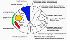
|
|
|
What is the clinical presentation of anterior cord syndrome?
|
Loss of motor function, pinprick and light touch sensation below the level of the lesion with preservation not posterior column function including some touch, position and vibratory sense.
Typically occurs following aortic surgery but may occur after severe hypotension, infection, myocardial infarction, vasospasm and aortic angiography |
|
|
List the non traumatic aetiologies of spinal cord dysfunction?
|
Affecting the spinal cord or blood supply directly:
MS Transverse myelitis Spinal AVM/SAH Syringomyelia HIV myelopathy Other myelopathies Spinal cord infarction Spinal epidural abscess Spinal epidural hematoma Diskitis Neoplasm (metastatic or primary) |
|
|
What are the clinical findings of Guillain-Barre syndrome? What is Miller-Fisher syndrome?
|
Progressive symmetrical distal weakness
Signs and symptoms worse in the lower extremities associated with diminution of deep tendon reflexes, variable sensory findings and anal sphincter sparing Miller Fisher syndrome is a rare form of GBS characterized by the triad of ataxia, ophthalmoplegia and areflexia |
|
|
What is Saturday-night palsy?
|
Radial mononeuropathy associated with improper positioning of the arm during deep, commonly inebriated sleep. The radial nerve is trapped between the humeral shaft and a firm surface causing an external compression mononeuropathy
|
|
|
How does ulnar nerve palsy present?
|
If injured at the elbow, there will be loss of sensation on the palmar/dorsal aspect of the 4th and 5th digit as well as the hypothenar eminence. Motor deficits include inability to flex the wrist or FDP in the 4th and 5th digits. Also cannot abduct and adduct the fingers nor flex the metacarpophalangeal joints of D4 and D5
|
|
|
What are the four components of the neuromuscular unit? Describe the clinical characteristics of neuromuscular diseases for each component?
|
Anterior horn cells of the spinal cord - > motor neuron disease (most commonly ALS) results in decreased strength, increased reflexes, normal sensation and wasting
Peripheral nerve -> neuropathy: may have normal or decreased strength more prominent symptoms distally than proximally, decreased reflexes and decreased sensation, muscle wasting is present Neuromuscular junction ->strength is normal but fatigues, reflexes and sensation are normal and there is no wasting muscle -> myopathy: strength is decreased but more proximally than distally, there are normal reflexes and sensation and there is muscle wasting present |
|
|
What are the clinical signs of myasthenia gravis? What is Lambert Eaton myasthenia syndrome?
|
-ptosis, diplopia, blurred vision
-respiratory muscle weakness -dysarthria, dysphagia Lambert eaton is a myasthenic syndrome where patients have auto-antibodies that cause inadequate release of ACh resulting in weakness that improves with use. (these symptoms are the opposite of myasthenia gravis that gets worse with use) |
|
|
what is the "ice bag test"?
|
It is based on the principle that cooling decreases the symptoms of MG. To perform the test, measure the distance from the upper to the lower eye lid, then apply ice for 2 minutes and re-measure, the distance should be further after the ice is applied
|
|
|
What are the clinical symptoms of botulism?
|
Botulism blocks voluntary and autonomic functions. It causes a descending, symmetric, flaccid paralysis resulting in 1) diplopia, 2) dysarthria 3) dysphagia and generalized weakness
|
|
|
What is an aseptic meningitis?
|
A meningitis with a negative bacterial CSF culture
|
|
|
List 10 non-infectious causes of meningitis?
|
Rubella
Varicella NSAIDs TMP-SMX -carbamazepine Collagen vascular diseases Wegeners Leukemia brain abscess epidural abscess |
|
|
What are the most common causes of viral encephalitis?
|
arboviruses, HSV, HHV are the most common
|
|
|
What is the mortality of HSV encephalitis with and without acyclovir?
|
30% with treatment and 60-70% without treatment
|
|
|
What is the tropism of HSV in CNS infections?
|
Temporal lobes
|
|
|
Describe the kernig and brudzinski signs?
|
kernig - patient lying supine, unable to straighten leg to full knee extension when the hip is flexed at 90 degrees
brudzinski - attempt to flex neck results in flexion of the hips |
|
|
What is the Waterhouse-Friedrichson syndrome?
|
bilateral adrenal hemorrhage
|
|
|
In addition to a cell count, glucose, a gram stain and a culture, what other CSF analysis test could you order and why?
|
india ink stain - cryptococcal
cryptoccocal antigen lactic acid bacterial antigen test (increased in bacterial and tubercular meningitis acid fast stain |
|
|
What is the empiric treatment for meningitis?
|
ceftriaxone or cefotaxime and vanco +/- ampicillin
|
|
|
Discuss the indications for chemoprophylaxis for contacts of bacterial meningitis
|
Meningococcus
-Household contacts of bacteriologically confirmed cases -intimate non household contacts who have had mucosal exposure to the patient's oral secretions There is no indication for chemoprophylaxis in pneumococcal meningitis HIB - only for non-pregnancy household contacts where there are children younger than 4years old in the house |
|
|
How does a common peroneal mononeuropathy present?
|
weakness of the foot dorsiflexors resulting in foot drop. THe everters of the foot are also weak, but the inverters are strong
|
|
|
What is the differential diagnosis of mononeuropathy multiplex?
|
vasculitis
DM neoplasm infectious (Lyme or HIV) sarcoid toxic transient cryoglobulinemia mononeuropathy multiplex is an asymmetrical sensorimotor usually distal pattern of peripheral neuropathy |
|
|
What proportion of strokes are ischemic vs hemorrhagic?
|
Ischemic 80%
Hemorrhagic 20% |
|
|
What proportion of ischemic strokes are thrombotic vs embolic?
|
1/3 thrombotic
1/4 embolic (most commonly from mural thrombus due to a fib) |
|
|
What are the possible etiologies of hemorrhagic stroke?
|
Hypertensive hemorrhage
Amyloid angiopathy (elderly) AVM (bleed may be SAH or ICH) drugs: PCP, cocaine, anticoagulants tumors |
|
|
Define amaurosis fugax
|
Transient monocular blindness due to embolization from a proximal carotid artery plague to the ophthalmic artery
|
|
|
Define TIA
|
Brief episode of neurologic dysfunction caused by a focal disturbance of brain or retinal schema with clinical symptoms lasting <1hour and without evidence of infarction
|
|
|
What proportion of patients with TIA will get stroke within 3 months?
|
10% (and half of these will occur within the first 2 days)
|
|
|
What is the ischemic penumbra?
|
Area of brain surrounding the primary injury, which is preserved by a supply of blood from collateral vessels (this is the area of greates interest to investigators for possible salvage in both ischemic and hemorrhagic stroke)
|
|
|
What proportion of the brain is supplied by the anterior circulation?
|
80%
|
|
|
Where does the anterior circulation originate?
|
the carotid arteries
|
|
|
What areas does the anterior circulation supply?
|
-optic nerve
-retina -frontoparietal -anterior temporal lobe |
|
|
What areas does the anterior cerebral artery supply?
|
basal and medial aspects of the cerebral hemispheres
anterior 2/3 of the parietal lobe |
|
|
What areas does the middle cerebral artery supply?
|
putamen
part of the anterior limb of the internal capsule lentiform nucleus external capsule lateral surface of the cerebral cortex from the frontal lobe to the posterolateral occupital lobe |
|
|
What proportion of the brain is supplied by the posterior circulation?
|
20% of the brain
|
|
|
Where does the posterior circulation originate?
|
the vertebral arteries
|
|
|
What areas does the posterior circulation supply?
|
brainstem
cerebellum thalamus auditory and vestibular centres of the ear medial temporal lobe visual occipital cortex |
|
|
Differentiate Broca's and Wernicke's aphasia
|
Broca: expressive aphasia (patient can understand but cannot express)
Wernicke's: receptive aphasia (patient does not understand speech) |
|
|
What presenting symptoms are more typical of a posterior circulation CVA?
|
LOC
nausea vomiting vertigo ataxia visual field defects (deficits may involve both sides of the body as opposed to anterior circulation strokes in which neuro deficits are always limited to one side) |
|
|
What is the range of possible scores in the NIH stroke scale (NIHSS)?
|
0-42
|
|
|
What is considered a large stroke (by the NIH scale) warranting caution with the use of rTPA?
|
>20
|
|
|
What is the prognostic value of the NIHSS score?
|
Median NIHSS score in patient treated in the NINDS study: 14
Prognosis: 80% of patients with NIHSS <12-14 and 20% of patient with NIHSS >20-26 have a good or excellent outcome |
|
|
What are the early ischemic changes on CT head?
|
-hyperdense artery sign (acute thrombus in a vessel)
-sulcal effacement -loss of the insular ribbon -loss of gray-white interface -mass effect -acute hypodensity |
|
|
What are the most common sites for hypertensive ICH?
|
Putamen
Thalamus Cerebellum Pons Other cortical areas |
|
|
What is the formula for calculating the volume of an ICH on CT?
|
(A*B*C)/2
(C = number of 10mm slices) |
|
|
What are the NINDS recommended stroke evaluation targets?
|
Door to doctor: 10min
Dorr to CT completion: 25min Door to CT scan read: 45min Door to treatment: 60min Access to neurologic expertise: 15min (by phone or in person) Access to neurosurgical expertise: 2 hours (by phone or in person) |
|
|
What are the inclusion criteria for fibrinolysis in acute ischemic stroke?
|
-Age >/=18 years
-Clinical diagnosis of ischemic stroke causing a measurable neurologic deficit -Time of symptom onset well established to be <180min before treatment would begin |
|
|
What are the exclusion criteria for fibrinolysis in acute ischemic stroke?
|
-evidence of intracranial hemorrhage on non contrast CT
-only minor or rapidly resolving stroke symptoms -high clinical suspicion of SAH even with normal CT findings -Active internal bleeding (GI or urinary bleeding in last 21d) -Known bleeding diathesis (including but not limited to platelets <100,000, patient has received heparin within 48hours and has an elevated aPTT greater than upper limit of normal for lab, recent use of anticoagulant and elevated PT >15s) -within 3 months of intracranial surgery, serious head trauma or previous stroke -within 14 days of major surgery of serious trauma -recent arterial puncture at a noncompressible site -lumbar puncture within 7days -history of ICH, AVM or aneurysm -witnessed seizure at stroke onset -recent AMI -on repeated measurements SBP >185mmHg or DBP >110mmHg at time of treatment requiring aggressive treatment to decreased BP to within these limits |
|
|
When should HTN be treated in the context of acute ischemic stroke?
|
-markedly elevated BP
-fibrinolytic therapy is planned -specific medical indications: AMI, AD, hypertensive encephalopathy, severe left ventricular failure |
|
|
What is the management of acute hemorrhagic stroke?
|
IV nitroprusside/labetalol if SBP >180 or DBP >105
Hyperventilation + mannitol for increased ICP Seizure prophylaxis Neurosurgery consult (surgery is more efficacious in patients with cerebellar hemorrhage) |
|
|
What is the ABCD2 score?
|
Age >60 (1 point)
SBP >140 and/or DBP >90 (1 point) Clinical features: unilateral weakness (2 points), speech disturbance without weakness (1 point), other (0 points) Duration of symptoms: <10min (0 points), 10-59min (1 point), >60min (2 points) Diabetes (1 point) |
|
|
What is the significance of the ABCD2 score
|
The higher the score the higher the risk of stroke at 2, 7, and 90 days
High risk (score 6-7) 8% - 2 day risk Moderate risk (score 4-5) 4% low risk (score 0-3) 1% |
|
|
What are "crescendo TIAs"?
|
3 or more TIAs within 72hours
|
|
|
What are predisposing factors to stroke in patients 15-45years of age
|
Increased clotting: OCP, pregnancy, antiphospholipid antibodies, protein C and S deficiencies, polycythemia
Fibromuscular diseases, migraine Cocaine, PCP, amphetamine carotid and vertebral artery dissections |
|
|
What are signs of anterior cerebral artery stroke?
|
mainly affects frontal lobe function:
-altered mentation and impaired judgement and insight -primitive grasp and suck reflexes -bowel and bladder incontinence may be features -paralysis and hypesthesia of the lower limbs on the contralateral side -leg weakness>arm weakness -apraxia or clumsiness of gait |
|
|
What are signs of a middle cerebral artery stroke?
|
-contralateral motor and sensory disturbances
-worse in the arm and the face than the leg -homonymous hemianopsia (blindness in one half on the same side of both eyes) -agnosia (inability to recognize sugjects) -aphasia if it is dominant -patient looks at the destructive lesion (stroke) but away from the irritative lesion (seizure focus) |
|
|
What are signs of a posterior circulation stroke?
|
-cranial nerve deficits
-cerebellar involvement -involvement of the neurosensory tract -involvement of the reticular activative system (emesis center and responsible for consciousness) -patients frequently present with nausea and vomiting and may have LOC -vision and thought processing may be impaired (alexia, visual agnosia, visual neglect, homonymous hemianopsia, cranial nerve III palsy) -vertebrobasilar arteru insufficiency (vertigo, diplopia, nystagmus, weakness, paralysis) -crossed deficits: motor deficits on one side and sensory loss on the other |
|
|
Which strokes may have pinpoint pupils?
|
-thalamus
-pontine |
|
|
What are poor prognostic indicators in hemorrhagic strokes?
|
-decreased LOC on arrival
-IVH -ICH volume >40cc |
|
|
What is the CHADS2 score?
|
Congestive heart failure (any history) (1)
Hypertension (prior history) (1) Age >75 (1) Diabetes Mellitus (1) Stroke (ischemic or TIA) (2) |
|
|
What are signs of CN III palsy?
|
Eyes deviated down and out
ptosis pupil dilation |
|
|
What are signs of CN IV palsy?
|
Can't look to the floor (or medial)
|
|
|
What are signs of CN VI palsy?
|
Can't look lateral
|
|
|
What does gaze preference suggest?
|
brainstem or cortical involvement
|
|
|
What is a sign of PCA aneurysm?
|
Pupil dilation
|
|
|
What indicates lesion of the vasa vasorum in DM2 and HTN
|
CN III lesion without dilatation
|
|
|
What is the differential diagnosis of stroke?
|
EDH
SDH Carotid dissection Aortic dissection into the carotids brain tumor abscess air embolism temporal arteritis Polyarteritis nodosa SLE Wernicke's encephalopathy Bells palsy Venous sinus thrombosis |
|
|
What is the triad of Wernicke's encephalopathy
|
ataxia
ophthalmoplegia confusion |
|
|
Where do venous sinus thromboses occur?
|
In the superior saggital sinus or the lateral sinus
|
|
|
What are risk factors for venous sinus thrombosis?
|
-trauma
-infection -hypercoagulable state -low flow state -dehydration -drugs (androgens, ecstasy, oral contraceptives) -compression of the venous sinus -pregnancy or postpartum state |
|
|
How is venous sinus thrombosis diagnosed?
|
MRI/MRV
|
|
|
What are signs of venous sinus thrombosis on contrast CT?
|
empty delta sign (lack of full contrast)
|
|
|
What are signs of venous sinus thrombosis on non-contrast CT?
|
delta sign:dense triangle in superior sagittal sinus
|
|
|
What is the treatment of a venous sinus thrombosis?
|
LMWH - even in those who demonstrate hemorrhage
|
|
|
What are specific BP therapies in patients with ischemic stroke who are not candidates for thrombolysis?
|
DBP>140mmHg -> sodium nitroprusside 0.5ug/kg/min (goal is 10-20% reduction)
SBP >220, DBP >120, MAP >130 -> labetalol 10-20mg SBP<220, DBP<120, MAP <130 -> no therapy unless medical condition |
|
|
What are the indications to treat blood pressure in acute ischemic stroke in a patient who is a thrombolytic candidate?
|
If SBP >185 or DBP >110 the ngive labetalol, if BP not reduced and maintained below 185/110 then patient should not be treated with TPA
|
|
|
What does Rosen's recommend for management of HTN in Spontaneous ICH?
|
Treat if SBP>160-180 or DBP >105
Rosens recommends nitroprusside though it can increase hemorrhage/ICP through vasodilation Alternatives: labetalol and nicardipine |
|
|
What are complications of interventricular hemorrhage or hemorrhage into the posterior fossa?
|
Hydrocephalus which may require insertion of a ventricular catheter
|
|
|
What are the American stroke association guidelines for treatment of spontaneous ICH?
|
If SBP >200mmHg or MAP >150mmHg consider aggressive reduction
If SBP >180mmHg or MAP >130mmHg there is a possibility of elevated ICP. If elevated ICP maintain CPP of >/=60mmHg If SBP >180mmHg or MAP >130mmHg and there is not elevation in ICP then consider modest reduction in BP to MAP of 110mmHg or target BP of 160/90 using intermittent or continuous IV medications |
|
|
When is it possible to discharge an ischemic stroke patient?
|
Multiple previous strokes and already thoroughly evaluated
Mild new stroke Completed stroke days to weeks after event |
|
|
Where is the motor cortex?
|
At the end of the frontal lobe
|
|
|
Where is the sensory cortex?
|
At the front of the parietal lobe
|
|
|
What are symptoms of anterior cerebral artery infarction?
|
Sensory and motor symptoms in the lower extremity with sparing of the hands and face
Also may have akinetic mutism and transcortical mortor aphasia, motor hemineglect |
|
|
What are typical symptoms of an MCA stroke?
|
hemiparesis
facial plegia sensory loss contralateral to the affected side |
|
|
What are presenting features of a posterior cerebral artery infarction?
|
classically visual field defects (contralateral homonymous hemianopsia and unilateral cortical blindness)
Motor function is typically minimally affected |
|
|
What is the presentation of vertebrobasilar infarction?
|
-multiple simultaneous symptoms
-commonly vertigo -headache -nausea -visual disturvances -oculomotor palsies, ataxia, sensory disturbance, limb weakness, oropharyngeal dysfunction The hallmark is crossed neurologic deficits (ipsilateral cranial nerve deficits with contralateral motor weakness) |
|
|
What are the signs of basilar artery occlusion?
|
quadriplegia
coma locked-in syndrome (complete muscle paralysis except for upward gaze) |
|
|
What are presenting features of cerebellar infarction?
|
vertigo
gait instability limb ataxia headache dysarthria nausea vomiting cranial nerve abnormalities |
|
|
What is particular about cerebellar infarction?
|
-there may be rapid deterioration secondary to increased brainstem pressure caused by cerebellar edema
-the most important factor influencing outcomes is the presence of obstructing hydrocephalus |
|
|
What are lacunar infarcts
|
pure motor or sensory deficits caused by infarction of small penetrating arteries and are commonly associated with chronic hypertension and increasing age
|
|
|
What is a partial complex seizure?
|
Involves altered cognition
Cognition is defined as involving at least 2 of 5 features -perception -attention -emotion -memory -executive function |
|
|
What is a spasm?
|
A specific debilitating seizure syndrome that occurs in infants
|
|
|
What is myoclonus?
|
rhythmic shock like muscle contractions typical for specific seizure syndromes
|
|
|
What is the Jacksonian march?
|
seizure with ipsilateral motor signs that spread contiguously in a stepwise fashion
|
|
|
What are nonconvulsive generalized seizures?
|
absence (typical or atypical)
myoclonic tonic atonic (drop attack) |
|
|
What is the definition of status epilepticus?
|
Continuous seizure activity lasting more than 5 minutes or more than two discrete seizures without intervening recovery of consciousness
|
|
|
What are common etiologies of status epilepticus?
|
Metabolic disturbances
Infectious processes Withdrawal symptoms CNS lesions Intoxication |
|
|
What are triggers for seizures?
|
awakening
sleep deprivation emotional or physical stress alcohol menses specific sensory stimuli (flashing lights) medication noncompliance |
|
|
What intoxications can cause seizures?
|
Bupropion
Camphor Clozapine Cyclosporine Flumazenil Imipenem Isoniazid Lead Lidocaine Lithium Metronidazole Theophylline TCA |
|
|
What CNS lesions can cause seizures?
|
acute hydrocephalus
anoxic or hypoxic insult AVM brain mets CVA chronic epilepsy eclampsia head trauma ICH neoplasm neurosurgery posterior reversible leukoencephalopathy remote structural injury |
|
|
Which withdrawal syndromes can cause seizures?
|
Alcohol
Antiepileptic drugs Baclofen Barbiturates Benzodiazepines |
|
|
What metabolic disturbances cause seizures?
|
hepatic encephalopathy
hypocalcemia hypoglycemia or hyperglycemia hyponatremia uremia |
|
|
What infectious processes can cause seizures?
|
CNS abscess
encephalitis meningitis |
|
|
What is an immediate post-traumatic seizures?
|
Occurs within 24 hours of injury
|
|
|
What is an early post-traumatic seizures?
|
Occurs within 1 week of injury
|
|
|
What is late post-traumatic seizure?
|
Occurs greater than 1 weeks after injury
|
|
|
What are the recommendations for antiepileptic drugs post trauma?
|
they are recommended for prophylaxis against post traumatic seizures occurring within the first 7 days after severe brain injury. The have not been shown to be effective in preventing late post-traumatic seizures
|
|
|
What is Todd's paralysis
|
a focal motor deficit that may persist up to 24 hours post seizure
It is associated with a high likelihood of an underlying structural cause |
|
|
Which patients with seizures require CT head
|
new focal deficit
persistent altered mental status fever recent trauma persistent headache PMH of cancer anticoagulant use suspicion or known history of AIDS age>40 years partial complex seizure |
|
|
What proportion of patients diagnosed with having seizures are found to have been misdiagnosed?
|
20-25%
|
|
|
What is the differential diagnosis of a seizure?
|
Psychogenic
Hyperventilation Breath holding Toxic and metabolic states TIA narcolepsy movement disorders |
|
|
What is the management of a seizure?
|
Left lateral decubitus position
place bite block Benzodiazepines Phenytoin |
|
|
Why is lorazepam the drug of choice for the initial management of epilepsy?
|
terminates seizures rapidly and ahs a long duration of action (4-6hours)
this is different from the management of ETOH withdrawal seizures |
|
|
What is the maximum rate of infusion for phenytoin?
|
50mg/min
|
|
|
What is the onset of action of phenytoin
|
10-30 minutes
|
|
|
What is the dose of propofol in status epileptics?
|
1-3mg/kg bolus followed by an infusion of 1-15 mg/kg/hr
|
|
|
How does barbiturate coma work for status epileptics?
|
suppresses all brainstem function. The preferred agent is pentobarbital. Patients require intubation, ventilatory support, continuous cardiac monitoring, invasive hemodynamic monitoring
|
|
|
What is the treatment for refractory ictus?
|
Isoflurane anesthesia
|
|
|
What are the diagnostic criteria for migraine?
|
->/=5 attacks fulfilling the other criteria
-attache lasts 4-72hours -headache has >/=2 of the following (unilateral, pulsating, moderate to severe pain intensity, aggravation by or causing avoidance of routine physical activity) -during headache >/=1 of the following (nausea and/or vomiting, photophobia and phonophobia) -not attributable to another disorder |
|
|
Define status migranosus
|
Severe migraine headache that persists longer than 72 hours
|
|
|
What is the management of moderate to severe headache?
|
Dihydroergotamine 1mg IV or IM
Sumatriptan 6mg SC Metoclopramide 10mg IV Prochlorperazine 10mg IV or IM |
|
|
What is the management of refractory migraine/status migranosus?
|
Dihydroergotamine 1mg IV q8h
Dexamethasone 10mg IV to reduce recurrence 24-72h after treatment (NNT = 9) |
|
|
What are contraindications to the use of dihydroergotamine?
|
Pregnancy
Breast-feeding Poorly controlled hypertension Coronary artery disease Peripheral vascular disease Recent intake of drug in the triptan class (they last 24hours) |
|
|
What are triggers of cluster headache and typical features?
|
Triggers
ingestion of alcohol stress climatic change Clinical presentation several episodes within 24 hours each HA lasts from a few minutes to 2 hours unilateral sharp, stabbing pain in the eye pani is in the territory of the trigeminal nerve may awaken patient from sleep 30% have partial Horners (ptosis and miosis) |
|
|
What is the treatment of cluster HA?
|
Sumatriptan 6mg SC given as early as possible
High flow oxygen 7-10L/min If no response: intranasal lidocaine prednisone 60mg QD x 10 days followed by a 1 week taper |
|
|
What are the RF associated with nontraumatic SAH?
|
Hypertension
Smoking Excessive alcohol consumption sympathomimetic drugs intracranial vascular anomaly (berry aneurysm, AV malformation, etc.) |
|
|
What is the most common cause of nontraumatic SAH?
|
80% have a ruptured saccular aneurysm (=berry aneurysm)
~5% of the population harbour a berry aneurysm |
|
|
What is the Hunt and Hess SAH grading scale?
|
Grade 0 - unruptured aneurysm, asymptomatic
Grade 1 - asymptomatic or minimal headache and slight nuchal rigidity Grade 2 - moderate or severe headache, nuchal rigidity, no neurologic deficit other than cranial nerve palsy Grade 3 - drowsiness, confusion or mild focal deficit Grade 4 - stupor, moderate to severe hemiparesis Grade 5 - deep coma, decerebrate posturing, moribund appearance Grade 1 and 2 -> good prognosis Grade 4-5 -> poor prognosis |
|
|
What is the sensitivity of CT head for the diagnosis of spontaneous SAH?
|
>90% for hemorrhage less than 24 h old
~50% by the end of the first week |
|
|
How do you differentiate a traumatic tap from SAH?
|
-the method of comparing the rbc count in the first and last tube has been shown to be unreliable
-the patient's csf should be spun and the supernatent observed for xanthochromia -the yellowish pigmentation of xanthochromia is secondary to metabolism of hemoglobin to pigmented molecules of oxyhemoglobin and bilirubin and is a process that takes approx 12h -order vascular imaging when the level of clinical suspicion of SAH is high and CT head is negative and CSF is negative for xanthochromia |
|
|
What is the management of nontraumatic SAH?
|
ABCs
nimodipine 60mg PO q4h to reduce the risk of poor outcome Antiemetics Analgesics Treat seizures with benzodiazepines (seizure prophylaxis is controversial) neurosurgery consult BP control to be discussed with neurosurgery (likely will place a ventriculostomy if antihypertensive drugs are required, use labetalol) |
|
|
What is the management of carotid/vertebral dissection?
|
Supportive
CT angiography Vascular surgery consult Early anticoagulation followed by antiplatelet therapy |
|
|
What is the clinical presentation of cerebral venous sinus thrombosis?
|
Headache, nausea, vomiting, seizures, decreased LOC that may progress to coma.
With cavernous sinus thrombosis -> ocular findings including orbital pain, proptosis and EOM paralysis |
|
|
What is the presentation of trigeminal neuralgia?
|
Unilateral
Brief electric shock like pains in the distribution of >/=1 division of the trigeminal nerve may be triggered by trivial stimuli may occur spontaneously individual attacks are brief, lasting a few seconds to <2minutes |
|
|
What are the diagnostic criteria for idiopathic intracranial hypertension
|
-alert patient with neuro exam that yields normal findings or demonstrates any of the following findings (papilledema, enlarged blind spot, visual field defects)
-increased CSF pressure (>20 cmH20 in non obese and >25cm H20 in obese patients) -normal csf chemistry -intracranial disease ruled out by appropriate investigations -no metabolic, toxic or hormonal cause |
|
|
What are the HA redflags
|
Fever
First Worst thunderclap AMS Trauma Immunocompormised child Focal exam Nuchal rigidity Syncope Anticoagulated Elderly Seizure Change from usual Progressive and unresolving HA never changes side Neoplastic history New HA in someone over 50 Triggered by exertion, sex, or valsalva |
|
|
What is the mnemonic for migraine headache
|
POUND
Pulsatile hOurs 4-72hrs Unilateral Nausea and Vomiting Disabling |
|
|
What is the differential diagnosis of migraine
|
Ruptured aneurysm
AVM tumor GCA cerebral vascular disease |
|
|
What is the purpose of the Hunt and Hess classification
|
It predicts outcome post SAH
Grade I or II = good prognosis Grade IV or V = do poorly |
|
|
What is HHH therapy for SAH
|
Hypertension
Hypervolemia Hemodilution |
|
|
What is the classic triad of a brain timor?
|
Sleep disturbance
Severe pain Nausea and Vomiting |
|
|
What is Giant Cell Arteritis?
|
Inflammatory process of small and medium-sized arteries
|
|
|
What are the American College of Rheumatology Criteria for Temporal Arteritis?
|
Onset of disease at the age of 50 years or older
New HA Temporal artery abnormalities (tenderness to palpation or decreases in pulsation) ESR >50mm/hr Abnormal findings on temporal artery biopsy |
|
|
What is the treatment of temporal arteritis?
|
Prednisone 60-120mg/day
|
|
|
What is the most frequent cause of stroke in patients<45years
|
Carotid or vertebral artery dissection
|
|
|
What are the causes of carotid and vertebral artery dissection?
|
Sudden neck movement or trauma
(neck torsion, chiropractic manipulation, MVC, coughing, minor fall) |
|
|
What is the classic triad of carotid dissection?
|
Unilateral HA
Ipsilateral Horner's Contralateral hemispheric findings |
|
|
What is the classic presentation of vertebral dissection?
|
relatively young person
severe, unilateral posterior HA and neurologic findings |
|
|
How do you diagnose carotid/vertebral dissection?
|
CT
MRI MRA catheter angiography |
|
|
What is the treatment for carotid and vertebral artery dissection?
|
Early anticoagulation followed by antiplatelet therapy
|
|
|
What are the other names for idiopathic intracranial hypertension?
|
Pseudotumor cerebri
Benign intracranial hypertension |
|
|
What is the pathophysiology of idiopathic intracranial hypertension?
|
Increased brain water content and decreased csf outflow
|
|
|
What are the signs of idiopathic intracranial hypertension?
|
Papilledema
Visual Field defects (enlarged blind spot followed by decrease in peripheral vision) Occasionally VI palsy |
|
|
What are the diagnostic criteria for delirium?
|
-clouding of consciousness with decreased ability to focus, sustain or shift attention
-a cognitive change or perceptual disturbance that is not better accounted for by a preexisting established evolving dementia -development of the disturbance over hours to days with a tendency to fluctuate during the course of the day -evidence from the HPI, PE, or lab studies that disturbance is caused by a general medical condition, medication, or other substance exposure, substance withdrawal or multiple etiologic disorders |
|
|
What are the causes of delirium?
|
I WATCH DEATH
Infectious Withdrawal Acute metabolic Trauma CNS disease Hypoxia Deficiencies Environmental Acute vascular Toxins/drugs Heavy metals |
|
|
What are the diagnostic criteria for dementia?
|
A. Multiple cognitive deficits manifested by both of the following:
1) memory impairment 2) one or more: aphasia, apraxia, agnosia, disturbance in executive functioning (planning, organizing, sequencing, abstracting) B. The cognitive deficits cause significant impairment in social or occupational functioning C. The deficits do not occur exclusively during the course of delirium |
|
|
What are the reversible causes of demetia?
|
Hypothyroidism
Alcohol-related Vitamin B12 deficiency Infectious (syphilis, AIDS) Trauma (SDH) |
|
|
What is the MMSE
|
Total: 30
Orientation: year, season, date, day, month (5) city, state, country, hospital, floor (5) registration name 3 objects (3) attention and calculation serial sevens backwards or spell WORLD backward (5) recall ask for 3 objects (3) Language name pencil and watch (2) repeat no ifs and or buts (1) three stage command (3) read and obey "close your eyes" (1) write a sentence (1) Copy the design (1) |
|
|
Differentiate delirium and acute psychosis with regards to onset, VS, prior psych history, course, psychomotor activity, involuntary activity?
|
delirium
acute onset typically abnormal VS uncommon psych history rapid/fluctuating course variable psychomotor activity possible asterixis or tremor acute psychosis acute onset normal VS usually psychiatric history stable course variable psychomotor activity absent involuntary activity |
|
|
Differentiate delirium and acute psychosis with regards to orientation, attention, concentration, hallucinations, delusions, speech and course
|
delirium
impaired orientation impaired attention impaired concentration visual and/or auditory transient poorly organized delusions pressure slow speech, possibly incoherent typically resolves acute psychosis occasional impaired orientation attention may be disorganized concentration is impaired primarily auditory hallucinations systematized delusions speech is usually coherent responds to therapy but recurs commonly |
|
|
What is the diagnostic evaluation for delirium?
|
cbc
SMA10 CXR/urinalysis/LP CT head anion gap lactic acid ammonia ECG TSH/T4 B12 Folic acid RPR |
|
|
Describe the presentation of normal pressure hydrocephalus
|
dementia, ataxia and urinary ataxia
(usually <60yrs) |
|
|
What is the clinical function of CN I?
|
sense of smell
|
|
|
What types of trauma can injure CN I?
|
Skull fracture
Shear injury interrupting olfactory fibers traversing the cribiform plate |
|
|
What types of tumor can injure CN I?
|
Frontal lobe masses compressing the nerve
|
|
|
What is the clinical function of CN III?
|
Extraocular function via motor fibers to levator palpebrae, superior rectus, medial rectus, inferior rectus and inferior oblique muscles
Pupillary constriction via parasympathetic fibers |
|
|
What are causes of injury to CN III?
|
Trauma: herniation of the temporal lobe through the tentorial opening causing compression and stretch injury
Ischemic: especially in diabetes microvascular ischemic injury to nerve causes EOM paralysis but usually is pupillary sparing Vascular: intracranial aneurysms may press on the nerve, leading to dysfunction Myasthenia gravis can lead to atraumatic ocular muscle palsy |
|
|
What is the clinical function of CN IV?
|
motor supply to the superior oblique muscle
|
|
|
What are the pathologic features of CN IV injury?
|
inability to move eye downwards and inwards
|
|
|
What causes CN IV dysfunction?
|
trauma is the most common cause of injury
|
|
|
What is the clinical function of CN V?
|
Motor supply to the muscles of mastication and to the tensor tympani
Sensory to face, scalp, oral cavity |
|
|
What are the pathologic features of CN V dysfunction?
|
Partial facial anesthesia
Episodic lancinating facial pain |
|
|
What is the clinical function of CN VI?
|
Motor supply to the lateral rectus muscle
|
|
|
What are the pathologic features of CN VI injury?
|
Inability to move affected eye laterally
Diplopia on attempting lateral gaze |
|
|
What are causes of injury to CN VI?
|
tumor: lesions in the cerebellopontine angle
Any lesion vascular or otherwise in the cavernous sinus may compress the nerve elevated ICP because of its position and long intracranial length. |
|
|
What is the clinical function of CN VII?
|
Motor supply to the muscles of facial expression
Parasympathetic stimulation of the lacrimal, submandibular and sublingual glands Sensation to the ear canal and tympanic membrane |
|
|
What are the pathologic features of CN VII?
|
LMN:Hemifacial paresis - leaves entire side of face paralyzed
UMN: leaves forehead musculature functioning Abnormal taste Sensory deficit around the ear intolerance to sudden loud noises |
|
|
What are causes of CN VII injury?
|
LMN:
infection (viral) lyme disease bacterial infection extending from otitis media UMN: stroke tumor |
|
|
What is the clinical function of CN VIII?
|
hearing and balance
|
|
|
What are the pathologic features of CN VIII?
|
Unilateral hearing loss
tinnitus vertigo and unsteadiness |
|
|
What are the causes of CN VIII injury?
|
tumors: acoutic neuroma
mimics: meniere's, perilymphatic fistula |
|
|
What are the clinical functions of CN IX?
|
General sensation to the posterior 1/3 of the tongue
Taste for posterior 1/3 of the tongue Motor supply to the stylopharungeus |
|
|
What are the causes of CN IX injury?
|
Brainstem lesion
Glossopharyngeal neuralgia |
|
|
What are the clinical functions of CN X?
|
Motor supply to the striated muscles of the pharynx, larynx and tensor palatine
Motor to the smooth muscles and glands of the pharynx, larynx, thoracic, and abdominal viscera Sensory to larynx, trachea, esophagus, thoracic and abdominal viscera |
|
|
What are the pathologic features of CN XI injury?
|
downward and lateral rotation of the scapula and shoulder drop
|
|
|
What are the presentations of UMN and LMN injury to CN XII?
|
LMN - toward the lesion
UMN - away from the lesion |
|
|
What are the causes of trigeminal neuralgia?
|
Idiopathic
Compression by vessel/AVM/tumor |
|
|
What is the differential diagnosis of trigeminal neuralgia?
|
-dental infection
-sinus disorder -OM -acute glaucoma -TMJ disease -herpes zoster |
|
|
What is the medication of choice in trigeminal neuralgia?
|
Carbamazepine
week 1: 100mg BID week 2: 100mg TID max 1200 mg/day |
|
|
What is the treatment of peripheral CN VII lesions?
|
patch the eye
lacrilube f/u with neurology and ophthalmology steroids - thought to reduce edema of nerve which is confined in facial canal (pred 1mg/kg/d 7-10 d, best if started within 24 hours but can be given within 1 week valacyclovir - 1000mg BID x 10d best within 24hrs but can start within 1 week |
|
|
What is Ramsey Hunt syndrome?
|
Unilateral facial paralysis with herpetiform rash (pinna, external auditory canal, TM, soft palate, oral cavity, face and neck down to shoulder and vesticulocochlear dysfunction
Pain worse than in Bell's palsy, lower incidence of complete recovery |
|
|
What is the most common cause of bilateral VII paralysis?
|
Lyme or infectious mono
|
|
|
What injury typically causes facial nerve injury?
|
temporal bone fracture with nerve transection
|
|
|
What is the hallmark of acoustic neuroma?
|
asymmetrical sensorineural hearing loss
|
|
|
How do you differentiate sensorineural and conductive hearing loss?
|
Weber test - tuning fork on vertex, localizes to same side in conductive and opposite side in sensorineural hearing loss
Rinne test - place tuning fork on mastoid process then to ear once no longer heard if conductive then not able to hear after mastoid test in sensorineural the ability to sense the tuning fork is diminished at mastoid and ear |
|
|
Which CN can present with diabetic cranial mononeuropathy?
|
CN III, IV or VI
|
|
|
What is the pathophysiology of diabetic cranial mononeuropathy?
|
ischemia to the intraneural arteries of the CN. mostly the central portion of the nerve is affected because the peripheral portions have a collateral blood supply
|
|
|
What is the management of diabetic cranial mononeuropathy?
|
patch affected eye
analgesics antiplatelets tends to improve in 3-6 moths and resolves in 1 year. |
|
|
What are the major dural sinuses?
|
Superior sagittal
Inferior sagittal Straight sinuses Lateral sinuses Sigmoid sinuses |
|
|
What is the gold standard for the diagnosis of cerebral venous thrombosis?
|
MRI and MRV
|
|
|
What is the management of cerebral venous thrombosis?
|
Admit to NICU
Systemic anticoagulation to PTT of 80-100secs Consider catheter-based thrombolysis if decreased LOC, elevated ICP or rapid deterioration on neurologic examination |
|
|
What is Uhthoff's phenomenon?
|
MS exacerbations are worse when the body temperature is increased
|
|
|
What are the csf findings in MS?
|
Pleocytosis (>5lymphocutes/hpf )
High IgG Oligoclonal IgG bands on electrophoresis |
|
|
What are the reasons for emergency MRI?
|
Cauda
Transverse myelitis cancer and back pain SAH of cord |
|
|
Where does the spinal cord end?
|
L1/L2
|
|
|
What nerves give rise to brachial plexus?
|
C5-T1
|
|
|
What nerves give rise to the lumbosacral plexus?
|
L2-S3
|
|
|
What artery supplies the anterior 2/3 of the spinal cord?
|
Anterior spinal artery
|
|
|
Where does the artery of Adamkiewicz originate?
|
Between T8 and L4
|
|
|
What is the lateral corticospinal tract?
|
A descending tract that controls voluntary movement on the ipsilateral side (decussates in the medulla)
|
|
|
What are the posterior columns?
|
Ascending tracts that carry proprioceptive and vibratory sense on the ipsilateral side of the cord
|
|
|
What are the lateral spinothalamic tracts?
|
Ascending tracts that carry pain/temp sensation on the contralateral side of the body (decussate immediately)
|
|
|
What are the deficits in Brown Sequard symdrome?
|
Motor loss ipsilateral to cord lesion
Position and vibration loss ipsilateral to the cord lesion Pain and temperature sensation loss contralateral to the cord lesion |
|
|
What are the deficits in anterior cord syndrome?
|
Motor loss or weakness below cord level
Loss of pain and touch sensation, vibration and position sense preserved |
|
|
What are the deficits in transverse cord syndrome?
|
Loss of sensation below the level of the cord injury
Loss of voluntary motor function below cord level Sphincter control lost |
|
|
What is spinal shock?
|
Loss of muscle tone and reflexes with complete cord syndrome during the acute phase.
Usually <24hrs but up to weeks |
|
|
What are the deficits in central cord syndrome?
|
Variable sensory deficits
Upper extremity weakness distal>proximal |
|
|
What is the cause of central cord syndrome?
|
hyperextension from squeezing/pinching of spinal cord anterior and posterior by inward bulging of the ligamentum flavum
|
|
|
Which patient populations classically get central cord syndrome?
|
Elderly with degenerative arthritis and spinal stenosis or any patient with cervical narrowing
|
|
|
What are the mechanisms for Brown-Sequard spinal cord syndrome?
|
Penetrating injury
Spinal cord tumor Spinal EDH Vascular malformations Cervical spondylosis DDD Radiation injury Spinal instrumentation |
|
|
What are the mechanisms for anterior cord syndrome?
|
Post aortic surgery
Severe hypotension Infection MI vasospasm aortic angiography cervical hyperflexion with cord contusion from protrusion of bony fragments |
|
|
What is conus medullaris syndrome?
|
Increased motor tone/reflexes
sphincter impaired distrubance in urination/sexual dysfunction |
|
|
Differentiate conus medullaris syndrome from cauda equina syndrome?
|
Conus medullaris
rare usually bilateral symptoms UMN pattern tend to have overflow incontinence Cauda equina progressive symptoms decreased DTR (LMN pattern) urinary retention |
|
|
What are the findings on strength, DTR, sensation and muscle wasting in myelopathy?
|
Strength normal to decreased
DTR increased Sensation normal to decreased Wasting no |
|
|
What are the findings on strength, DTR, sensation and muscle wasting in motor neuron disease (ALS)
|
Strength decreased
DTR increased sensation normal Wasting yes |
|
|
What is myelopathy?
|
Cord pathology
|
|
|
What are the findings on strength, DTR, sensation and muscle wasting in neuropathy (like GBS)?
|
Strength normal to decreased
DTR decreased Sensation Decreased Wasting yes |
|
|
What is transverse myelitis?
|
Acute or subacute spinal cord dysfunction due to inflammation
|
|
|
What are the causes of transverse myelitis?
|
Post-viral
Infectious Autoimmune Idiopathic |
|
|
What is a syringomyelia?
|
A cavitary lesion within the spinal cord which is usually chronic progressive
|
|
|
What are the clinical features of syringomyelia?
|
HA
neck pain sensory abnormalities in a "cape-like" distribution over the shoulders and arms |
|
|
What is a spinal epidural abscess?
|
An infection of the adipose tissue of the dorsal epidural space
|
|
|
How do you diagnose a spinal epidural abscess?
|
MRI
LP is relatively contraindicated |
|
|
What is the management of a spinal epidural abscess?
|
Refer for decompressive surgery
3rd gen ceph, IV vanco and PO rifampin |
|
|
What is diskitis?
|
Infection of the nucleus pulposus with secondary involvement of the cartilagenous end plate and vertebral body
|
|
|
Which cancers typically metastasize to the spine
|
Lung
Breast Lymphoma |
|
|
What is hypokalemic periodic paralysis
|
Rare neuromuscular disorder related to a defect in muscle ion channels characterized by episodes of painless muscle weakness which is precipitated by heavy exercise, fasting or high-carbohydrate meals
|
|
|
What are the components of the neuromuscular unit?
|
Anterior horn cells of the spinal cord
Peripheral nerve Neuromuscular junction Muscle |
|
|
What is a myelopathy?
|
Disorder that involves the spinal cord
|
|
|
What is a radiculopathy?
|
Disorder that involves the nerve roots
|
|
|
What is a myopathy?
|
Disorder that involves the muscles
|
|
|
What types of receptors are found at the motor synapses?
|
Nicotinic
|
|
|
How do you grade motor strength?
|
5 = normal
4 = weak but able to resist examiner 3 = moves against gravity but unable to resist examiner 2 = moves but unable to resist gravity 1 = flicker but no movement 0 = no movement |
|
|
Distinguish between UMN and LMN involvement with regards to DTR, muscle tone, atrophy, fasciculations, babinski
|
UMN
increased DTR increased muscle tone no atrophy no fasciculations babinski present LMN decreased DTR decreased muscle tone atrophy present fasciculations present babinski absent |
|
|
What are indications for intubation in neuromuscular disease?
|
FVC < 15cc/kg or
max negative inspiratory force <15mmHg (<20cmH20) unable to count from one to 25 in one breath |
|
|
What muscles weaken before respiratory drive is lost?
|
The ability to lift the head (neck muscles)
|
|
|
What are typical motor neuron diseases?
|
ALS
Poliomyelitis |
|
|
What is ALS?
|
Degeneration of motor neurons without sensory involvement
|
|
|
What is poliomyelitis?
|
Degeneration of the anterior horn cells resulting in LMN disease without sensory involvement
|
|
|
How does neuropathy present?
|
Distal muscle weakness>proximal muscle weakness with or without decreased DTR and decreased sensation
|
|
|
How do diseases of the neuromuscular junction present?
|
Progressive motor fatigability
|
|
|
What is myasthenia gravis?
|
Autoantibodies against post synaptic nicotinic ACh receptors (therefore no muscarinic symptoms)
|
|
|
What is botulism?
|
Disorder where toxin binds irreversibly to the presynaptic membrane of peripheral and cranial nerves inhibiting ACh release
|
|
|
What are the features of a Lambert-Eaton myasthenic syndrome?
|
Weakness that is more pronounced at the beginning of muscle use which improves with repetition as more ACh is released
|
|
|
What are features of myopathies?
|
Generalized symmetric weakness with or without muscle tenderness
|
|
|
What findings are often the first in myasthenia gravis?
|
Ocular findings (ptosis worse at the end of the day, diplopia or blurred vision)
|
|
|
How do you diagnose myasthenia gravis?
|
-test serum for ACh receptor antibody
-edrophonium (tensilon test) where you measure teh distance from the upper to the lower eyelid before and after IV edrophonium (AChE blocking agent) -ice test (cooling decreases the symptoms of MG)positive test is improvement of 2mm in distance between upper and lower eyelid after an ice pack is applied for 2 min |
|
|
What is in the differential for myasthenic crisis?
|
Cholinergic crisis
|
|
|
How do you differentiate a myasthenic crisis from a cholinergic crisis?
|
Control the airway
Hold medications -> if the patient improves than symptoms are from a cholinergic crisis |
|
|
What is the management of a myasthenic crisis?
|
CONTROL the airway
Cholinesterase inhibitors (inhibit ACh degradation) -Pyridostigmine -Neostigmine There is no big benefit from pyridostigmine or neostigmine in MG crisis when compared to steroids or plasmapheresis. Most patients with MG crisis have failed AChE inh and need a rest from their meds Thymectomy Immunomodulatory therapy (IVIG or plasma exchange) |
|
|
What drugs may exacerbate MG?
|
Things that worsen sodium entry or calcium flow into the cell
BB CCB Lidocaine Procainamide Aminoglycosides Tetracycline Clindamycin Phenytoin Neuromuscular blockers Corticosteroids Thyroid replacement Cholinesterase inhibitors |
|
|
In what disease do you get Lambert Eaton Myasthenic syndrome?
|
Small cell lung CA
|
|
|
What is the pathophysiology in Lambert Eaton myasthenic syndrome?
|
Autoantibodies to presynaptic membrane causes inadequate ACh release
|
|
|
What is the treatment for lambert eaton
|
Plasmapheresis and IVIG
|
|
|
What are the clinical features of botulism?
|
Blocks both voluntary and autonomic functions
no sensory deficit or pain Classically present with a descending symmetric paralysis with bulbar muscles and CN usually affected first |
|
|
What is the differential diagnosis of acute ascending flaccid motor paralysis?
|
tick paralysis
GBS electrolyte disturbance polymyositis |
|
|
What is polymyositis?
|
Weakness of the proximal>distal muscles with pain and tenderness
Associated with viral infections, sarcoid and hypereosinophilic syndromes |
|
|
What are the skin findings in dermatomyositis?
|
Periorbital heliotrope
erythema and swelling on the extensor surfaces of the joints photosensitive facial rash |
|
|
What are the clinical features of GBS?
|
symmetrical ascending motor neuropathy
Decreased/absent reflexes Mild sensory involvement autonomic dysfunction |
|
|
What are the clinical features of ciguatoxin?
|
Diarrhea, abdominal pain, nausea, vomiting
Painful paresthesias ataxia altered hot/cold perception myalgias bradycardia hypotension |
|
|
What are the categories of infectious causes of aseptic meningitis
|
Viruses
Bacteria fungi Parasites Ricketssiae |
|
|
What is aseptic meningitis?
|
All cases of meningitis with negative bacterial CSF culture
|
|
|
What are the categories of noninfectious causes of aseptic meningitis?
|
Postinfectious/postvaccinial
Drugs Systemic disease Neoplastic disorders Inflammation of neighbouring structures Miscellaneous |
|
|
What viruses cause CNS infections?
|
Arboviruses (westnile)
Herpes virus (EBV, CMV, HSV, VZ) Enteroviruses (coxsackie, polio) Retroviruses (HTLV and HIV) Paramyxovirus (measles, mumps) Rabies |
|
|
What are risk factors for CNS abscesses?
|
IVDU
local contiguous and systemic infections neurosurgery cranial trauma HIV/immunocompromised OM/sinusitis Strep, anaerobes and staph |
|
|
What is meningitis?
|
Inflammation of membranes of the brain or spinal cord
|
|
|
What is encephalitis?
|
Inflammation of the brain
|
|
|
What is myelitis?
|
Inflammation of the spinal cord
|
|
|
What are chronic/slow infections of the CNS?
|
Subacute sclerosing panencephalitis
Progressive multifocal leukoencephalopathy prion medidated spongiform encephalopathy neurocystercircosis |
|
|
What are host factors that predispose to meningitis?
|
Age<5years or >60years
Male gender Low socioeconomic status Crowding Splenectomy SCD Black race alcoholism and cirrhosis diabetes immunologic deficits recent colonization dural defect contiguous infection household contact with meningitis patients thalassemia major bacterial endocarditis IVDU VP shunt malignancy |
|
|
In an immunocompetent patient what factors exclude meningitis?
|
Absence of fever, stiff neck, MS changes
|
|
|
What is kernig's sign?
|
Inabilty to extend knee when hip flexed
|
|
|
What is brudzinski sign?
|
flexing the neck results in flexion of the hips
|
|
|
What are immediate complications of bacterial meningitis?
|
Coma
Loss of airway reflexes seizures cerebral edema vasomotor collapse DIC Respiratory arrest dehydration pericardial effusion death others |
|
|
What are delayed complications of bacterial meningitis?
|
Seizure disorder
focal paralysis subdural effusion hydrocephalus intellectual deficits sensorineural hearing loss ataxia blindness bilateral adrenal hemorrhage death others |
|
|
What is the prognosis in meningitis?
|
Listeria 40% mortality
Pneumoncoccal 20-25% mortality Meningococcal <20% community acquired gram negative <20% |
|
|
What are contraindications to LP?
|
Infection of the skin or soft tissues at the puncture site
Likelihood of brain herniation (?INR >1/4, plt <50) |
|
|
What are indications for CT pre-LP?
|
Markedly depressed sensorium
Focal neuro deficit Papilledeam Surgery (in the past week) Head trauma (?impaired cellular immunity) |
|
|
What is the normal csf opening pressure in the lateral recumbent position?
|
5-20cm H20
|
|
|
in what situations when there be false opening pressure elevations?
|
Patient tense
Patient obese Marked muscle contraction |
|
|
What are normal findings on cell count of adult csf?
|
</= 1 PMN
</= 5 wbc/mm3 </= 1 eosinophils |
|
|
What are typical findings in bacterial meningitis?
|
Usually >500cells/mm3 sometimes >10,000cells/mm3
preponderance of PMN leukocytes low glucose high protein |
|
|
What are typical findings in viral meningitis?
|
Usually <500cells/mm3
nearly 100% of cells are mononuclear |
|
|
What would be the normal peripheral cell count in a traumatic LP?
|
1 wbc per 700rbc
|
|
|
What is xanthochromia?
|
Yellowish discoloration of centrifuged csf specimen due to release of pigments from lysed rbc
|
|
|
What is an abnormal csf to serum glucose ration in normoglycemic and hyperglycemic subjects?
|
normoglycemic <0.5
hyperglycemic <0.3 |
|
|
What csf finding suggests fungal meningitis?
|
greatly elevated protein >1000mg/dL
|
|
|
What is the utility of testing for lactic acid in the csf?
|
normal lactic acid suggests viral cause while lactic acid will be elevated in bacterial meningitis and this elevation will be seen prior to the decrease in glucose
|
|
|
when is csf antigen testing useful?
|
If antibiotics were given prior to LP
|
|
|
What does xanthochromia imply?
|
The presence of rbcs in the spinal fluid for 4 hours prior to LP it can occasionally be caused by a traumatic tap
|
|
|
What are the gram stain characteristics of staphylococci?
|
gram positive cocci
singles, doubles, tetrads, clusters |
|
|
What are the gram stain characteristics of streptococcus pneumoniae
|
gram positive cocci
paired diplococci |
|
|
What are the gram stain characteristics of streptococci other than strep pneumo?
|
gram positive cocci in pairs and chains
|
|
|
What are the grain stain characteristics of listeria monocytogenes?
|
gram positive rods single or chains
|
|
|
What are the gram stain characteristics of neisseria meningitidis
|
gram negative cocci
negative paired diplococci with a kidney or coffee bean appearance |
|
|
What are the gram stain characteristics of haemophilus influenza?
|
gram negative coccobacilli: pleomorphic bacilli
|
|
|
What are the gram stain characteristics of enterobacteriaceae (incl ecoli)?
|
gram negative rods
|
|
|
What are the gram stain characteristics of pseudomonas aeruginosa?
|
gram negative rods
|
|
|
What is the differential diagnosis of chronic meningitis?
|
TB meningitis
Fungal CNS infection Tertiary syphilis Listeria HIV, CMV, EBC, HSV, VZV Borrelia burdorferi CNS neoplasm Lupus cerebritis CNS vasculitis Sarcoidosis Chem or drug induced meningitis NSAID Intrathecal agents |
|
|
What is empiric therapy for bacterial meningitis in <1mo?
|
Cefotaxime and ampicillin
or ampicillin and aminoglycoside org: strep agalactiae, e coli, listeria |
|
|
What is empiric therapy for bacterial meningitis 1-23 months?
|
third generation cephalosporin and vancomycin
org strep pneumo, n meningitides, s agalactiae, hflu, ecoli |
|
|
What is empiric therapy for bacterial meningitis 2-50yr?
|
Third generation cephalosporin and vanco
org: strep pneumo, n meningitidis |
|
|
What is empiric therapy for bacterial meningitis >50yr?
|
Third gen cephalosporin
vanco ampicillin org: s pnemo, n meningitides, l monocytogenes, aerobit gram negative |
|
|
What is empiric therapy for bacterial meningitis in basilar skull fracture?
|
Third gen ceph
vanco |
|
|
What is empiric therapy for bacterial meningitis in penetrating trauma?
|
Cefepime and vanco
(b/c of pseudomonas) |
|
|
What is empiric therapy for bacterial meningitis post-neurosurgery?
|
cefepime and vanco
or vanco and meropenem b/c of pseudomonas |
|
|
What is empiric therapy for bacterial meningitis in patients with a csf shunt?
|
vanco plus cefepime (b/c of pseudomonas)
|
|
|
What is the role of steroids in meningitis?
|
Adults may benefit with pneumococcus
children may benefit with HIB and can consider for pneumococcal but guidelines say that unless the organism is known there is little role for dex steroids decrease the penetration of vanco |
|
|
What is the treatment for fungal meningitis?
|
Amphotericin
It is rarely life-threatening and require prolonged therapy therefore initiation of therapy is rarely indicated in the ED |
|
|
What is the risk of transmission to meningococcal meningitis to household contacts?
|
5%
|
|
|
What is prophylaxis for meningococcal meningitis? Who is it given to?
|
Rifampin
household contacts and intimate contacts Healthcare workers do not need prophylaxis unless mucosal exposure to secretions (mouth to mouth, ETI, nasotracheal suctioning) Alth: cipro or ceftriaxone |
|
|
What is HIB meningitis prophylaxis and who is it given to?
|
Rifampin
Non-pregnancy household contacts and children <4years |
|
|
Is post-exposure prophylaxis required for pneumococcal meningitis?
|
No
|
|
|
What are the most common causes of encephalitis?
|
Arboviruses
HSV |
|
|
What is the treatment of encephalitis?
|
Acyclovir 10mg/kg q8h
|