Reading...
![]()
Play button
![]()
Play button
![]()
Use LEFT and RIGHT arrow keys to navigate between flashcards;
Use UP and DOWN arrow keys to flip the card;
H to show hint;
A reads text to speech;
76 Cards in this Set
- Front
- Back
|
Where is the enlargement for the arms on the spinal cord?
|
C4-T1
|
|
|
Where is the lumbosacral enlargement?
|
L2-S2
|
|
|
Lateral horn
|
Visceral motor neurons
Seen in T2, T10, L1 Not seen in S4- coming out of lumbosacral enlargement so larger horn |
|
|
What is cytoarchitecture?
|
cell shape
cell size density and distribution of cell bodies intensity of stainings |
|
|
What did Bror Rexed create?
|
He was the guy who made Rexed's Lamina, which are lines and roman numerals for each area of the gray matter
- generally continuous at every level except laminas 6 & 7 |
|
|
What do laminas 1-6 correspond to?
|

dorsal horn
|
|
|
What is lamina 7?
|
- intermediate zone, heterogenous
- in T10- IML (intermedial lateral)- visceral motor neurons, sympathetic - in T10- IMM (intermedial medial) - parasympathetic at S4 level - nucleus dorsalis (of Clark's nucleus) here - ventral spinocerebellar tract (VST) located on surface of ventral horn) |
|
|
Nucleus dorsalis of Clark's nucleus
|
-group of cell bodies getting input from muscle spindles and sends output to cerebellum
- input we don't become consciously aware of - spinocerebellar tract |
|
|
Laminas 8&9
|
- in ventral horn
- lamina 9 for motor neurons - lamina 8 has some interneurons |
|
|
Lamina 10
|
surrounds central canal
|
|
|
funiculus/funiculi
|
-divide white matter surrounding gray matter of spinal cord
- lateral, dorsal, ventral |
|
|
fasciculus
|
tract of funiculi
|
|
|
Spino-thalamic Pathway Function
|
-involved in pain/nociception, temperature, and crude touch
|
|
|
Dorsal column/Medial Lemniscus Pathway Function
|
-Fine, 2-point, discriminative touch
|
|
|
Corticospinal Pathway Function
|
voluntary movement
|
|
|
Spinothalamic Pathway Location from S1
|
ascends in ventral portion of lateral funiculus
|
|
|
Dorsal Column/Medial Lemniscus Pathway Location from S1
|
- ascends from dorsal funiculus/dorsal column called fasciculus gracilis
|
|
|
corticospinal pathway location from S1
|
-descending in dorsal portion of lateral funiculus
- axon from ventral horn to muscle - interneuron btw corticospinal tract neuron and motor neuron - corticospinal tract in ventral lateral funiculus |
|
|
How does the corticospinal tract descend?
|
goes ventrally after decussating dorsally
|
|
|
Ascending Posterior Pathways (1- Cuneatus)
|
Joint Capsule->Primary Neuron->Nucleus Cuneatus (in dorsal funiculi of lower medulla)->Internal Arcuate Nucleus->Medial Lemniscus (Medial and ventral)->ventral posterior lateral nucleus of the thalamus->Cortex
|
|
|
Ascending Posterior Pathway (2- gracilis)
|
Dorsal Root Ganglion->Primary Neuron->Nucleus Gracilis (in dorsal funiculi of lower medulla)->Internal Arcuate Nucleus->Medial Lemniscus (Medial and ventral)->ventral posterior lateral nucleus of the thalamus->Cortex
|
|
|
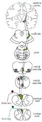
Ascending Lateral Spinothalamic Tract
|
Pain Receptor->Primary Neuron->Lateral Spinothalamic Tract below medulla->peripheral and medial fibers which ascend in lateral funiculi and branch out to reticular formation (in upper and lower medulla and pons) and periaqueductal gray (in inferior colliculus)->Ventral Posterior Lateral Nucleus Thalamus->Cortex
|
|
|
Ascending Anterior Spinothalamic Tract
|
Touch Receptor->Primary Neuron->Anterior Spinothalamic Tract below medulla decussates and ascends anteriorly until it hits the medulla when it almost merges with lateral spinothalamic tract where it branches to reticular formation->Ventral Posterior Lateral Nucleus Thalamus->Cortex
|
|
|
Ascending Posterior Spinocerebellar Tract
|
Position Movement Sensations Below C8->Primary Neuron which goes through dorsal funiculi in S1 and ascends and bends in L3->in L3, synapses on Clarke's column->Posterior Spinocerebellar Tract which ascends in dorsal portion of lateral funiculi->inferior cerebellar peduncle->Cerebellum
|
|
|
Ascending Cuneocerebllar Tract
|
Position Movement Sensations Above C8->Primary Neuron which goes through dorsal funiculi in C7 and ascends to dorsal portion of lateral funiculus->in lower medulla, synapses on exterior cuneate nucleus->Cuneocerebellar Tract which ascends in dorsal portion of lateral funiculus->inferior cerebellar peduncle->Cerebellum
|
|
|
Descending Corticospinal Tract
|

Muscle->Neuromuscular Junction->Axon->Anterior Motor Neuron in Dorsal horn of spinal cord gray matter-> 2 paths:
1) synapses on terminal fiber and goes up anterior corticospinal tract which ascends anteriorly up to the primary motor area as well as branching off at the lower medulla to decussate and join the lateral corticospinal tract 2) synapses on terminal fiber->lateral corticospinal tract, which descends to anterior horn motor neuron |
|
|
medial longitudinal fasciculus (MLF)
|
- descending fibers
- vestibular cell groups - direction that eyes should move |
|
|
external/lateral cuneate nucleus
|
- brings info from upper limbs
|
|
|
medial lemniscus
|
- info from lower limbs (hindlimbs)
|
|
|
What is the top of the ventricle called?
|
Pallium
|
|
|
What does the pallium evolve into?
|
cortices
|
|
|
How many layers is the neocortex?
|
6
|
|
|
Where does the nucleus basalis innervate?
|
Innervates cortex, compromised in Alzheimer's, cholinergic
|
|
|
What is the indusium griseum?
|
the hippocampal rudiment; rostral dorsal part that doesn't develop much
|
|
|
Where does the olfactory bulb arise from?
|
placode
|
|
|
What does the subpallium give rise to?
|
nuclei
|
|
|
What do gene patterns corroborate as far as the alar and basal plates?
|
corroborates alar/basal segments in caudal part of brain as well as neuromeres (repeating segments vertically instead of horizontal)
|
|
|
How are the basal and alar plates divided in the telencephalon?
|
- basal portion smaller compared to alar portion
|
|
|
Where does the sulcus limitans end?
|
optic chiasm
|
|
|
What is the space between the fornix and the third ventricle choroid plexus called?
|
transverse cerebral fissure
|
|
|
In what shape does the temporal lobe form and how does that effect the way other structures are shaped in development?
|
It is horeshoe shaped and makes the lateral ventricles, hippocampal formation, fornix, caudoputamen, and stria terminalis this shape too
|
|
|
Where do commisural fibers (anterior commissure and corpus callosum) cross?
|
cross in or dorsal to lamina terminalis
|
|
|
What is the fornix?
|
-efferent tract of hippocampal formation arching over the thalamus
- the tract passes by the anterior commisure and ends mostly in the mammilary bodies (fibers of fornix cross in caudal extension of the lateral terminalis) |
|
|
lateral lemniscus
|
auditory pathway from superior olivary nucleus
|
|
|
superior olivary nucleus
|
auditory relay center in pontine tegment
|
|
|
What tracts end or innervate in the inferior cerebellar peduncle?
|
1) Olivo-cerebellar tract
2) Dorsal Spino-Cerebellar Tract 3) Cuneocerebellar Tract |
|
|
What tracts end or innervate the middle cerebellar peduncle?
|
Ponto-cerebellar cells
|
|
|
What tracts end or innervate the middle cerebellar peduncle?
|
1) Ventral Spino-Cerebellar Tract
2) Brachium conjunctiva |
|
|
Main accessory nucleus of V
|
fine touch from face
|
|
|
descending tract of V
|
pain, temp, crude touch
|
|
|
brachium conjunctiva
|
crosses at midbrain tegmentum and most get to thalamus
|
|
|
2 parts of the substantia nigra
|
1) substantia nigra pars compacts- pigmented cells
2) substantia nigra pars reticulata |
|
|
pyramidal cells
|
cell body triangular/pyramidal, 10-50 micron diameter, some giant
|
|
|
Betz cells
|
100-150 microns
only in primary motor cortex type of pyramidal cell |
|
|
internal pyramidal cell layer
|
descending connections to;
striatum red nucleus cranial nerve nuclei pontine nuclei (corticopontine fibers) reticular formation spinal cord |
|
|
external pyramidal layer
|
output to corpus callosum (contralateral)
- commissure fibers |
|
|
external granular layer
|
association fibers
project to cortex on same side |
|
|
internal granular layer
|
doesn't leave cortex
mostly excitatory variety of places to end in cortex high concentration of granular cells |
|
|
multiform layer
|
spindle/fusiform cells here
have 2 dendrites in different directions dendrites branch at layer IV axons out to thalamus |
|
|
specific relay nuclei of the thalamus
|
afferent
terminate heavily on layer IV on granule cells axon branches lots in layer IV allows communication btw thalamus and cortex |
|
|
non-specific nuclei of thalamus
|
diffuse pattern of termination
don't necessarily terminate in every layer or same way afferent raphe nuclei (serotonin), LC (NE), nucleus basalis (ach), VTS (especially frontal- DA) |
|
|
association commissural fibers
|
99% of fibers in cortex
go to layers II, III (branch diffusely here) afferent |
|
|
Korbinian Brodmann
|
found 52 areas of regions in cortex based on differences of cell densities
|
|
|
Lorento de No
|
saw vertical columns and spaces among layers
student of Cajal elementary units of cortex each cell type contained in each column |
|
|
Mountcastle
|
found all cells in column respond similaryly to same stimulus
showed bar of light to cats in certain orientations that would begin firing; for a particular column there was a particular bar of light orientation they respond to (seen in auditory and visual cortex) |
|
|
Where is the nucleus accumbens?
|
btw striatum and septal nuclei rostrally, near rostral end of caudate and putamen (where they touch)
|
|
|
Where is the cortex thickest?
|
primary motor cortex (4.5mm thick)
|
|
|
Where is the cortex thinnest?
|
primary visual cortex (1.5mm)
|
|
|
What is consistent about the cortex' most superficial layer?
|
- near pia
- cell-poor - mostly dendrites or axons - called molecular layer |
|
|
Where are the main projections cells?
|
- deeper than molecular layer
- usually have dendrites in molecular layer (apical dendrites) |
|
|
gyrus
|
bulge up
|
|
|
sulcus
|
fold in
|
|
|
fissure
|
deep groove
|
|
|
Where does the posterior pituitary gland arise from?
|
neural tube
|
|
|
where does the anterior pituitary gland arise from?
|
placode
|
|
|
What does the epithalamus consist of?
|
habenular commisure, posterior commissure, pineal gland
|