Reading...
![]()
Play button
![]()
Play button
![]()
Use LEFT and RIGHT arrow keys to navigate between flashcards;
Use UP and DOWN arrow keys to flip the card;
H to show hint;
A reads text to speech;
20 Cards in this Set
- Front
- Back
|
Definition of the Trigger Zone
|
The spike initiation zone in a neuron
|
|
|
Continuous conduction
|
Conduction in an unmyelinated axon
The leading edge of a propagating action potential moves to the next patch of membrane w/ sufficient v-gated channels |
|
|
At the peak of the AP, what is the relationship btwn the influx of Na+ and efflux of K+?
|
Na+ influx = K+ efflux
|
|
|
The structural similarities of v-gated K+, Na+ and Ca2+ channels (4)
|
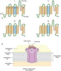
• Each channel has 4 identical subunits clustered around a central pore
• Each of the 4 subunits has 6 transmembrane-spanning segments • Segment 4 of each subunit has a charged component that acts as a voltage sensing element for the channel • Each subunit has a pore loop that acts as a selectivity filter |
|
|
Absolute vs Relative Refractory periods:
• responsive other impulses/stimuli? • State of v-gated Na+ channels • State of v-gated K+ channels |
Absolute period:
• Never other impulses/stimuli • All are open • gradually opening Relative Refractory periods: • Only responsive to strong impulses/stimuli • Na+ channels are inactivated • a large amount are open and are slow to inactivate |
|
|
How Na+ and K+ channels are distributed in unmyelinated axons and skeletal muscle fibers
|
They are distributed uniformly
|
|
|
In myelinated axons, where v-gated Na+ and K+ channels are found
|
v-gated Na+ channels are found at:
• the initiation zone (the initial segment of the axon) • the pre-seynaptic terminals • nodes of Ranvier v-gated K+ channels are found: • at the paranodal & juxtaparanodal portions of peripheral and centrally myelinated axons • in presynaptic terminals • the initiation zone |
|
|
Impulses in myelinated axons:
• only occur at this region • are repolarized at these regions |
Impulses in myelinated axons:
• nodes of Ranvier • paranodal and juxtaparanodal position |
|
|
An important point about how impulses are propagated in myelinated and unmyelinated axons
|
They are propagated without decrement
|
|
|
Factors limiting the conduction velocity of unymyelinated axons (2)
|
1. They have small diameters, decreasing the effectiveness of current spread along the axon's interior
2. The axonal membrane must be driven to threshold sequentially |
|
|
Rule of thumb to find the conduction velocities of unmyelinated and myelinated axons
|
Unmyelinated: the value (m/s) is ~2x the fiber diameter (μm)
Myelinated: ~6x |
|
|
The advantage of myelination of axons
|
• With impulses only being generated at nodes of Ranvier, there is an increase in the conduction velocity
• Because impulses are confined to nodes, energy is saved • Large velocity can be achieved in fibers w/ relatively small diameters |
|
|
Two toxins of v-gated Na+ channels and their mode of action
|
Tetrodotoxin (TTX) and Saxitoxin (STX)
They bind to the outside (extracellular site) of the channel |
|
|
Local Anesthetics:
• how they get into the cell • what they do once they get inside the cell • The parts of the AP they block • The hierarchy of fibers they block (3) |
Local Anesthetics:
• They cross the cell membrane in an uncharged form • Enter the Na+ channel and block the entry site • Initiation and conduction • 1st - small myelinated axons (A δ) 2nd - unmyelinated axons (C-fibers) 3rd - large myelinated axons |
|
|
Define the safety factor and what it means for myelinated axons
|
It reflects that the current flowing from one active node to the next is
exceeds the requirement to excite the next node. Myelinated axons have a large safety factor, ~5x, and the excitation of its node occurs in <20μs. |
|
|
Three types of impairment caused by demyelination and a brief description of each
|
• Frequency-related block:
Once the current reaches the demyelinated region, it dissipates such that future nodes cannot reach threshold • Total conduction block: The severity of the demyelination is such that an active node cannot depolarize the demyelinated region sufficiently • "Cross talk": In the demyelinated regions, a dissipated current may depolarize an adjacent hyperexcitable axon and generate impulses in both directions on that axon |
|
|
The most common type of impairment caused by demyelination in diseases
|
Total conduction block
|
|
|
Multiple Sclerosis:
• is relatively rare outside the US except for those with these types of genes • Lesions in the spinal cord result in this group of sx • its effect on cranial nerves |
"Scandinavian genes"
|
|
|
Guillain-Barr syndrome:
• mode of action • etiology • when it becomes life threatening |
Guillain-Barr syndrome:
• autoimmune d/o against Schwann cells • usually, onset is several wks following a pre-disposing respiratory or intestinal infection • is life threatening when the diaphragm and other respiratory muscles become paralyzed |
|
|
Congential neuropathies:
• age of onset • state of neurons |
Congenital neuropathies:
• infancy - 2yrs • loss of of myelination or dysmyelination of neurons |