Reading...
![]()
Play button
![]()
Play button
![]()
Use LEFT and RIGHT arrow keys to navigate between flashcards;
Use UP and DOWN arrow keys to flip the card;
H to show hint;
A reads text to speech;
37 Cards in this Set
- Front
- Back
|
Pattern of degeneration for an axon
|
Starts proximal to injury and spreads distally(anterogradely/Wallerian)
2-1 |
|
|
First morphological change in neural injury
|
Swelling and fragmentation of neurofilaments and accumulation of mitochondria at nodes of Ranvier
followed by axonal swelling due to water uptake and mitochondrial breakdown. This is then followed by separation of myelin lamellae and fragmentation of myelin sheath. 2-1 |
|
|
Factors determining neurohistologic changes in degenerating axon
|
age, axon size, lesion type
2-1 |
|
|
Perikaryal response to axonal injury is called? Dependent on?
|
Axon reaction and depends on location and type of injury.
2-2 |
|
|
Morphological changes following axotomy...
|
1.) Increase in cell body size due to water uptake
2.) eccentric nucleus 3.) Chromatolysis - loss of basophilic staining of Nissl substance 4.) Increase nucleolus size 5.) Synapses on axotomized neurons withdraw their contact from the soma/dendrites and glial processes become interposed between the boutons and the neuronal surface. 2-2 |
|
|
Type I Synapse
|
AKA Asymmetrical synapse
excitatory, wide synaptic cleft(300 A), large active zone(1-2 um^2), round vesicles, a thick postsynaptic density and substantial dense material in the synaptic cleft. uses glutamic acid 3-1 |
|
|
Type II Synapse
|
AKA Symmetrical Synapse
inhibitory, narrower synaptic cleft(200 A), smaller active zone(less than 1 um^2), flattened vesicles, pre and postsynaptic membranes of more nearly equivaleent thickness and less dense material in the cleft. GABA-ergic 3-1 |
|
|
Dale's Law
|
Whatever combination of transmitters/modulators used by a neuron, the same ones will be present in all of its synapses
3-2 |
|
|
The post synaptic receptors will always perfectly match the presynaptic neurotransmitters released. (T/F)
|
False, frequent examples of receptor-transmitter mismatch appear in the body, possible explanation is extrasynaptic receptors.
3-2 |
|
|
Which neurotransmitters are mentioned that can be both ionotropic and metabotropic?
|
GABA, Ach
3-2 |
|
|
Electrical Synapse
|
Close membrane apposition(3.5nm), channels carry most ions and small molecules, post-synaptic response has same polarity but smaller, bidirectional and FAST communication(<0.1ms delay), enables synchronization of many coupled neurons
3-4 |
|
|
Chemical Synapses
|
Wide Cleft(20-40nm), post-synaptic response differs in time and polarity, unidirectional and SLOW communication(>1 ms delay), amplifies or diminishes signal, strength of transmitted signal may be modifiable
3-4 |
|
|
Time sequence of Chemical Synapse
|
1.) Creation/filling of neurotransmitter vesicles
2.) action potential invades presynaptic terminal 3.) depolarization leads to Ca+2 influx 4.) Ca+2 mediated vesicle fusion 5.) NT release 6.) NT diffusion 7.) NT diffusion, NT reuptake or degradation 3-4 |
|
|
Methods of amplification by chemical synapse
|
ionotropic - one transmitter opens pore to MANY IONS
metabotropic - each transmitter molecule activates MANY G-protein molecules, each G-protein molecule activates MANY effector enzymes, each enzyme molecule catalyzes MANY 2nd messengers, each enzyme opens pores to MANY ions. 3-4 |
|
|
PSC vs PSP
|
post synaptic current charges the membrane to create a Post synaptic potential
3-5 |
|
|
Excitatory currents/potentials vs inhibitory
|
excitatory tends to be influx of sodium, inhibitory tends to be influx of Cl- or K+ efflux
3-5 |
|
|
PSP(C) reversal potential
|
the membrane potential at which there is no net transmembrane current flow
3-5 |
|
|
Synaptic Transmission - molecular level
|
ion channels, NT receptors, auxiliary intra-membranes and cytoplasmic molecules that couple NT receptors to intracellular effectors, Reuptake molecules, NT enzymes
3-6 |
|
|
Synaptic Transmission - cellular level
|
Which specific neurons and which of their most proximal synaptic connections may mediate a behavior or the behavioral effects of a given drug
3-6 |
|
|
Synaptic Transmission - systems level
|
link the neurons that make and release a NT into synaptic junctions to the possible effects of this release at the behavioral level
3-6 |
|
|
Synaptic Transmission - behavioral level
|
integrate neurons with the physiological expression of a learned, reflexive or spontaneously generated behavioral response
3-6 |
|
|
3 Major Categories of Neurologic Symptoms
|
Cognition, Locomotion, Sensation
4-2 |
|
|
Cardinal Manifestations of Neurologic Disease
|
1.) Alterations in Consciousness
2.) Abnormalities of Intellect and Behavior 3.) Impaired Motility 4.) Disorders of Stance and Gait 5.) Pain 6.) Disorders of Special Senses 7.) Abnormal Eye Movements and Pupillary Reactions 8.) Changes in Sleep 9.) Autonomic Disorders 4-4 |
|
|
Alterations in Consciousness can be caused by...
|
reduction in blood flow leading to syncope, neuronal discharge usually manifested by seizure, protracted alteration in the metabolic function of the RAS and/or its cerebral connections leads to altered consciousness that ranges from lethary to stupor to coma.
4-4 |
|
|
Abnormalities of Intellect and Behavior can be caused by...
|
reflects both diffuse and focal disease in the cerebral cortex. Examples include irritability of hypoxia, apathy of dementia, etc.
4-4 |
|
|
Autonomic Disorders manifest what type of symptoms?
|
inability to regulate blood pressure, maintain bladder control, erectile dysfunction, regulate water metabolism or appetite
4-5 |
|
|
List Major points of Neurological Exam
|
1.) Mental Status and Higher Cerebral Function
2.) Cranial Nerves 3.) Motor 4.) Sensation 5.) Coordination 6.) Reflexes 7.) Autonomic Function 8.) Head, Neck and Spine 9.) Palpable or Tender peripheral nerves |
|
|
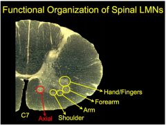
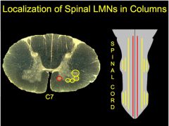
Lecture 5
|
|
|
innervation of Deltoid
|
C5*, C6
Lecture 5 |
|
|
Innervation of Bicep
|
C5,C6*
Lecture 5 |
|
|
Innervation of Triceps
|
C6,C7*,C8
Lecture 5 |
|
|
Innervation of Flexor Digitorum Profundus
|
C7,C8*,T1
Lecture 5 |
|
|
Innervation of Thenar, Hypothenar, Interossei
|
C8,T1*
Lecture 5 |
|
|
Crossed neuronal deficit(contralateral facial deficit, ipsilateral corporeal deficit) is usually indicative of damage in what area?
|
The midbrain/brain stem area.
Lecture 4 |
|
|
Which is the only lower motor neuron which innervates a contralateral muscle?
|
The trochlear motor nucleus at the caudal midbrain
Lecture 5 |
|
|
Muscle spindles sense...
|
an increase in muscle length
Lecture 5 |
|
|
golgi tendon organs sense...
|
a decrease in muscle length. Or muscle shortening.
Lecture 5 |