Reading...
![]()
Play button
![]()
Play button
![]()
Use LEFT and RIGHT arrow keys to navigate between flashcards;
Use UP and DOWN arrow keys to flip the card;
H to show hint;
A reads text to speech;
70 Cards in this Set
- Front
- Back
- 3rd side (hint)
|
This component of the upper motor neurons is responsible for planning, initiating and directing voluntary movements.
A. Basal ganglia B. Brainstem centers C. Cerebellum D. Motor cortex |
D. Motor cortex
|
|
|
|
This component of the upper motor neurons is responsible for basic movements and postural control.
A. Basal ganglia B. Brainstem centers C. Cerebellum D. Motor cortex |
B. Brainstem centers
|
|
|
|
This is responsible for gating proper initiation of movement.
A. Basal ganglia B. Brainstem centers C. Cerebellum D. Motor cortex |
A. Basal ganglia
|
|
|
|
This is responsible for sensory motor coordination of ongoing movement.
A. Basal ganglia B. Brainstem centers C. Cerebellum D. Motor cortex |
C. Cerebellum
|
|
|
|
Like lower motor neurons, you can map out upper motor axons in the spinal cord. In general the lateral axons are from the [ brainstem / motor cortex ] and ventral-medial axons are from [ brainstem / motor cortex ].
|
lateral axons are from motor cortex; ventral-medial axons are from the brainstem
The upper motor neurons in the cerebral cortex will cross the midline in the medulla at the pyramidal deccussation and then descend in the lateral columns to get to lower motor neurons. The upper motor neurons in brainstem on the other hand will descend in the anterior-medial white matter of the spinal cord and give off bilateral projections to both sides of the spinal cord. |
|
|
|
The upper motor neurons from the [ brainstem / cerebral cortex ] will cross the midline in the medulla at the pyramidal deccussation and then descend in the lateral columns to get to lower motor neurons. The upper motor neurons from the [ brainstem / cerebral cortex ] on the other hand will descend in the anterior-medial white matter of the spinal cord and give off bilateral projections to both sides of the spinal cord.
|
cerebral cortex axons crosses at pyramidal deccusation ; brainstem axons descend in anterior-medial white matter of the spinal cord
|
|
|
|
Which tract is described:
voluntary control of limb, head and neck muscles A. Lateral corticospinal and corticobulbar tracts B. Rubrospinal tract C. Anterior corticospinal tract D. Vestibulospinal tract E. Reticulospinal tract F. Tectospinal tract |
A. Lateral corticospinal and corticobulbar tracts
|
|
|
|
Which tract is described:
controls upper limbs and postural muscles A. Lateral corticospinal and corticobulbar tracts B. Rubrospinal tract C. Anterior corticospinal tract D. Vestibulospinal tract E. Reticulospinal tract F. Tectospinal tract |
B. Rubrospinal tract
|
|
|
|
Which tract is described:
controls movements of bilateral axial muscles A. Lateral corticospinal and corticobulbar tracts B. Rubrospinal tract C. Anterior corticospinal tract D. Vestibulospinal tract E. Reticulospinal tract F. Tectospinal tract |
C. Anterior corticospinal tract
|
|
|
|
Which tract is described:
controls positioning of head and neck and controls balance A. Lateral corticospinal and corticobulbar tracts B. Rubrospinal tract C. Anterior corticospinal tract D. Vestibulospinal tract E. Reticulospinal tract F. Tectospinal tract |
D. Vestibulospinal tract
|
|
|
|
Which tract is described:
controls automatic posture and gait-related movments A. Lateral corticospinal and corticobulbar tracts B. Rubrospinal tract C. Anterior corticospinal tract D. Vestibulospinal tract E. Reticulospinal tract F. Tectospinal tract |
E. Reticulospinal tract
|
|
|
|
Which tract is described:
controls coordination of head and eye movements A. Lateral corticospinal and corticobulbar tracts B. Rubrospinal tract C. Anterior corticospinal tract D. Vestibulospinal tract E. Reticulospinal tract F. Tectospinal tract |
F. Tectospinal tract
|
|
|
|
90% of the corticospinal tract deccusates where and becomes what while the rest 10% deccusates where and becomes what?
|
90% deccusates at the pyramidal deccusation and becomes the lateral CS tract; 10% do NOT deccusate (just descend down) as the anterior CS tract and then has bilateral projections near its end.
|
|
|
|
Where are the cells of origin for
A. lateral corticospinal tract B. anterior corticospinal tract C. corticobulbar tract |
all originate in the same area: Pre-central gyrus (primary motor cortex) and to a lesser extent the premotor area.
|
|
|
|
True or False:
The corticospinal tracts and corticobulbar tract originate from the same area - the primary motor cortex area of the post-central gyrus. |
Half true and half false. Yes it originates from the same are the primary motor cortex but primary motor cortex is located in the PRE-CENTRAL GYRUS.
|
|
|
|
Betz cells are some of the neurons that form the
A. corticospinal tract B. rubrospinal tract C. vestibulospinal tract D. reticulospinal tract |
A. corticospinal tract
|
|
|
|
Regarding cytoarchitecture, layers _____ project to other cortical areas, layer _____ projects to subcortical areas, layer ____ projects to thalamus.
|
Layers II and III project to other cortical areas.
Layer V projects to subcortical areas. Layer VI projects to thalamus |
|
|
|
Cells of origin for the corticospinal tracts start in which cytoarchitectural layer?
|
Layer 5 of the primary motor cortex
|
|
|
|
Detail the lateral corticospinal tract.
(Ex. Starts, passes, crosses, etc.) |
The axons of these first order neurons immediately travel through the posterior limb of the internal capsule. All corticospinal and corticobulbar tract axons descend to the pons and are still ipsilateral. Remember back to the MRI sections , you saw a lot of pontine fiber bundles and then other myelinated stuff? That was the corticospinal tract! Once they get to medulla, they begin to cross in the pyramidal decussation. So, that 90% of the corticospinal tract cross at the pyramidal decussation and become the lateral corticospinal tract. So that upper motor neuron will be influencing muscles on the contralateral side of the body after crossing the pyramidal decussation. It travels down that side of the body and eventually synapse onto a lower motor neuron or an interneuron.
|
|
|
|
Detail the anterior/ventral corticospinal trac.
(Ex. Starts, passes, crosses, etc.) |
The axons of these first order neurons immediately travel through the posterior limb of the internal capsule. All corticospinal and corticobulbar tract axons descend to the pons and are still ipsilateral. 90% cross at pyramidal deccusation and become the lateral CS tract. The other 10% of the corticospinal tract don’t cross but instead descend in the anterior corticospinal tract. Once they get to the level they want, they bifurcate and innervate interneurons on both sides of the spinal cord in the ventral horn.
|
|
|
|
Corticobulbar tract axons will begin the primary motor cortex just like the corticospinal tract but where in the primary motor cortex and where else can the neurons reside?
|
lateral inferior portion of the primary motor cortex. Cingulate cortex too.
Lower portion of the face receives input from the contralateral primary motor cortex whereas the upper portion of the face receives input form the bilateral cingulate gyrus. |
|
|
|
Upper motor neurons controlling the lower face reside in the ________ and project [ bilaterally / contralaterally / ipsillaterally ] and upper motor neurons controlling the upper face appear to reside in the ________ and project [ bilaterally / contralaterally / ipsillaterally ] .
|
for lower face: from motor cortex, project contralaterally.
for upper face: from cingulate gyrus, project bilaterally |
|
|
|
Regarding the corticobulbar tract, If a lesion is in the primary motor cortex, which of the following would occur?
A. lower portion of contralateral face loses movement ability B. lower and upper contralateral half of the face loses movement ability C. upper contralateral half of the face loses movement ability |
A. lower portion of contralateral face loses movement ability
|
|
|
|
Regarding the corticobulbar tract, If a lesion is in the posterior limb of the internal capsule, which of the following would occur?
A. lower portion of contralateral face loses movement ability B. lower and upper contralateral half of the face loses movement ability C. upper contralateral half of the face loses movement ability |
A. lower portion of contralateral face loses movement ability
|
|
|
|
Regarding the corticobulbar tract, If a lesion is in the nerve itself or pons, which of the following would occur?
A. lower portion of contralateral face loses movement ability B. lower and upper contralateral half of the face loses movement ability C. upper contralateral half of the face loses movement ability |
B. lower and upper contralateral half of the face loses movement ability
|
|
|
|
True or False:
The corticobulbar tract has both crossed and non-crossed portions. |
True!
Crossed = face example Non-crossed = 10% that doesn't deccuaste at the pyramidal deccusation |
|
|
|
Other areas of the frontal lobe also contribute to motor function. Match each area to its contribution.
1. Broca's area 2. premotor cortex 3. frontal eye fields 4. supplemental motor area A. planning of movements, generation of patterns or programs of movement B. programming complex motor sequences C. important for eye movements (saccades and pursuit movements) D. speech production |
1. Broca’s area – D. speech production
2. premotor cortex – A. planning of movements, generation of patterns or programs of movement 3. frontal eye fields – C. important for eye movements (saccades and pursuit movements) 4. supplemental motor area – B. programming complex motor sequences |
|
|
|
True or False:
Prefrontal cortex is involved in "strategy" for movement but is not considered a part of the motor cortex. |
True
|
|
|
|
The motor humunculus is different from the sensory humunculus...which one are the hands more represented?
|
Motor
|
|
|
|
Does one upper motor neuron control one muscle?
|
NO. A single upper motor neuron can control several muscles. Upper motor neurons control general classes of movements (not single muscles).
|
|
|
|
The only pyramidal system in the old fashioned distinction of pyramidal versus extrapyramidal system is ...
A. lateral corticospinal tract B. anterior corticospinal tract C. rubrospinal tract D. tectospinal tract |
A. lateral corticospinal tract
|
|
|
|
Which tract has its axons that decussate in the ventral tegmental decussation?
A. lateral corticospinal tract B. anterior corticospinal tract C. rubrospinal tract D. vestibulospinal tract E. reticulospinal tract F. tectospinal tract |
C. rubrospinal tract
|
|
|
|
Which tract inhibits extensors and excites flexors?
A. lateral corticospinal tract B. anterior corticospinal tract C. rubrospinal tract D. vestibulospinal tract E. reticulospinal tract F. tectospinal tract |
C. rubrospinal tract
|
|
|
|
Which tract excites extensors and flexors, though a bit moreso flexors?
A. corticospinal tract B. rubrospinal tract C. vestibulospinal tract D. reticulospinal tract E. tectospinal tract |
A. corticospinal tract
|
|
|
|
Regarding the vestibulospinal tract, the centralized pathway for stabilizng gaze, head and posture, what is VCR and VSR?
|
Vestibulo-cervical reflex (VCR) – mediated by medial vestibular nuclei. VCR regulates head position by reflex activity of neck muscles in response to stimulation of semi-circular canals
Vestibulo-spinal reflex (VSR) – mediated by lateral vestibular nuclei. Axons mono-synaptically activate ipsilateral extensor MNs (of anti-gravity muscles) and disynaptically inhibit flexor MNs |
|
|
|
True or False:
Vestibulospinal tract originates in either the medial or lateral vestibular nuclei. Where it comes from determines its function. If it starts in the medial vestibular nuclei, it’ll be an upper motor neuron that will project only into the cervical spinal cord. |
True
|
|
|
|
True or False:
Vestibulospinal tract originates in either the medial or lateral vestibular nuclei. Where it comes from determines its function. If it starts in the medial vestibular nuclei, it’ll be an upper motor neuron that will project only into the cervical spinal cord. |
True
Vestibulo-cervical reflex (VCR) – mediated by medial vestibular nuclei. --VCR regulates head position by reflex activity of neck muscles in response to stimulation of semi-circular canals Vestibulo-spinal reflex (VSR) – mediated by lateral vestibular nuclei. --Axons mono-synaptically activate ipsilateral extensor MNs (of anti-gravity muscles) and disynaptically inhibit flexor MNs |
|
|
|
Which tract is inhibited by cerebellar inputs?
A. corticospinal tract B. rubrospinal tract C. vestibulospinal tract D. reticulospinal tract E. tectospinal tract |
C. vestibulospinal tract
specifically the lateral vestibular nucleus of the vestibulospinal tract |
|
|
|
Which tract runs the entire length of the brainstem but doesn't form distinct nuclei and is actually an amorphous haze of neurons that are scattered longitudinally?
A. corticospinal tract B. rubrospinal tract C. vestibulospinal tract D. reticulospinal tract E. tectospinal tract |
D. reticulospinal tract
|
|
|
|
Which tract is responsible for initiating feedforward adjustments to stabilize posture during movements (such as picking up a keg on the floor)?
A. corticospinal tract B. rubrospinal tract C. vestibulospinal tract D. reticulospinal tract E. tectospinal tract |
D. reticulospinal tract
Motor neuron in premotor cortex fires to tell his soleus muscle “hey he’s going to pick it up”. So note the first muscle that fires is the soleus muscle – postural muscle. Then the biceps contract to pickup the six pack – that voluntary movement. It still throws off the balance a little bit though, so there are additional postural firings just after picking up. |
|
|
|
Which tract has pontine and medullary divisions?
A. corticospinal tract B. rubrospinal tract C. vestibulospinal tract D. reticulospinal tract E. tectospinal tract |
D. reticulospinal tract
|
|
|
|
Of the reticulospinal tract's two subdivisions, which is responsible for the
A. general excitatory drive to both extensors and flexors (but stronger to extensors)? B. inhibitory input to extensors and flexors? |
A. The pontine reticular formaiton.
B. Medullary reticular formation |
|
|
|
The motor cortex can [ excite / inhibit ] the pontine reticular formation and can [ excite / inhibit ] the medullary reticular formation.
|
inhibits pontine ; excites medullary
|
|
|
|
Name symptoms of lower motor neuron syndrome. (6)
|
Weakness or paralysis, decreased superficial reflexes, hypoactive deep reflexes, decreased tone, fasciculations and fibrillations, severe muscle atrophy.
|
|
|
|
Hypoactive deep reflexes are part of the signs and symptoms for [ lower / upper ] motor neuron syndrome.
|
lower
|
|
|
|
Decreased tone is part of the signs and symptoms for [ lower / upper ] motor neuron syndrome.
|
lower
|
|
|
|
Fasciculations and fibrilations are part of the signs and symptoms for [ lower / upper ] motor neuron syndrome.
|
lower
|
|
|
|
Severe muscle atrophy is part of the signs and symptoms for [ lower / upper ] motor neuron syndrome.
|
lower
|
|
|
|
Spasticity is part of the signs and symptoms for [ lower / upper ] motor neuron syndrome.
|
upper
|
|
|
|
Increased tone is part of the signs and symptoms for [ lower / upper ] motor neuron syndrome.
|
upper
|
|
|
|
Hyperactive deep reflexes is part of the signs and symptoms for [ lower / upper ] motor neuron syndrome.
|
upper
|
|
|
|
Clonus (muscular spasms involving repeated movments) is part of the signs and symptoms for [ lower / upper ] motor neuron syndrome.
|
upper
|
|
|
|
Babinksi's sign is part of the signs and symptoms for [ lower / upper ] motor neuron syndrome.
|
upper
|
|
|
|
Loss of fine voluntary movements are part of the signs and symptoms for [ lower / upper ] motor neuron syndrome.
|
upper
|
|
|
|
Regarding terms normally used to describe weakness, which of the following indicate weakness/ partial paralysis.
A. Paresis B. -plegia C. Paralysis D. Palsy |
A. Paresis
ex. hemiparesis (weakness of one side of the body) |
|
|
|
Regarding terms normally used to describe weakness, which of the following indicate no movement.
A. Paresis B. -plegia C. Paralysis D. Palsy |
B. -plegia AND C. paralysis
ex. hemiplegia (no movement of one side of the body) or leg paralysis (no movement of the leg) |
|
|
|
Regarding terms normally used to describe weakness, which of the following is an imprecise term for wekaness or no movement.
A. Paresis B. -plegia C. Paralysis D. Palsy |
D. Palsy
ex. facial palsy: weakness or paralysis of facial muscles |
|
|
|
Regarding terms normally used to describe weakness, which of the following indicate the location of "one side of the body"
A. Hemi- B. Para- C. Mono- D. Di- E. Quadri- or Tetra- |
A. Hemi-
ex. hemiplegia (no movement of one side of the body) |
|
|
|
Regarding terms normally used to describe weakness, which of the following indicate the location of "both legs"
A. Hemi- B. Para- C. Mono- D. Di- E. Quadri- or Tetra- |
B. Para-
ex. Paraparesis (weakness of both legs) |
|
|
|
Regarding terms normally used to describe weakness, which of the following indicate the location of "one limb"
A. Hemi- B. Para- C. Mono- D. Di- E. Quadri- or Tetra- |
C. Mono-
ex. Monoparesis (weakness of one limb) |
|
|
|
Regarding terms normally used to describe weakness, which of the following indicate the location of "both sides of the body equally affected".
A. Hemi- B. Para- C. Mono- D. Di- E. Quadri- or Tetra- |
D. Di-
ex. Facial diplegia (symmetrical facial weakness) |
|
|
|
Regarding terms normally used to describe weakness, which of the following indicate the location of "all four limbs"
A. Hemi- B. Para- C. Mono- D. Di- E. Quadri- or Tetra- |
E. Quadri- or tetra-
ex. quadriplegia / tetraplegia (paralysis of all four limbs) |
|
|
|
Regarding damage to descending motor pathways, this is " an initial period of hypotonia following upper MN injury (eg. loss of control AND loss of reflex activity). After days function returns to some spinal cord circuitry."
A. clonus B. muscle tone C. spasticity D. spinal shock |
D. spinal shock
|
|
|
|
Regarding damage to descending motor pathways, this is "increased resistance to passive movement following damage to higher center (probably caused by removal of suppressive influences from higher centers)"
A. clonus B. muscle tone C. spasticity D. spinal shock |
C. spasticity
|
|
|
|
Regarding damage to descending motor pathways, this is the "resting level of tension in a muscle (dependent on reflex activity between muscle spindles and lower motor neurons)".
A. clonus B. muscle tone C. spasticity D. spinal shock |
B. muscle tone
|
|
|
|
Regarding damage to descending motor pathways, this is "a rhythmic pattern of muscle contractions due to the alternate stretching and unloading of muscle spindles in spastic muscles".
A. clonus B. muscle tone C. spasticity D. spinal shock |
A. clonus
|
|
|
|
What does a positive Babinski's sign entail and what does it indicate?
|
Babinski's entails fanning of toes (extensor plantar response) when plantar area is stroked. It is indicative of an upper motor neuron disease.
|
|
|
|
True or False:
Unilateral lesions of the anterior-medial motor system tracts generally produce no obvious defects. |
True
b/c most project bilaterally or innervate spinal interneurons that project bilaterally! |
|
|
|
True or False:
Lesions of the lateral motor systems while preserving the anterior-medial system leads to decerebrate rigidity. |
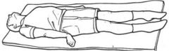
True.
|
Patient has no control of limbs and arms. May or may not have eye movements. Decerebrate rigidity is due to a cut off of the motor cortex and red nucleus.
|
|
|
Lesions of only the corticospinal tracts and preservation of rubrospinal tracts and the anterior-medial system leads to
A. cerebrate rigidity B. Horner's syndrome C. midbrain animal |
C. midbrain animal
You have lost all voluntary control of muscles and no fine motor skills yet you don’t have decerebrate rigidity. Righting reflex is still intact (the body's ability to reflexively right itself after being put in an abnormal position) persists in mid-brain animal. |
|