Reading...
![]()
Play button
![]()
Play button
![]()
Use LEFT and RIGHT arrow keys to navigate between flashcards;
Use UP and DOWN arrow keys to flip the card;
H to show hint;
A reads text to speech;
8 Cards in this Set
- Front
- Back
|
The student will be able to explain and account for differences in brain-injury patterns caused by high- and low-velocity missiles.
(o) |
k.e.= ½ m v2
so the faster the traveling bullet (military weapon) will be more likely to kill you than a slow moving bullet (civilian weapon) |
|
|
please define a concussion
(o) |
Diffuse, physiological and microscopic insult, with or without diffuse axonal injury
Usually defined as involving a period of confusion or unconsciousness, even if just brief Numerous common associated symptoms, e.g. headache, dizziness, irritability, emotionality, poor concentration |
|
|
highschool football player gets hit on the head in the second half of the game and dies. Why did this happen? this is an example of?
* |
You look back and see that the kid got hit on the head in the 1st half but it was dismissed or not realized
the SECOND IMPACT SYNDROME says that the brain is waaayyyy more vulnerable to a 2nd injury this is more common in younger ppl |
|
|
Little Johnny gets hit in the head during a game and gets knocked out. Mom and Dad bring him to you and ask if he can play the next game in 2 days. What do you tell them
|
have to be at least a week of no symptoms before they can resume play
|
|
|
pleas define a brain contusion..where do they tend to occur?
(o) |
Focal hemorrhages within brain tissue
Big enough to see with naked eye in a pathology specimen or as an area of intensity (whiteness) on a CT scan Tend to occur around edges of brain, particularly at base of cerebrum and in anterior temporal lobes |
|
|
describe a subdural hematoma including the location, and appearance on CT initially and chronically
(o) |
Traumatic hemorrhage from veins inside dura but outside brain
This behaves like an actual space, so, acutely, blood runs from anterior to posterior pole The hematomas appear initially white (X-ray dense) on CT Over time, the hematomas get diluted and resorbed, and become less X-ray dense |
|
|
describe an epidural hematoma including the location, and appearance on CT initially and chronically...what is the cause?
(o) |
Traumatic hemorrhage from meningeal arteries inside skull but outside dura
This is a potential space through which the fresh blood dissects from edge to edge, creating a lens-shaped hematoma that does not extend from pole to pole (the subdural does) The hematomas appear white (X-ray dense) on CTs acutely, but become less dense as they dilute and resorb |
|
|
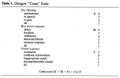
please describe the Glasgow Coma scale, including the 3 aspects, and scoring ranges
|
|