Reading...
![]()
Play button
![]()
Play button
![]()
Use LEFT and RIGHT arrow keys to navigate between flashcards;
Use UP and DOWN arrow keys to flip the card;
H to show hint;
A reads text to speech;
78 Cards in this Set
- Front
- Back
- 3rd side (hint)
|
Special Considerations......
|
Cranium
No lymphatic system Unique circulation Limited immunologic surveillance Blood-brain barrier Watershed area More General Thoughts: Age of the pt Immune status of pt Time of yr |
|
|
|
What is Meningitis?
Inflammation of leptomeninges There is: 1. acute 2. subacute 3. chronic |
What is Encephalitis?
|
|
|
|
Inflammation of the brain regions
there is: 1. Diffuse 2. Locatized |
What is a CNS abscess?
Infectious mass withing the parenchyma. central necrosis and capsule formations |
|
|
|
What are the main ways that pathogens can enter the CNS?
|
1. Hematogenous spread
2. Direct Impantation 3. Local Extention 4. Peripheral Nervous System |
|
|
|
example of hematogenous spread?
|
Arterial and venous spread
|
|
|
|
example of direct implantation spread?
|
almost always traumatic
|
|
|
|
Example of local extension
|
Air sinus (mastoid, frontal) or tooth
|
|
|
|
Example of peripheral nervous system?
|
Certain viruses......
rabies herpes zoster |
|
|
|
There are 5 basic categories of infections we are going to learn:
|
1. Acute bacterial or viral infections that involve leptomeninges and CSF
2. Acute bacterial infections of subdural spaces or CNS parenchyma 3. Chronic bacterial infections of the brain and meninges 4. Acute, subacute or chronic viral infection of the brain 5. Fungal and parasitic infections |
|
|
|
There are different CSF lab findings for different forms of meningitis......
|
Given the lab values tell me the type of Meningitis.
|
|
|
|
<5 lymphocytes
Glucose 45-85 Proteins 15-45 Pressure 70-180 |
Normal CSF
know we are looking at cerebral spinal fluid (CSF) to Dx meningitis |
|
|
|
Up to 90,000 Neutrophils
glucose is decreased <45 proteins are increased >50 Pressure is markedly elevated |
Purulent
Bacterial |
|
|
|
100-1,000 most lymphocytes
glucose is nl proteins are increased >50 pressure is slightly elevated |
Aspectic
Viral |
|
|
|
100-1,000 most lymphocytes
glucose is decreased <45 proteins are increased >50 Pressure is Moderately elevated |
Granulomatous
mycobacterial fungal |
|
|
|
What is the most common cause of acute meningitis*** in the given population?
Neonates |
e. coli
group B strep |
|
|
|
What is the most common cause of acute meningitis*** in the given population?
Infants and young children |
H. influenza
but decreased bc of immunizations |
|
|
|
What is the most common cause of acute meningitis*** in the given population?
Adolescents and young adults |
Neisseria meningitidis
|
|
|
|
What is the most common cause of acute meningitis*** in the given population?
Elderly |
Strep pneumo
Listeria monocytogenes |
|
|
|
What is the most common cause of acute meningitis*** in the given population?
Viral causes..... |
enteroviruses
(echo, coxsackie, polio) |
|
|
|
What are the clinical signs of Acute Meningitis?
|
Headache, fever, nuchal rigidity, cloudy sensorium, coma, death
Viral is self-limited and has low mortality |
|
|
|
What are the Gross findings of acute meningitis - Bacterial***
|
Purulent leptomeningeal inflammation, cloudy CSF, vascular engorgement, may extend to cortex
|
|
|
|
What are the Gross findings of acute meningitis - Viral***
|
Mild leptomeningeal inflammation, brain swelling
|
|
|
|
What are the microscopic findings of acute meningitis?
Bacterial*** |
PMN’s fill subarachnoid space; Gram-stain shows organism; vessels are involved by PMN’s
|
|
|
|
What are the microscopic findings of acute meningitis?
Viral*** |
Very little except lymphocyte infiltrate of leptomeninges
|
|
|
|
What the heck is Leptomeninges?
|
In medicine, leptomeninges (literally thin meninges) is a term used to refer to the pia mater and arachnoid mater, two of the membranes that surround the brain and the spinal cord
|
|
|
|
What is the sequelae of bacterial acute meningitis?
|
Hydrocephalus, leptomeningeal fibrosis (may cause cranial nerve impairment)
|
|
|
|
What is the sequelae of viral acute meningitis?
|
Very little, if any
|
|
|
|
What the heck does sequelae mean?
|
a pathological condition resulting from a disease, injury, or other trauma.
|
|
|
|
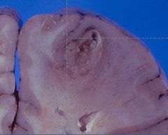
What may be seen on the meninges from acute meningitis?
|
The yellow-tan clouding of the meninges seen here is due to an exudate from acute meningitis
|
|
|
|
What are clinical signs of meningitis?
|
Clinical signs may include:
headache, neck stiffness (from irritation of spinal nerve roots), fever, and clouded consciousness. |
|
|
|
Here is another example of an acute meningitis from bacterial infection
|
The cerebrospinal fluid (CSF) in such cases typically has a low glucose, high protein, and many PMN's. A gram stain should be done to identify organisms.
|
|
|
|
Picture time
|
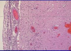
Microscopically, a neutrophilic exudate is seen involving the meninges at the left, with prominent dilated vessels. There is edema and focal inflammation (extending down via the Virchow-Robin space) in the cortex to the right.
This edema can lead to herniation and death. Resolution of infection may be followed by adhesive arachnoiditis with obliteration of subarachnoid space leading to obstructive hydrocephalus.*** This acute meningitis is typical for bacterial infection.***** |
|
|
|
picture
|
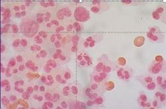
A gram stain reveals gram negative diplococci within a neutrophil, typical for Neisseria meningitidis.
|
|
|
|
Acute Focal Suppurative Infections Clinical Signs
|
Predisposing conditions:
Acute bacterial endocarditis, right to left shunts, chronic pulmonary sepsis Mastoiditis, paranasal sinusitis, acute otitis, open fracture, previous neurosurgery Might also see: Signs of increased intracranial pressure; focal neurologic deficits |
|
|
|
What are the gross findings of Acute focal suppurative infections?
|
CT/MRI; ring-enhancing lesion
Central region of liquefactive necrosis with fibrous capsule (often marked cerebral edema) |
|
|
|
What are the micro findings of acute focal suppurative infections?
|
Granulation tissue, collagen capsule, zone of reactive gliosis
|
|
|
|
What is the Sequelae of acute focal suppurative infections?
|
Increased intracranial pressure and progressive herniation can be fatal
Rupture of abscess can cause ventriculitis, meningitis, venous sinus thrombosis Surgery and antibiotic therapy can reduce mortality to less than 10% |
|
|
|
Acute brain swelling in the closed cranial cavity is serious
|

Swelling of the left cerebral hemisphere has produced a shift with herniation of the uncus of the hippocampus through the tentorium, leading to the groove seen at the white arrow.
|
|
|
|
next picture
|
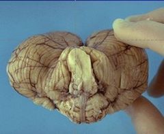
Acute cerebral swelling can also often produce herniation of the cerebelllar tonsils into the foramen magnum. Note the cone shape of the tonsils around the medulla in this cerebellum.
|
|
|
|
Next Picture
|
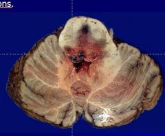
The end result of herniation is compression and Duret hemorrhages, as seen here in the pons.
|
|
|
|
What can cause abscesses in the brain?
|
This is a cerebral abscess. There is a liquefactive center with yellow pus surrounded by a thin wall. Abscesses usually result from hematogenous spread of bacterial infection, but may also occur from direct penetrating trauma or extension from adjacent infection in sinuses.
|
|
|
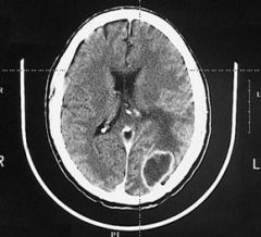
This computed tomographic (CT) scan of the head in transverse view demonstrates What????
|
This computed tomographic (CT) scan of the head in transverse view demonstrates an abscess in the brain in a patient who had septicemia.
|
|
|
|
What kind of stain is used to analyze cerebral abscess?
EXAM**** |
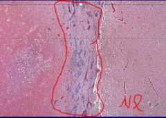
This trichrome stain demonstrates the light blue connective tissue in the wall of an organizing cerebral abscess.
Normal brain is at the right and the center of the abscess at the left. |
|
|
|
What is this a picture of?
|
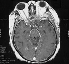
This magnetic resonance imaging (MRI) scan of the head in transverse (axial) view demonstrates a small abscess in the brain in a patient who had septicemia.
|
|
|
|
what is marked dilation of the cerebral ventricles?
|
Hydocephalus
|
|
|
|
What can cause Hydrocephalus?
|
Hydrocephalus can be due to lack of absorption of CSF or due to an obstruction to flow of CSF.
|
|
|
|
What are 3 causes of Chronic Bacterial meningocephalitis?
|
Tuberculosis (mycobacterium tuberculosis)
Neurosyphilis (treponema pallidum) Lyme Disease (borrelia burgdorferi) |
|
|
|
What is this?
Gross – fibrinous exudate at base of brain and encasing cranial nerves, white granules scattered over the leptomeninges |
Tuberculosis
|
|
|
|
What is this?
Micro – mononuclear cell mixture; may see granulomas, may see an intraparenchymal mass (tuberculoma) |
Tuberculosis
|
|
|
|
What is the clinical symptoms of Tb?
|
Clinical – headache, malaise, mental confusion, vomiting
|
|
|
|
What is this?
Gross – mononuclear meningitis involving base of brain and frontal lobe cortex |
Neurosyphilis
|
|
|
|
What does this explain?
Micro – perivascular inflammatory infiltrate of plasma cells and lymphocytes; may see gummas |
Neurosyphilis
|
|
|
|
What are the clinical signs of Neurosyphilis?
|
Clinical – insidious, progressive loss of mental and physical functions, mood changes, terminating in dementia; tabes dorsalis results from damage to dorsal roots
|
|
|
|
What does this describe?
Gross – variable, meningitis |
Lyme Disease
|
|
|
|
What does this describe?
Micro – vasculitis, granulomas, focal proliferation of microglial cells |
Lyme Disease
|
|
|
|
What are the clinical symptoms of Lyme disease?
|
Clinical – (B. burgdorferi transmitted by Ixodes tick) variable symptoms which include meningitis, polyneuropathies, facial nerve palsies
|
|
|
|
There are a number of different viruses that cause Viral meningoencephalitis....what are some?
|
Arthropod-borne (St. Louis, California, Eastern and Western equine, Venezuelan)
Herpes Simplex (HSV-1) HSV-2 Varicella-Zoster (Herpes Zoster) Cytomegalovirus Poliomyelitis Rabies HIV **Just as a note these are in order from 1 to 8 |
|
|
|
How will a viral meningoencephalitis present?
|
Clinical – variable, mental status change, fever, headache, may progress to coma
|
|
|
|
What are the gross findings of Viral meningoencephalitis?
|
Gross – variable, from not much to vascular congestion and intense edema
|
|
|
|
What are the GENERAL things seen on micro for viral meningoencephalitis
|
perivascular infiltrates, microglial nodules, neuron loss, neuronophagia
|
|
|
|
What are the SPECIFIC things seen on micro for viral meningoencephalitis?
|
Herpes-temporal lobe necrosis
Rabies-Negri bodies in hippocampal and Purkinje neurons HIV-microglial nodules and multinucleated giant cells KNOW THESE SPECIFICS***** |
|
|
|
The hemorrhages seen here in the temporal lobe are due to......
|
Herpes simplex virus infection.
|
|
|
|
What will you see micro for herpes viral infection?
|
Viral infections produce mononuclear cell infiltrates microscopically
|
|
|
|
What are fungal and Parasitic Meningoencephalitis?
|
Fungal – Usually only late in disease and primarily in immunocompromised
|
|
|
|
What is the most common cause of Fungal Meningoencephalitis?
|
Most common – Candida, Aspergillus, Mucor, Criptococcus
|
|
|
|
What areas fungal species are seen in endemic areas?
|
In endemic areas – Histoplasma, Coccidioides, Blastomyces following pulmonary or cutaneous
|
|
|
|
What are the 3 main patterns of Fungal and Parasitic Meningoencephalitis
|
Three main patterns
Chronic meningitis – with AIDS--Toxoplasmosis Vasculitis – Mucor, Aspergillus Parenchymal – Candida, Criptococcus |
|
|
|
What about Parasitic (protozoal)
Fungal and Parasitic Meningoencephalitis |
Parasitic (protozoal)
Offenders include malaria, toxmoplasmosis, amebiasis, trypanosomiasis, typhus, Rocky Mountain Spotted Fever, cysticercosis, echinococcosis |
|
|
|
What parasitic infection is of most concern?
|
Toxoplasmosis is of most concern because of involvement with AIDS (up to 30%)
See abscess, necrosis, chronic inflammation CT/MRI shows ring-enhancing lesion |
|
|
|
Disseminated infections can be seen in immunocompromised hosts. Such infections can include fungi.
|
Seen here are branching hyphae of Aspergillus invading a cerebral vessel. Aspergillus likes to invade vessels and produce hemorrhage and thrombosis.
|
|
|
|
Transmissible spongiform encephalopathies are Prion Diseases
|
What is Kuru?
Prion Host Human Subacute Spongiform Encephalopathy (SSE); Fore Tribe – New Guinea; consuming infected brains |
|
|
|
What is Creutzfeldt-Jakob Disease
|
Prion Dz
Host-human SSE Genetic prediposition |
|
|
|
What is Gerstmann-Straussler
|
Prion disease
Host-human SSE |
|
|
|
What is Fatal Familial Insomnia
|
Prion disease
host-human SSE |
|
|
|
What is Scrapie?
|
Prion dz
host-sheep SSE - from scraping their wool off on fences |
|
|
|
What are the clinical signs of Prion diseases?
|
Clinical
occurs in middle-aged to elderly Rapid progressive dementia Death within 6-12 months |
|
|
|
What are the gross signs of prion diseases?
|
Gross – not much
|
|
|
|
What are the micro signs of prion disease?
|
Micro – spongiform change of cerebral cortex and deep gray matter (caudate, putamen)
|
|