Reading...
![]()
Play button
![]()
Play button
![]()
Use LEFT and RIGHT arrow keys to navigate between flashcards;
Use UP and DOWN arrow keys to flip the card;
H to show hint;
A reads text to speech;
31 Cards in this Set
- Front
- Back
|
What is the perikaryon?
|
Neuronal cell body or soma
|
|
|
What is a neurite?
|
Cell projection from perikaryon such as an axon or dendrite
|
|
|
Name 5 fxns of glial cells
|
1. Regulate chemical milieu in extracellular space 2. Phagocytosis 3. Myelination 4. Line the ventricles 5. Repair in the case of injury
|
|
|
What is the average wt of the brain and what is its apprx. % of body weight? What is its O2 consumption?
|
1400 g, 2%, 20%
|
|
|
What are nuclei?
|
Groups of cell bodies (the CNS equivalent of ganglia)
|
|

Where is the corpus callosum? Where is the internal capsule? What are there functions?
|
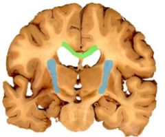
Corpus callosum (green) is a major pathway for axons crossing btw. the cerebra hemispheres. Internal capsule (blue) is the major pathway btw. cerebral hemispheres and more caudal structures (brainstem, etc)
|
|
Identify. What is it's function?
|
Thalamus. Major relay and processing center for sensation and motor function.
|
|
Identify. What is the fxn?
|
Hypothalamus. Primary regulator of ANS.
|
|
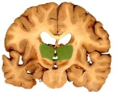
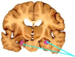
Identify and explain the function of the basal ganglia.
|
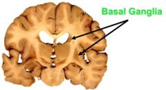
Refers to a group of nuclei in the brain that are involved in motor processing.
|
|

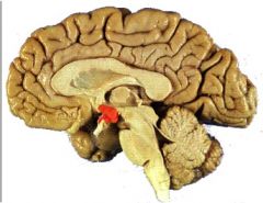
Identify and describe the function of the hippocampal formation.
|
It is involved in the consolidation of memory.
|
|
|
What is the fxn of the cerebellum?
|
Uses complex sensory information to unconsciously modulate motor activity such as coordination.
|
|
|
Describe some functions of the brainstem.
|
Provides processing of sensation such as hearing and taste. Interface for CNs. Contains neurons for both motor and parasympathetic output. Controls resp, HR and other autonomic functions.
|
|
|
What nuclei are contained in the midbrain?
|
Nuclei for eye movement and control through CN III and IV.
|
|
|
What nuclei are contained in the pons?
|
Communication between the cerebrum and cerebellum. Nuclei for CN V, VI, VII and VIII.
|
|
|
What major nuclei are found in the medulla?
|
Consciousness (reticular formation), autonomic control of HR and breathing and also CN IX, X, XI and XII.
|
|
|
What embryonic layers do the meninges develop from?
|
Mesoderm and neural crest
|
|
|
When does a primitive layer first surround the fetal NS and when do the meninges have the adult pattern?
|
1 mo, 3 mo
|
|
|
What forms from embryological ectomeninx? Endomeninx?
|
Dura, leptomeininges (arachnoid and pia)
|
|
|
How does a congenital dermal sinus defect occur and what are the assoc. pathologies?
|
Failure of ectoderm to dissociate completely from neuroectoderm leaving a channel btw. surface of skin and dura or subarachnoid. Spina bifida/recurrent meningitis.
|
|
|
What is pachymeninx?
|
Aka dura mater
|
|
|
What can you mechanically separate the dura matter around the brain into?
|
Periosteal dura (outer layer) and meningeal dura
|
|
|
What is the falx cerebri? Tentorium cerebelli?
|
Dural septa that lies in the longitudinal fissure; Lies axially in the transverse fissure.
|
|
|
What is the falx cerebelli? Diaphragma sella?
|
Dural septa lying in the midline of cerebellum hemispheres; Forms roof of the hypophyseal fossa encircling the infindibulum.
|
|
|
Where do vessels in the cranium generally originate from and run? What can happen if these break?
|
Interal carotid. Btw. periosteum and periosteal dura. Epidural hematoma.
|
|
|
Describe innervation of the tentorium, anterior, middle and posterior cranial fossa assoc. dura.
|
Tentorial N. (from opthalmic N.), ant & middle - trigeminal N, and C2-C3 for posterior.
|
|
|
What histological difference occurs at the dural border cell layer and what is the clinical significance?
|
Lack of dense connective tissue may lead to dissection of layer in the case of bleeding and a resultant subdural hematoma (bleed btw. dural and arachnoid layers).
|
|
|
What meningeal layer is avascular and not innervated?
|
Arachnoid mater
|
|
|
What are arachnoid villi and where are they located? What are arachnoid granulations? Pacchionian bodies?
|
Specializations of the dura-arachnoid interface that occur in dural sinuses to allow for the drainage of CSF. Collections of villi. Calcifications of arachnoid granulations in geriatrics.
|
|
|
When does the pia enter the brain? What space is this assoc. with?
|
Pia may accompany a vessel into the brain or spinal cord. It will be surrounded by a space containing interstitial fluid, Virchow-Robin space. (leukemia cells can enter brain via this space)
|
|
|
What cells are most likely involved in meningiomas?
|
Clusters of arachnoid cells in the villi. Often at points where a vessel or N transversus the dura.
|
|
|
In what meningeal layer is bacterial meningitis most likely to occur?
|
Subarachnoid space
|