Reading...
![]()
Play button
![]()
Play button
![]()
Use LEFT and RIGHT arrow keys to navigate between flashcards;
Use UP and DOWN arrow keys to flip the card;
H to show hint;
A reads text to speech;
76 Cards in this Set
- Front
- Back
|
Describe brain laterailty in terms of organization
|
-bilateral anatomical symmetry: 2 hemispheres essentially same shape
-unilateral functional differences: left for language, right for emotions/music etc -contralateral sensorimotor control: left controls rt movement, vice versa |
|
|
Describe structural and functional organization in the brain
|
-sensory and motor systems have specialized nerve cells for their own sensations
|
|
|
Describe topographical organizaion in the brain
|
-precentral gyrus: motor
-postcentrl gyrus: sensory -auditory and visual cortices -subcortical: interal capsule, BG, thalamus |
|
|
Describe these brain sections:
-coronal -sagital/midsagital -horizontal |
c--vertical slice, separates anterior and posterior
s--vertical slice, seperates right and left (midsag, exact middle) h-horizontal slice, seperates top and bottom |
|
|
From BODY perspective, describe:
-caudal -rostral -anterior -posterior -dorsal -ventral |
caud-toward the head
rost-toward the tailbone ant/vent-toward font of bodu post/dors-toward back |
|
|
From the CEREBRAL perspective, describe:
-caudal -rostral -anterior -posterior -dorsal -ventral |
caud/post--back of brain/head
rost/ant--front of head dorsal--top of brain ventral--botton of brain |
|
|
From the BRAINSTEM perspective, describe:
-caudal -rostral -anterior -posterior -dorsal -ventral |
caud--coccygeal end of spinal cord
rost-toward brain dors-back of BS/SC vent-toward front of body |
|
|
Main structures in the CNS
|
Brain: cerebrum, brainstem (midbrain, pons, medulla), cerebellum
Spinal cord |
|
|
Main structures in the PNS
|
motor and sensory nerves
|
|
|
Name the 6 layers of the cerebral cortex
|
1. molecular (interconnections)
2. external granular (cortex) 3. external pyramidal (cortex) 4. internal granular (thal. & subcort.) 5. internal pyramidal (Betz cells, motor cortex, BS, cerebellum, SC) 6. multiform (thalamus) --MEgEpIgIpM |
|
|
Name the primary vessicles, and what they mature into (brain divisions)
|
1. Prosencephelon--> Telencephalon, Diencephalon
2. Mesencephalon--> Mesencephalon 3. Rhombencephalon--> Metencephalon, Myelencephalon |
|
|
Gross anatomical structures that arise from...
...telencephalon ...diencephelon ...mesencephalon ...metencephalon ...myelencephelon |
tele: cerebrum, limbic lobe, BG
dien: thalamus, hypothal. mesen: midbrain structures mete: cerebellum, pons myel: medula oblongtata |
|
|
Describe embryonic development during 3rd week gestation
|
-neuro structures laid down
-neuroectodermal layer--> CNS & PNS -neural tube--> BS & SC -neural crest--> sensory ganglia, autonomic nerves |
|
|
What are the major lobes of the brain?
|
Frontal, parietal, occipital, temporal
|
|
|
what are the functions of the...
...precentral gyrus ...postcentral gyrus |
pre: motor homunculus
post: sensory homunculus |
|
|
Name the classification of neurons
|
-unipolar (T-shaped, only one process)
-bipolar (peripheral process and central process) -multipolar (many dendrites, one axon) -golgi type I (LONG axon) -golgi type II (short axon) |
|
|
Describe locations of...
...unipolar ...bipolar ...multipolar ...Golgi I ...Golgi II |
Uni: posterior root ganglia, cranial nerves, CNS & PNS connections
Bi: retina, sensory cochlear, vestibular ganglion Multi: CNS nerve tracts, PNS |
|
|
What is myelin? What cells produce it in the CNS and PNS?
|
-fatty sheath insulating nerve for better impulse conduction
-CNS: oligodendroglia cells -PNS: Schwann cells |
|
|
Factors influencing neural regeneration in CNS and PNS
|
CNS: poor, can't cross scars, may be influenced by growth hormones
PNS: good, 3-4 days, endoneurial membrane and neurilemma help sprout protien |
|
|
Describe:
--astrocytes --oligodendrocytes --microglea --ependyma --Schwann cells |
ast: CNS, grey/white matter; form lining around ext. surface of brain & blood vessels
--olig: CNS, produce myelin --micro: CNS, engulf debris --epend: Ventricular Cavity; lining of ventricular surface --Schwann: PNS; produce myelin |
|
|
Describe major componants of a single neuron
|
-cell body (soma)
-dendrites (receptors, transmit toward cell body) -axons (effector, transmit away from cell body) |
|
|
Describe...
...Excitatory Impulses (EPSPs) ...Inhibitory Impulses (IPSPs) |
E: lowers post-synaptic membrane potential
I: Makes post-synaptic membrane hyperpolarized (inside more negative) |
|
|
Describe the function and location of acetylcholine, as well as pathologies resulting from a breakdown
|
-CNS & PNS
-reticular formation, forbrain, cortex -controls circadian cycles, excitation/inhibition, stereotypical movements -MG, Alzheimers, myasthenia gravis can result if not enough |
|
|
Describe the function and location of Dopamine, as well as pathologies resulting from a breakdown
|
-cortex, midbrain (substantia nigra)
-controls voluntary movement -Parkinson's disease can result if not enough |
|
|
Describe the function and location of Norepinephrine, as well as pathologies resulting from a breakdown
|
-pons, medulla, limbic, thalamus, cortex
-conrols sleep, sustained memroy and vigilance |
|
|
Describe the function and location of Serotonin, as well as pathologies resulting from a breakdown
|
-GI tract, blood, BS, subcortical and cortical
-controls arousal, sleep, pain control -depression can result from low levels |
|
|
Describe the function and location of GABA, as well as pathologies resulting from a breakdown
|
-CNS (inhibitory)
-Huntinton's chorea can result from low levels |
|
|
Describe the function and location of Glutamate, as well as pathologies resulting from a breakdown1
|
-major excitatory
-creates links between neurons for learning and long term memory |
|
|
Describe the function and location of Noradrenaline, as well as pathologies resulting from a breakdown
|
-excitatory
-induces physical and mental arousal, elevated mood -produced in locus coreuleus |
|
|
Describe the meninges, what is in the space between them, and how they are layered
|
-covers CNS from top of brain to tip of spinal cord
-between the two inner "diapers" is cerebrospinal fluid -3 layers |
|
|
Where is the efferent system in the cortex, and what comprises the tracts?
|
-direct activation pathways: pyramidal system (coticobulbar and cortico spinal tracts)
-indirect activation pathway: extrapyramidal system -final common pathway (FCP): LMN system, cranaial and spinal nerves go to striated muscle |
|
|
Where is the afferent system and what comprises the system tracts?
|
-post-central gyrus
-dorsal column-medial lemniscal system -antero-lateral system |
|
|
Decribe what these conditions are any why they result:
--anencephaly --cranium bifidum --spina bifida --hydrocephalus --microsephalus |
an: missing cerebral hemispheres (defect of neural tube inutero)
CB: protrusion of brain & SC (inc. fusion of rostral neural tube) SB: post. vertebral arches don't fuse, post. skin epithelialized (caudal neural tube doen'st fuse) hyd: enlarged cerebral ventricles, too much CSF micro: small brain, MR (environment, genes, radiation) |
|
|
Describe functions of...
...nissl substance ...golgi apparatus ...mitochondria ...neurofibrils ...microtubules ...lysosomes ...centrioles ...librofuscin ...melanin |
nissl: protien synthesis
golgi: cell membrane production mito: chemical energy neurofib: cell transport, cytoskeleton makeup mirco: cell transport, motor movement lyso: cleaning waste centr: cell division lipo: metabolic by-product mel: formation of dopa |
|
|
Describe what happens in a synapse
|
-presynaptic cell transmits neurotransmitter via terminal bouton to synaptic cleft
-postsynaptic cell receives it and generated impulse |
|
|
Describe...
...hyperpolarized ...depolarized ...repolarized ...polarized ...absolute refactory period |
hyp: period after impulse where potential < -70mV
dep: impulse becomes less negative as Na+ added rep: impulse becomes more negative as K+ added pol: at -70mV ARP: period during hyperpolarization |
|
|
Describe the process of nerve excitability
|
1. stimuli to nerves
2. Na+ depolarizes cell 3. action potential elicited 4. K+ repolarizes cell 5. cell becomes hyperpolarized briefly 6. cell returns to resting potential |
|
|
Describe how Na+ and K+ influence nerve conduction
|
Na+: rush into cell to depolarize it before action potential
K+: rush into cell to repolarize after action potential |
|
|
What are...
...epicritic senses ...protopathic senses |
e: discriminate slight differences in senses
p: sensing pain/pressure/temperature in non-localized fashion |
|
|
Describe the following kinds of neurological breakdowns:
-neoplastic -vascular -degenerative -motoric -bacterial |
n: slow presenation
v: of the blood vessels d: progressively worsening m: movement disorder b: caused by bacteria |
|
|
Functions of the cerebral hemisphere
|
-mental functions, multimodal processing, initiate motor acts, language/vision/hearing, organization/planning, memory, executive functions
|
|
|
Functions of the Limbic Lobe
|
emotional behavior, body regulation, survival functions
-driven by hormones |
|
|
Functions of the Basal Ganglia
|
-motoric precisions, initiation of movement dynamics/force/speed, templates for redundant verbal/motor acts, new verbal/motor training
-CSF by lateral ventricles |
|
|
Functions of the Thalamus
|
sensory/motor gateway, 30+ nuclei fused together (independant of brain), interconnection b/w cortex and subcortical
|
|
|
Functions of the Hypothalamus
|
hormone secretion (regulates behavior), ANS functions, fight/sex/thirst/body temp
-3rd ventricle gives CSF |
|
|
Functions of the midbrain
|
-made of tegmentum, red nucleus, substantia nigra, reticular formation, cranial nuclei
-funnels/monitors CNS/PNS sentory and motor input/output -cerebral aqueduct gives CSF |
|
|
Functions of the Pons
|
sensory and motor pathways, cranial nerve nuclei
|
|
|
Functions of the Cerebellum
|
control circuit, feedback used to regulate body
info from SC->BS->cortex (and back) -4th ventricle gives CSF |
|
|
Functions of the Medulla
|
pyramidal tracts and ascending sensory tracts, major point of decussation, cranial nerve nuclei
(no ventricles) |
|
|
In general, where are the sig. structures for s/l/h in each major lobe?
|
-Brocca's area: Left Frontal lobe
-Wernicke's: Left Temporal -Heschels gyrus: Left upper temporal lobe (near frontal) -supramarginal/angular gyri: parietal lobe |
|
|
Name major tracts that connect neuronal areas and cerebral cortex
|
-association fibres: superior longitudinal fassiculus, cinglulum, superior occipito-frontal fassiculus, inferior long.fassic, uncinate fasic.
-commisural fibres: corpus collosum -projection fibres: corona radiata (decending motor tract of pyr. syst.) |
|
|
Describe the connection of the corpus collosum
|
connexts L&R hemispheres via assiciation areas, NOT the primary sensory, motor, auditory or visual centers
|
|
|
Describe the tracts of the internal capsule
|
Anterior limb: anterior thalamic radiation, corticopontine
Genu: coticobulbar, coticoreticular Posterior limb: corticospinal, frontopontine, superior thalamic, corticotectal, corticorubral, corticoreticular |
|
|
what are the major cortical sulci?
|
-central sulcus
-lateral fissure -parieto-occipital |
|
|
Structures of the parietal lobe
|
-post-central gyrus/sensory cortex
-superior and inferior parietal lobules -supramarginal and angular gyri |
|
|
structures of the frontal lobe
|
-primary motor cortex
-premotor cortex -prefrontal cortex |
|
|
Major structures of the occipital lobe
|
visual cortex
|
|
|
major structures of the temporal lobe
|
-angular gyrus
-Heschel's gyrus -Wenicke's area |
|
|
where does the Corona Radiata merge?
|
-subcortially near BG/Thal. region, thru internal capsule
|
|
|
Characteristics of an axon
|
-aka nerve fiber
-efferent (motor), transmit AWAY from body -extend long distances -collaterals (extensions) -terminal boutons release neurotransmitters |
|
|
Characteristics of a dendrite
|
-afferent (receptive)
-transmit TOWARD cell body -short with branches |
|
|
Brain injury effects
|
-axonal retrograde: injury of axon
-Wallerian degeneration: axon cut, inflames, degenerates, atrophies -Chromatolysis: degeneration, discoloration -neuroglial response: increase of # of nerve cells and cell size |
|
|
Lesion classification by levels
|
-supratentorial: brain
-posterior fossa: cerebellum, BS, medula -spinal: below medulla -peripheral: final common path of nerve |
|
|
Supratentorial level
|
-derivitives of telen/diencephalons
-cerebral hemispheres, BG, thal., hypthal, cranial nerves I & II |
|
|
Posterior fossa level
|
-structures b/w tentorium cerebelli and foramen magnum
-midbrain, pons, medulla, cerell., cran. nerves III->XII |
|
|
Spinal level
|
-below foramen magnum, in vertebral column
-dorsal root ganglia (sensory), ventral root ganglia (motor) |
|
|
peripheral level
|
-cranial/peripheral nerves
-ANS & ganglia, striated muscles |
|
|
Efferent Tract:
|
-from motor cortex-->OUT
-motor homunculus, internal capsule, BS, SC (H, IC, BS, SC) |
|
|
Afferent Tract
|
-from periphery-->IN!
-dosal spinal ganglia, contralateral parietal lobes, sensory homunculus (DSG, P, H) |
|
|
1. Dorsal
2. Rostral, anterior 3. Ventral 4. Caudal, posterior 5. Dorsal |
Label
|
|
|
1. molecular
2. external granular 3. external pyramidal 4. internal granular 5. internal pyramidal / Betz cells 6. multiform |
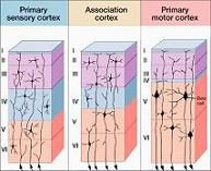
Label
|
|
|
1. dendrites
2. cell body 3. axon 4. nodes of ranvier 5. myelin sheath 6. terminal boutons |
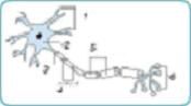
Label
|
|
|
1. neurons
2. oligodendrocyte 3. axon 4. microclian cell 5. ependymal cell 6. astrocyte 7. capillary |
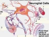
Label
|
|
|
Describe the following terms and if they're CNS or PNS
-Nucleus -Tract -Fasiculus -Ganglion -nerve |
nuc: mass of neurons (CNS)
trac: bundle of parallel axons a/common origin and termination (CNS) fas: several // running tracts (CNS) gang: collection of neurons (PNS) nerve: bundle of axons (PNS) (optic nerve=CNS) |
|
|
Describe the 6 layers of gray matter
-mollecular -ext. granular -ext. pyramidal -int. granular -int. pyramidal -multiform |
I-terminal denrdites and axons from cortex form interconnections
II-small granular interneurons recieve input from cerebrum III-small pyramidal neurons with projections to other cereb. regions IV-small granular interneurons receive input from thal. and other subcort. nuclei V-Lg pyramidal cells (Betz cells) axons project to BS, cebell., and SC VI-fusifrom neurons with projections to thalamus |
|
|
Important Brodmann areas:
-primary sensory cortex -primary motor cortex -primary visual cortex -prim. auditory cortex -2ndary association cortex -Wernicke's -Broca -Supramarginal gyrus -angular gyrus |
-1, 2, 3 (post central gyrus)
-4 (precentral gyrus) -17 (med. occipital lobe) -41, 42 (Heschl's gyrus) -5, 7 (superior parietal lobule) -22 (superior temporal gyrus) -44 (lower 3rd frontal convulution -40 -39 |