Reading...
![]()
Play button
![]()
Play button
![]()
Use LEFT and RIGHT arrow keys to navigate between flashcards;
Use UP and DOWN arrow keys to flip the card;
H to show hint;
A reads text to speech;
45 Cards in this Set
- Front
- Back
|
Name 2 regions in the CNS with neuromelanin.
|
Substantia nigra
Locus Ceruleus |
|
|
How is central chromatolysis different from acute neuronal injury in terms:
1. cause 2. reversibility 3. histology |
Central chromatolysis:
due to axonal injury reversible enlarged cell body with nissl substances peripherally; eccentric nucleus and large nucleolus Acute neuronal injury due to ischemia, hypoglycemia, infection, or toxin Irreversible forms red neurons (shrinkage and eosinophila of cell body, pyknotic nucleus, disappearance of nuclelous and nissl substance) |
|
|
Name 2 intraneuronal inclusions with tau protein. 1 with alpha-synuclein.
What are common diseases associated with each inclusion |
inclusions with tau protien
1. Neurofibillary tangles - seen in aging, AD, PD, progerssive supranuclear palsy, Down Syndrome, dementia 2. Pick body - Pick disease or FTD inclusion with alpha-synuclein 1. Lewy body - seen in PD, LBD (lewy body dementia) |
|
|
GFAP stains?
|
astrocytes
|
|
|
Name 5 functions of astrocytes.
|
1. Metabolic buffer or detoxifier
2. nutrient supplier 3. electrical insuator for neuron 4. BBB 5. repair and scar formation |
|
|
Define gliosis
|
hypertrophy and hyperplasia of astrocytes in response to injury
|
|
|
What are Alzheimer type II cells?
Describe its histology |
hypertrophy of protoplasmic astrocytes
large nucleus and chromatin clearing. |
|
|
What are rosenthal fibers?
Describe their histology |
degenerated astrocytes.
appear as thick, elonaged eosinophilic strcuctures |
|
|
What are corpora amylacea?
How are they different from Lafora bodies? |
Corpora amylacea are lamellated basophilic bodies containing glycogen and protein.
due to aging Lafora bodies are basophilic intracytoplasmic bodies in neurons. PAS postive and seen in myoclonic epilepsy pts. |
|
|
What are rod cells?
Microglial nodules? |
Rod cells are reactive microglia due to neurosyphilis
Microglial nodules are reactive microglia that are clustered. This is due to viral infections |
|
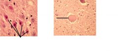
the lines are pointing at pathologic neurons.
the Left neurons are called? due to? The right neuron is due to? |
Left - Red neuron due to acute neuronal injury
Right - due to axonal injury; called central chromatolysis |
|
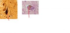
These are pathologic neurons with intraneuronal bodies.
What bodies are present in the Left neuron? right? |
Left - neurofibrillary tangle
Right - Lewy body |
|
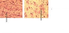
The above pictures show gliosis.
What type of gliosis is shown in each picture? |
Left - fibrillary gliosis
Right - gemistocystic gliosis |
|
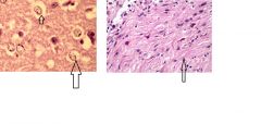
The arrows are pointing at special types of astrocytes.
Name them. What is the common cause of the astrocytes on the Left pic? |
Left - Alzheimer's type II cells due to hyperammonia
right - rosenthal fibers (degenerated astrocytes |
|

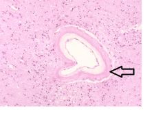
This is a slide of brain.
Name the objects indicated by the arrows. |
corpora amylacea
|
|
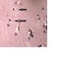
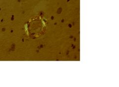
the arrows are pointing at what type of glial cells?
Which histological feature signifies the identity of this cell? |
Oligodendrocytes
Halo cytoplasm around the nucleus |
|
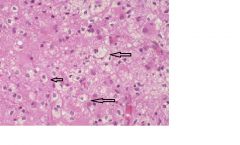
This is a slide of CNS.
What type of cells are the arrows pointing at? What 2 pathophysiology can develop such cells? |
Foamy macrophages.
due to infarct or demyelination |
|
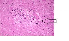
The arrow is indicating what kind of nodule?
common cause? |
microglial nodue due to viral infection
|
|
|
Name 3 areas in the cerebrum that ar emost susceptible to generalized (global) hypoxia.
|
1. Sommer sector of hippocampus
2. Watershed area of infarction (b/t areas of ACA and MCA) 3. layers III and V of cortex |
|
|
How can you differentiate b/t acute and subacute phases of cerebral infraction histologically?
|
acute - Red neurons, neutrophils
subacute - foamy macrophages, reactive gliosis, vascular proliferation, liquefactive necrosis, and early cavitation. |
|
|
What type of plaques cause "pale" infarct? "red" infact?
|
Pale infarcts are commonly due to atherosclerotic plauqes.
Red infarcts are commoly due to embolism. |
|
|
What regions of the brain are affected by lacunar infarct?
Which arteries are occluded? |
Deep regions of the brain (basal ganglia and thalamus)
Occlusion of small penetrating arteries. |
|
|
What is the most common type of aneurysm that leads to intraparenchymal hemorrhage in basal ganglia?
What is the cause of aneurysm development? |
Charcot-bouchard microaneurysm due to hypertension.
|
|
|
What disorder is commonly associated with intraparnchymal lobar hemorrhage in the brain?
|
Amyloid angiopathy due to deposition of beta-amyloids
|
|
|
What aneurysm is associated with lipohyalinosis of arterioles?
What does this cause? common site? |
Charcot-Bouchard aneurysm
Causes IP hemorrhage usually in the basal ganglia |
|
|
What type of aneurysm is most commoly associated with subarachnoid hemorrhage?
Where is the usual location? Which vessels (2 most common)? |
Saccular or berry aneurysm
bifurcation Anterior comm artery (40%) and MCA (34%) |
|
|
Fusiform aneurysm is common which arteries?
What is the cause? |
Vertebral and basilar arteries
Atherosclerosis of the vessel AKA atherosclerotic aneurysm. |
|
|
What is the pathophysiology of mycotic aneurysm?
What is the complication of this aneurysm? |
Infected emboli (bacterial or fungal) usually from cardiac source get in the vessels causing inflammatory rnx in the wall resulting in weakening and formation of aneurysm.
Rupture and cause intraparenchymal hemorrhage |
|
|
Name 4 types of vascular malformation.
|
arteriovenous malformation
Cavernous angioma venous angioma capillary telangiectasia |
|
|
What is AVM?
|
Arteriovenous malformation - clusters of arteries and veins
|
|

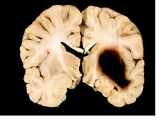
What kind of infarct causes necrosis in the above brain? How do you know?
|
Lacunar infarcts (pale)
There are cavitations in the thalamus and basal ganglia |
|
Where is the hemorrhage?
What is the cause? What type of aneurysm is most commonly formed in the above brain? |
Intraparenchymal hemorrhage in the basal ganglia.
Rupture of Charcot-Bouchard aneurysm due to HTN |
|
This is a slide of Charcot-Bouchard aneurysm. What is the arrow indicating?
|
Lipohyalinosis
|
|
The above picture is a section of artery in the cerebrum.
1. what kind of stain? What view? 2. What is this disorder called? 3. What type of hemorrhage can this result in? |
1. Congo red stain in apple-green birefringence view
2. Amyloid angiopathy 3. Intraparenchymal lobar hemorrhage |
|
|
Describe the CSF flow from the lateral ventricles to sinuses.
Be sure to describe the connection b/t 4th ventricle and cisterns |
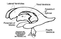
CSF produced from choroid plexus of all ventricles.
Lateral ventricle --> foramen of Monro --> third ventricle --> aqueduct of Sylvius --> 4th ventricle --> Foramina of Magendie (to cisterna magna cistern) and formina of Luschka (to pontine cistern) --> subarachnoid space --> arachnoid villi --> sinuses |
|
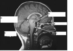
This is a sagittal MRI scan of the brain.
Identify the spaces indicated form A to E. |
A. Interpeduncular cistern
B. chiasmatic cistern C. Pontine cistern D. Superior or Quadrigeminal cistern E. Cisterna Magna |
|
|
What is the total volume of CSF?
What is the normal CSF pressure? |
140 ml
100-200 mmH20 or 15mmHg |
|
|
Differentiate communicating form non-communicating hydrocephalus.
|
Communicating hydrocephalus - hydrocephalus caused by a blockage after the 4th ventricle outlet formane or at arachnoid granulation
Non-communicating - obstruction prior to or at exit of the 4th ventricle outlet foramen. |
|
|
There is an obstruction at the foramen of Magendie. What kind of hydrocephalus is this?
|
non-communicating hydrocephalus
|
|
|
What is the normal cerebral perfusion pressure (CPP)?
How do you calculate this? |
normal CCP = 50 mmHg
CCP = MAP - ICP = 70 - 20 (mmHg) |
|
|
What are 2 common treatments for hydrocephalus?
|
ventricular shunt and third ventriculostomy
|
|
|
What are 3 components of blood-brain barrier?
|
1. tight juctions of the capillary endothelium
2. basement membrane 3. astrocytic endfeet |
|
|
What are 3 components of the blood-CSF barrier?
|
1. fenestrated capillary endothelium
2. basement membrane 3. tight junctions of the choroid plexus endothelium. |
|
|
List 3 types of cerebral edema. For each edema, describe where the edema is and indicate whether the BBB is intact or not.
|
1. Cytotoxic edema - intracellular edema with intact BBB
2. Vasogenic edema - extracellular edema with disrupted BBB 3. Interstitial edema - edema of the ventricles and there is an increased ICP that causes CSF to leak into the parenchyma of the brain |
|
|
Describe the Monro-Kellie doctrine.
|
There is a limited space in the head and if there is an increase in ICP something has to give to compensate for it.
Volume of skull = brain + SAS + vessels + ventricles |