Reading...
![]()
Play button
![]()
Play button
![]()
Use LEFT and RIGHT arrow keys to navigate between flashcards;
Use UP and DOWN arrow keys to flip the card;
H to show hint;
A reads text to speech;
47 Cards in this Set
- Front
- Back
|
What is the cerebral cortex embryologically derived from? |
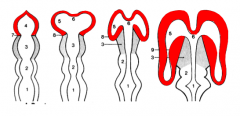
Telencephalon
|
|
|
Where does input to the Cerebral Cortex come via? Output to?
|
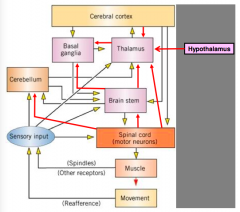
- Input all channels through Thalamus
- Output to Basal Ganglia, Thalamus, Brain Stem, and Spinal Cord |
|
|
How can the Cerebral Cortex be subdivided?
|
- Archicortex (3 layers; includes hippocampus and dentate gyrus)
- Paleocortex (includes olfactory cortex) - Neocortex (6 layers) |
|
|
What are the Hippocampus and Dentate Gyrus a part of?
|
Archicortex - component of the Cerebral Cortex
|
|
|
What is the Olfactory Cortex a part of?
|
Paleocortex - component of the Cerebral Cortex
|
|
|
What are the poles of the Cerebral Cortex?
|
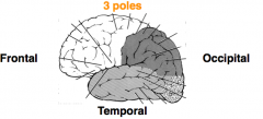
- Frontal
- Temporal - Occipital |
|
|
What are the lobes of the Cerebral Cortex?
|
- Frontal Lobe
- Parietal Lobe - Temporal Lobe - Occipital Lobe - Insula and Limbic Lobes = subdivisions |
|
|
What are the components of the Frontal Lobe of the Cerebral Cortex?
|

- Precentral gyrus
- Superior frontal gyrus - Middle frontal gyrus - Inferior frontal gyrus: Opercular, Triangular, and Orbital - Prefrontal cortex - Orbital gyrus - Gyrus rectus |
|
|
What are the components of the Parietal Lobe of the Cerebral Cortex?
|
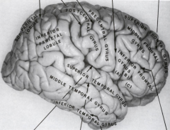
- Postcentral gyrus
- Superior parietal lobule - Inferior parietal lobule: Supramarginal gyrus and Angular gyrus - Precuneus - Paracentral lobule (posterior part) |
|
|
What are the components of the Temporal Lobe of the Cerebral Cortex?
|

- Superior temporal gyrus
- Middle temporal gyrus - Inferior temporal gyrus - Occipitotemporalgyrus or Fusiform gyrus - Parahippocampal gyrus - Hippocampus |
|
|
What are the components of the Occipital Lobe of the Cerebral Cortex?
|
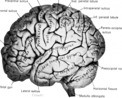
- Lateral Occipital gyrus
- Cuneus gyrus and Lingual gyrus - Occipitotemporal gyrus |
|
|
What are the components of the Limbic Lobe of the Cerebral Cortex?
|
- Cingulate Gyrus
- Parahippocampal Gyrus - Hippocampus |
|
|
What arteries supply the Cerebral Cortex with blood?
|
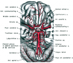
Components of Circle of Willis:
- Anterior Cerebral A. - Middle Cerebral A. - Posterior Cerebral A. - Anterior Communicating A. - Posterior Communicating A. |
|
|
What sinuses drain the Cerebral Cortex?
|
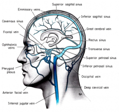
- Superior Sagittal Sinus
- Inferior Sagittal Sinus - Straight Sinus - Transverse Sinuses - Sigmoid Sinuses - Drain into Internal Jugular Veins |
|
|
What are the 6 cellular layers of the Neocortex (the majority of the cortex)?
|
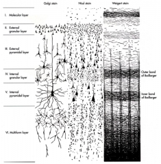
1. Molecular Layer
2. External Granular Layer 3. External Pyramidal Layer 4. Internal Granular Layer 5. Internal Pyramidal Layer 6. Multiform Layer |
|
|
What are the horizontal bands and vertical bundles in the Cerebral Cortex?
|

Myelinated axons (e.g., especially in visual cortex)
|
|
|
What are the major cell types in the Cerebral Cortex?
|
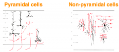
Pyramidal and Non-pyramidal
|
|
|
What are the structures that form functional units in the Cerebral Cortex?
|
Columns and Modules
|
|
|
What is the organization of the "Columns and Modules" of the Cerebral Cortex?
|
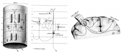
- Each column extends through the 6 layers that share similar functions
- Functional columns form modules in various cortical areas, esp. the primary somatosensory, visual, and auditory cortices - Columns of cortical neurons are interconnected within the same hemisphere and between the two hemispheres |
|
|
What are the important Brodmann's areas? Location?
|

- Areas 3, 1, 2: Postcentral Gyrus - Primary Somatosensory Cortex (RED)
- Area 4: Precentral Gyrus - Primary Motor Cortex (BLUE) - Area 17: Cuneus and Lingual Gyri - Primary Visual Cortex (GREEN) - Areas 41, 42: Transverse Gyri of Heschl - Primary Auditory Cortex (YELLOW) - Areas 44, 45: Inferior Fronal Gyrus - motor area of speech (Broca's area) - primarily in L hemisphere (NAVY) |
|
|
What is the location of Brodmann's Areas 3, 1, and 2? Features?
|

- Postcentral Gyrus
- Primary Somatosensory Cortex (RED) - Somatotopic organization (contralateral) - sensory homunculus - Increased representation of face and hand; lower limb is medial |
|
|
What is the location of Brodmann's Area 4? Features?
|

- Precentral Gyrus
- Primary Motor Cortex (BLUE) - Somatotopic organization (contralateral) - motor homunculus - Increased representation of face and hand; lower limb is medial |
|
|
What is the location of Brodmann's Area 17? Features?
|

- Cuneus and Lingual Gyri
- Primary Visual Cortex (GREEN) - Visuotopic organization - Central VF - most posterior - Peripheral VF - most anterior - Vertical meridian - at border of areas 17/18 - Horizontal meridian - bisect horizontally |
|
|
What is the location of Brodmann's Areas 41 and 42? Features?
|

- Transverse Gyri of Heschl
- Primary Auditory Cortex (YELLOW) - Tonotopic organization - Binaural representation |
|
|
What is the location of Brodmann's Areas 44 and 45? Features
|
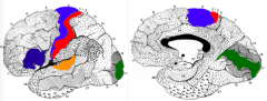
- Part of Inferior Frontal Gyrus
- Motor area of speech (Broca's Area) - mostly dominant in left hemisphere (NAVY) |
|
|
What happens if there is a lesion to the Postcentral gyrus? What Brodmann's Area is this?
|

Contralateral loss of somesthetic sensation - Areas 3, 1, and 2 (RED)
|
|
|
What happens if there is a lesion to the Precentral gyrus? What Brodmann's Area is this?
|

Contralateral spastic paralysis - Area 4 (BLUE)
|
|
|
What happens if there is a lesion to the Cuneus and Lingual gyri? What Brodmann's Area is this?
|

- Contralateral hemianopia
- If restricted to upper or lower banks of the Calcarine fissure --> contralateral inferior or superior quadrantanopia - Area 17 (GREEN) |
|
|
What happens if there is a lesion to the Transverse gyri of Heschl? What Brodmann's Area is this?
|

Bilateral lesions lead to loss of hearing
Areas 41 and 42 (ORANGE) |
|
|
What happens if there is a lesion to the Broca's area? What Brodmann's Area is this?
|

- Dominant side (left) - motor aphasia, Broca's aphasia, or expressive aphasia
- Non-dominant side (right) - difficulty expressing emotional aspect of language - Areas 44 and 45 (NAVY) |
|
|
What are the main functional areas of the Frontal Cortex?
|
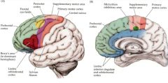
- Primary motor cortex: area 4
- Premotor cortex: area 6 - Supplementary motor cortex - Frontal eye field - Broca’s area - Limbic orbitofrontal cortex - Prefrontal cortex |
|
|
What happened to Phineas Gage?
|
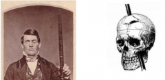
- Destroyed much of left prefrontal lobe
- Partial paralysis of left side of face - Loss of vision in left eye - Ptosis in left eye - Survived for 12 years - Major personality and mental changes |
|
|
What are the structures in the association cortical areas of the parietal lobe?
|
- Posterior parietal lobe: polymodal convergence - determining where something is
- Superior parietal lobule (areas 5 and 7) - Inferior parietal lobule (supramarginal gyrus - area 40; angular gyrus - area 39) |
|
|
What happens when there are lesions to the association cortical areas of the parietal lobe?
|
- Dominant (usually left) hemisphere --> astereognosis - inability to identify an object by touch without visual input (area 40) & Aphasia (inability to express or understand language) / Alexia (inability to read) / Agraphia (inability to write) (area 39)
- Non-dominant (usually right) hemisphere --> spatial distortion and contralateral neglect (ignore L side of body or draw only R half of picture) |
|
|
What are the components of the association cortical areas of the occipital lobe?
|
- Secondary visual cortex: area 18 or V2
- Association cortical areas: V3, V4, etc. - 32 visual areas in primate (occipital, parietal, and temporal) |
|
|
What happens if there are lesions to the association cortical areas of the occipital lobe?
|
Variety of visual deficits
|
|
|
What are the components of the association cortical areas of the temporal lobe?
|
Wernicke's Area - posterior part of superior temporal gyrus (area 22) - language comprehension (dominant on L)
|
|
|
What happens if there are lesions to the association cortical areas of the temporal lobe?
|
- Wernicke's area (dominant hemisphere, L) --> sensory aphasia, Wernicke's aphasia, or receptive aphasia
- Wernicke's area (non-dominant hemisphere, R) --> difficulty in comprehending the emotional aspect of language |
|
|
What kind of fibers connect different parts of the cortex to each other?
|
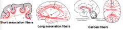
- Short Association Fibers - within same hemisphere
- Long Association Fibers - within same hemisphere - Callosal Fibers - via corpus callosum to opposite hemisphere |
|
|
What are the general functions of the cerebral cortex?
|
- Perception of special sensations - somatic, visual, auditory, olfaction (cortical maps for each of the major senses)
- Planning and execution of voluntary movements - Emotions and behavior - Mental functioning - Memory |
|
|
What can cause dysfunction of the cerebral cortex?
|
- Vascular hemorrhage, thrombosis, or tumor
- Tumors usually of glial origin |
|
|
A lesion to what part of the cerebral cortex would cause Contralateral Paralysis?
|
Primary Motor Cortex
|
|
|
A lesion to what part of the cerebral cortex would cause Contralateral Loss of Somatic Sensation?
|
Primary Sensory Cortex
|
|
|
A lesion to what part of the cerebral cortex would cause Contralateral Hemianopia
|
Primary Visual Cortex
|
|
|
A lesion to what part of the cerebral cortex would cause Astereognosis (inability to identify an object by touch without visual input)?
|
Supramarginal Gyrus (area 40)
|
|
|
A lesion to what part of the cerebral cortex would cause Alexia (inability to read) and Agraphia (inability to write)?
|
Lesion of the Angular gyrus (area 39)
|
|
|
A lesion to what part of the cerebral cortex would cause motor aphasia?
|
Lesion to Broca's area on L side (area 44 and 45) |