Reading...
![]()
Play button
![]()
Play button
![]()
Use LEFT and RIGHT arrow keys to navigate between flashcards;
Use UP and DOWN arrow keys to flip the card;
H to show hint;
A reads text to speech;
6 Cards in this Set
- Front
- Back
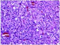
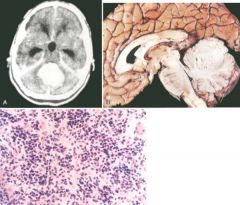
40-year-old man who presented with headaches and ataxia. What is the most likely diagnosis in this case?
a. Subependymal giant cell astrocytoma b. Pilocytic astrocytoma c. Arteriovascular malformation d. Hemangioblastoma e. Angioblastic glioma |
d. Hemangioblastoma
is a hypercellular, vascular tumor containing foamy interstitial cells, which represent the actual neoplastic element. Hemangioblastomas occur most commonly in the cerebellum and are often associated with von Hippel-Lindau syndrome. Subependymal giant cell astrocytoma is an intraventricular tumor (lateral ventricles) resembling gemistocytic astrocytoma that is usually seen in the context of tuberous sclerosis. Pilocytic astrocytomas are biphasic neoplasms composed of alternating areas of compact spindle cells and more loosely textured zones and often accompanied by Rosenthal fibers. |
|
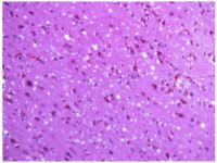
Microscopic examination of this hippocampus from a 68-year-old patient who suffered a cardiac arrest during surgery and survived on a respirator for 2 weeks would most likely show the following:
a. Neurofibrillary tangles in the dentate gyrus b. Red, pyknotic neurons (acute neuronal injury) in CA1 c. Granulovacuolar degeneration d. Hirano bodies e. Red, pyknotic neurons (acute neuronal injury) in CA2 |
b.
Acute hypoxic/ischemic injury is associated (if the patient survives at least six hours) with shrunken eosinophilic neurons. Certain populations of neurons are selectively vulnerable, especially large neurons in CA1 sector of the hippocampus, the Purkinje cells of the cerebellum, and neurons of layers 3 through 5 of the cerebral cortex. Neurofibrillary tangles, granulovacuolar degeneration, and Hirano bodies are neurodegenerative changes. CA2 is the so-called “resistant zone” of the hippocampus that is less vulnerable to hypoxia/ ischemia. |
|
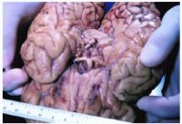
Which one of the following statements does NOT apply to the abnormality depicted in this gross dissection of the circle of Willis?
a. Associated processes include polycystic kidney disease and coarctation of the aorta. b. Common at the middle cerebral trifurcation. c. A familial inheritance pattern has been noted in fewer than 2% of intracranial aneurysms. d. This condition is common in infants and children. e. Hemorrhage and secondary infarcts are potential sequelae. |
Berry aneurysms
involving the circle of Willis are uncommon in children. The photomicrograph shows the basilar aspect of the brain with a berry aneurysm at the trifurcation. The risk of rupture, which produces hemorrhage into the subarachnoid space at the base of the brain, is greatest for females in the fifth decade. |
|
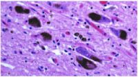
What is the most likely diagnosis based on this photomicrograph?
a. Multisystem atrophy b. Unverricht epilepsy c. Adult polyglucosan disease d. North American blastomycosis e. Parkinson disease |
. Lewy bodies
are inclusion bodies found in Parkinson disease and diffuse Lewy body disease. In pure Parkinson disease, Lewy bodies are located predominantly in the pigmented neurons in the substantia nigra (as depicted in the photomicrograph) and locus ceruleus, whereas in diffuse Lewy body disease, they are also found in the cerebral cortex. |
|
|
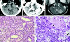
Medulloblastoma
isochromosome 17q or i(17q) Sonic hedgehog/patched pathway (involved in control of normal proliferation of cerebellar granule cells) |
|
|
Atypical teratoid / rhabdoid tumor
DD: PNET/medulloblastoma (no rhabdoid cells, no 22q11 deletions, IN1+) |