Reading...
![]()
Play button
![]()
Play button
![]()
Use LEFT and RIGHT arrow keys to navigate between flashcards;
Use UP and DOWN arrow keys to flip the card;
H to show hint;
A reads text to speech;
39 Cards in this Set
- Front
- Back
|
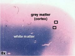
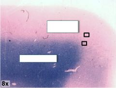
Figure 5-1 (cerebrum)
Cerebrum, fast blue stained. The distribution of superficial grey matter (cerebral cortex) and deeper white matter is demonstrated by fast blue staining which is specific for myelinated axons. |
|
|
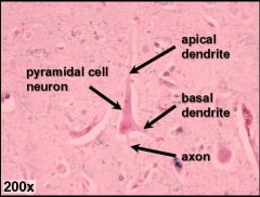
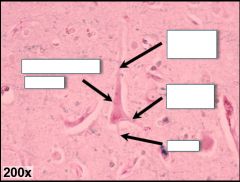
Figure 5-1 (cerebrum)
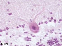
Pyramidal neurons, which can be found in the cortical grey matter, are large and roughly pyramid-shaped. They possess a prominent, dark staining nucleolus that is visible inside a pale nucleus |
|
|
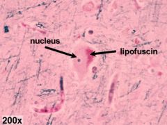
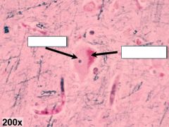
Figure 5-1 (cerebrum)
Lipofuscin (age pigment) is often visible as a darker, magenta colored inclusion within the cytoplasm of the soma. |
|
|
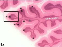
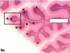
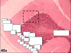
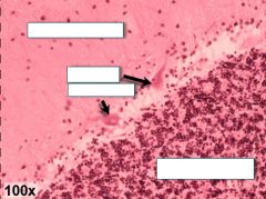
Figure 5-2 (cerebellum)
Cerebellum, H&E stained. Grey and white matter distribution is similar to the cerebrum. However, cerebellar cortex consists of three distinct layers of gray matter which are readily visualized using H&E stain. |
|
|
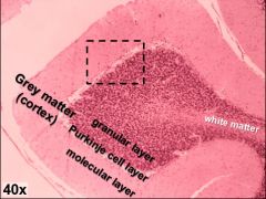
Figure 5-2 (cerebellum)
The innermost granular layer (G) contains a high density of small granular cell neurons. The somas of large Purkinje neurons are located in the Purkinje cell layer (P), while the extensive dendritic arborizations of Purkinje cells occupy most of the molecular layer (M). Dark staining nuclei of other neurons are also scattered throughout the molecular layer. |
|
|
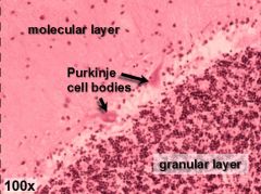

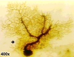
Figure 5-3 (Purkinje cell structure)
Routine staining protocols readily identify the large somas and proximal dendrites of Purkinje cells (top), but do not detect most of the structural complexity of the dendritic branches |
|
|

Figure 5-3 (Purkinje cell structure)
Figure 5-3 (Purkinje cell structure) Complete structural detail of neurons requires use of more selective staining protocols, such as the Golgi (silver) staining method (bottom), or fluorescent imaging (not shown). |
|
|
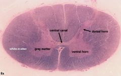
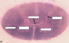
Figure 5-4 (spinal cord)
Cross section through spinal cord, cervical region. Fast blue stained. |
|

|
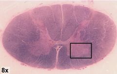
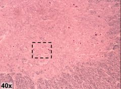
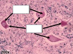
Figure 5-5 (motor neurons)
Cervical spinal cord, fast blue stained. Large multipolar motor neurons are easily visible throughout the ventral horn. Cell bodies and one or more dendrites are typically observed for most motor neurons. Aging pigment is also frequently present. The dense tissue surrounding neuronal cell bodies is a complex tangle of dendrites, axons, and glia, often referred to as neuropil. |
|
|
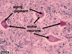
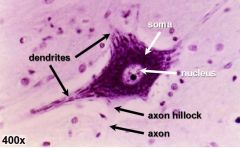
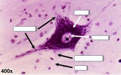
Figure 5-6 (motor neuron structure)
Motor neuron from spinal ventral horn, Nissl stained. This detailed image reveals Nissl bodies within the soma and proximal portions of several dendrites. Background staining reveals the structure of the axon and axon hillock, which are devoid of Nissl bodies. The cell nucleus is pale (also lacking Nissl bodies) and contains a dense nucleolus. |
|

|

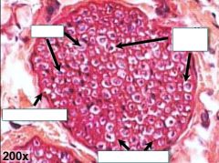
Figure 5-7 (peripheral nerve)
Cross section of peripheral nerve, stained with H&E (above) and OsO4 (below). Most fascicles contain both myelinated and non-myelinated axons. Observe the three layers of connective tissue that surround the whole nerve (epineurium), fascicles (perineurium), or individual axons (endoneurium). In some preparations, whorls of neurokeratin present in myelin sheaths are visible as an artifact. |
|
|
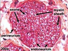
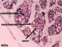
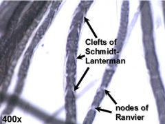
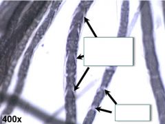
Figure 5-8 (teased peripheral axons)
Longitudinal sections of axons teased from a peripheral nerve and stained with OsO4. Distinguish nodes of Ranvier from clefts of Schmidt-Lanterman. The latter are found only in peripheral white matter. |
|
|
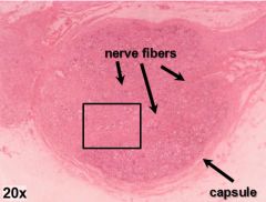
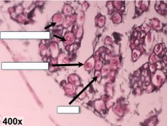
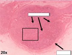
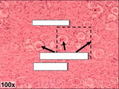
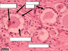
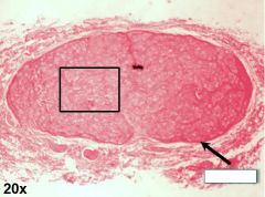
Figure 5-9 (dorsal root ganglion)
Dorsal root (spinal) ganglion, H&E stained. The large cell bodies of pseudounipolar sensory neurons possess centrally located nuclei and are surrounded by numerous glial satellite cells. Spoke-like neuro-keratin structures surround sensory cell bodies as artifacts of slide preparation. Observe the thick connective tissue capsule which surrounds the ganglion as well as the numerous nerve fibers passing through the ganglion interior. |
|
|
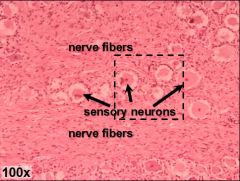
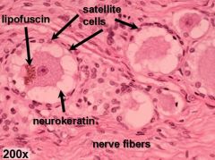
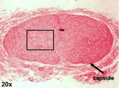
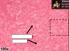
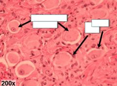
Figure 5-10 (sympathetic ganglion)
Sympathetic ganglion, H&E stained. Numerous cell bodies of sympathetic post-ganglionic neurons are visible throughout the ganglion. Note the eccentrically positioned nuclei, in contrast to the central nuclei of spinal ganglionic neurons. Satellite cells surround the cell bodies, but are fewer in number than in spinal ganglia. In addition to glial cells, the space around the multipolar neurons is densely packed with dendritic processes. |
|
|
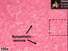
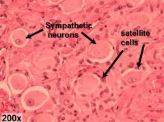
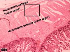
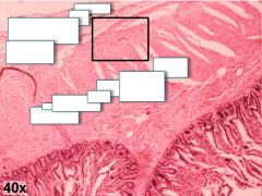
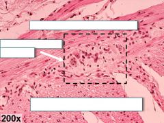
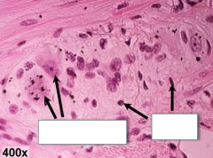
Figure 5-11 (parasympathetic ganglion)
Parasympathetic ganglion from Aurbach’s plexus in jejunum, H&E stained. Cell bodies of multipolar post-ganglionic neurons are visible as well as multiple glial satellite cells. These parasympathetic ganglia are small and lack a surrounding capsule. They can be found between the two layers of the muscularis externa within the wall of the GI tract. |
|
|
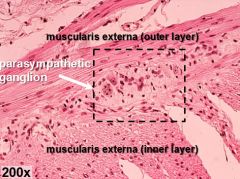
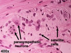
Figure 5-11 (parasympathetic ganglion)
Parasympathetic ganglion from Aurbach’s plexus in jejunum, H&E stained. Cell bodies of multipolar post-ganglionic neurons are visible as well as multiple glial satellite cells. These parasympathetic ganglia are small and lack a surrounding capsule. They can be found between the two layers of the muscularis externa within the wall of the GI tract. |
|

|
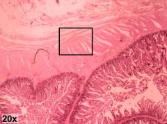
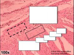
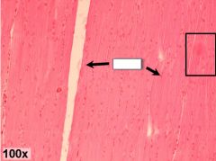
Figure 5-12 (smooth muscle)
Jejunum, H&E stained. The muscularis externa is composed of two sheets of smooth muscle, which are perpendicular to one another. Myocytes of the inner, or circular layer, are oriented perpendicular to the long axis of the gut, whereas cells of the outer, or longitudinal layer, are oriented parallel to this axis. Individual smooth myocytes are difficult to distinguish; however, the single centrally located nuclei are clearly visible |
|
|
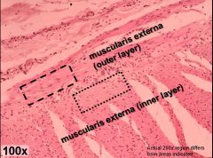
Figure 5-12 (smooth muscle)
Jejunum, H&E stained. The muscularis externa is composed of two sheets of smooth muscle, which are perpendicular to one another. Myocytes of the inner, or circular layer, are oriented perpendicular to the long axis of the gut, whereas cells of the outer, or longitudinal layer, are oriented parallel to this axis. Individual smooth myocytes are difficult to distinguish; however, the single centrally located nuclei are clearly visible |
|

|
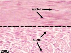
Figure 5-12 (smooth muscle)
Jejunum, H&E stained. The muscularis externa is composed of two sheets of smooth muscle, which are perpendicular to one another. Myocytes of the inner, or circular layer, are oriented perpendicular to the long axis of the gut, whereas cells of the outer, or longitudinal layer, are oriented parallel to this axis. Individual smooth myocytes are difficult to distinguish; however, the single centrally located nuclei are clearly visible |
|
|
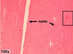
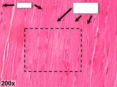
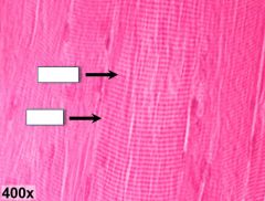
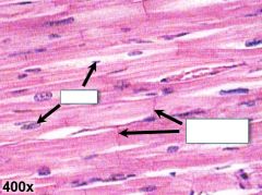
Figure 5-13a (skeletal muscle l.s.)
Skeletal muscle, longitudinal section, H&E stained. Skeletal myofibers are very large, elongated, multinucleated cells. Many nuclei lie along the periphery of the cells. Skeletal myofibers have a very regular arrangement, and many fiber-shaped cells can be seen side-by-side. At higher magnifications, their striated appearance can be resolved as an alternating pattern of A-bands and I-bands (dark and light bands, respectively). |
|
|
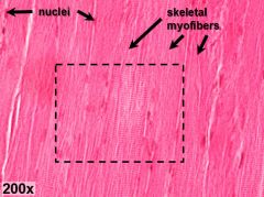
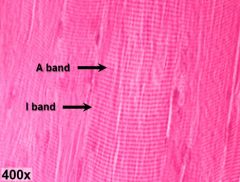
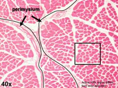
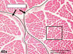
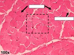
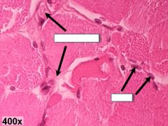
Figure 5-13b (skeletal muscle c.s.)
Skeletal muscle, cross section, H&E stained. Fascicles of muscle tissue (outlined in black above) are encased by sheaths of dense irregular connective tissue called perimysium. At higher magnifications connective tissue surrounding individual myofibers, or endomysium, can be observed. Multiple nuclei are frequently visible in the periphery of each myofiber. |
|
|
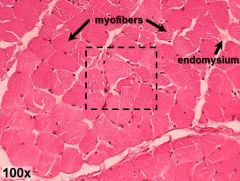
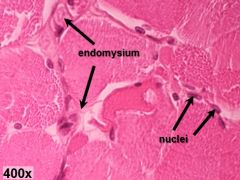
Figure 5-13b (skeletal muscle c.s.)
Skeletal muscle, cross section, H&E stained. Fascicles of muscle tissue (outlined in black above) are encased by sheaths of dense irregular connective tissue called perimysium. At higher magnifications connective tissue surrounding individual myofibers, or endomysium, can be observed. Multiple nuclei are frequently visible in the periphery of each myofiber. |
|

|
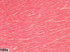
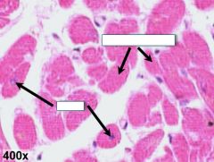
Figure 5-14 (cardiac muscle)
Cardiac muscle in longitudinal section (above) and cross section (lower right), H&E stained. Observe the less regular arrangement of the smaller, striated cardiomyocytes, as compared to skeletal myofibers. Many cells are branched as well as binucleate, with nuclei occupying a more central position compared with skeletal muscle cells. Intercalated discs can be observed at attachment sites between neighboring cardiac cells, when viewed in longitudinal section. |
|
|
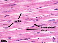
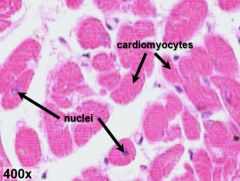
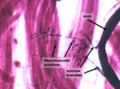
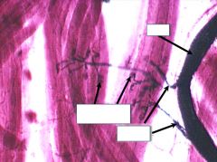
Figure 5-15 (neuromuscular junction)
Neuromuscular junctions between branching nerve terminals and skeletal muscle fibers. |