![]()
![]()
![]()
Use LEFT and RIGHT arrow keys to navigate between flashcards;
Use UP and DOWN arrow keys to flip the card;
H to show hint;
A reads text to speech;
84 Cards in this Set
- Front
- Back
|
Mycology lecture 6, Fungal Structure and Ultrastructure |
Mycology lecture 6, Fungal Structure and Ultrastructure |
|
|
BASICS -Essentially a tube Indeterminate length -Hyphae grow only at the tips -Extension zone -Older parts: autolysis(desctruction of cell through own enzymes) or heterolysis(apoptosis from surround hydrolytic enzymes) -Most higher Fungi: septa -Septa have pores; therefore ‘compartments’ -Reproductive areas |
BASICS -Surrounded by a complex wall -Thin at apex -Plasma membrane fixed tightly to wall |
|
|
ULTRASTRUCTURE: structures that are very small and must be viewed with electron microscopy or other means |
s |
|
|
The extreme hyphal tip, is the tip of the hypha. What accumulates here? |
A lot of membrane bound vesicles accumulate here. |
|
|
What do these vesicles show? |
Vesicles show differences in density |
|
|
What is lacking at these extreme hyphal tips? |
These extreme hyphal tips lack organelles |
|
|
Where are these organelles located? |
These organelles are located farther back |
|
|
What is the source of these vesicles? |
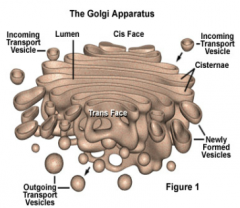
Their source is the Golgi bodies. |
|
|
What is responsible for the chitin in cell walls? |
Chitin synthase. |
|
|
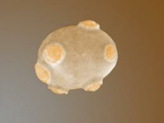
What is the Apical vesicle cluster(also called Spitzenkorper)? |
Its a cluster of vesicles that form at the Apex(hypha tip) |
|
|
What is in the centre of the Apical Vesicle Cluster? |
In the centre of the apical vesicle cluster there is a lack of vesicles. |
|
|
How do these vesicles get to the hyphae tip? |
Actin microfilaments, microtubules, myosin are though to bring vesicles from the golgi to the apex of the hyphae tip. |
|
|
What happens where there is a shift in the hyphae? |
There is a change in direction or branching occur |
|
|
What is located behind the apex? |
Behind the apex you will find mitochondria, it is rich in mitochondria. This is similar to plants/animals: cistern style |
|
|
What does the mitochondria do? |
Generates a proton-motive force that drives the hyphal tips to uptake nutrients |
|
|
Next we will be talking about the Subapical region of the hyphae, what does subapical mean? |
Subapical means right below the apex. |
|
|
s |
|
|
What is located further back in the hypha? |
Located further back in the hypha is the branched tubular vacuoles. |
|
|
What do the branched tubular vacuoles do? |
They expand through pores to other compartments and act as a transport system for the movement of metabolites(includes phosphates which are often in short supply) |
|
|
How are the nuclei distributed between the fungal groups? |
It varies between fungal groups, coenocytic vs. septate. |
|
|
What is the nuclei distribution for oomycota? |
Several nuclei in each oogonia |
|
|
What is the nuclei distribution for septate Fungi(ascomycota)? |
Two nuclei in each compartment. Can squeeze through pores, so concept is fluid. |
|
|
What is the nuclei distribution for septate fungi(Basidiomycota)? |
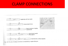
It can be both, monokaryon(one per compartment), or dikaryon(two per compartment) which will have a dolipore septum |
|
|
What do hyphal colonies develop from? |
They typically develop from a single spore. |
|
|
How do hyphal colonies develop from a single spore? |
They extend the germ tube; then begin to branch behind the tip. |
|
|
What does the typical colony form? |
A circle |
|
|
What happens to older hyphae? |
The older hyphae fuse back with other hyphae. |
|
|
What is this process called? |
Hyphal anastomosing |
|
|
What is the point of hypal anastomosing? |
To pool together protoplasm to produce other large structures. |
|
|
What do the hyphae do in nutrient rich conditions? |
The branching hyphae diverge from one another. |
|
|
What controls this behaivour? |
It is unknown, but speculated to be CO2 or growth metabolites |
|
|
What do the hyphae do in nutrient poor conditions? |
The centre hyphae grow towards each other and fuse only at the tips and only between individuals in the same species. |
|
|
When do oomycota only fuse at? |
At the time of the zygospore. |
|
|
What is a yeast? |
A single celled organism. |
|
|
Can yeast produce hyphae? |
Only some yeast can produce hyphae |
|
|
Are most anamorphs or telomorphs? |
Most yeasts are anamorphs |
|
|
How do yeast reproduce? |
By budding or fission |
|
|
What do they produce? |
Some produce asci, some produce basidia |
|
|
What is found in the yeast single cell? |
single nucleus, conspicious vacuole |
|
|
What is an example of a yeast that goes through budding? |
Saccharomyces cerveisaia |
|
|
Some yeasts are dimorphic, what does this mean? |
Depending on the environmental conditions they can either become hyphae or be yeast. |
|
|
What would a wetter environment produce? |
Yeast |
|
How do yeasts grow? |
Often bud from multiple locations on the surface of the cell. Small outgrowth at bud site. Gets longer and longer before forming a round shape. New wall material is synthesized. Nucleus migrates to bud and divides when the bud reaches adult size. Septum develops by a ring of chitin developing at bud site and expanding inward until a complete chitin plate is formed. Leaves a bud scar. We can use fluorescent dyes that attach to chitin to count the bud scars. |
|
|
Bud Scars: Saccharomyces: multipolar: -Always buds from new location -100 in theory; 40 in reality -Others: bipolar: -From the poles of the yeast cell -In theory immortal -Typical content of chitin in yeasts: 1-2% -All in the bud scars |
Taxonomy complicated by lack of distinguishing morphological characteristics -Molecular techniques now being used -Monophyletic (Ascomycetous types) -Different from fission yeast (Schizosaccharomyces) -Don’t bud; form segments that fragment -arthrospores |
|
|
Roles of hyphal walls -Structural -Determines style of growth: hyphal or yeast -How components are assembled and bonded -Interface with the environment -Protects against osmotic lysis -Regulates the pass of molecules through wall pores -Contains melanin: - Protection against UV - Lytic enzymes of other organisms |
s |
|
|
What are some physiological functions of the walls? |
They are binding sites for enzymes, since disaccharides and peptides need to be degraded. They are also used to interact with other organisms. |
|
|
What are the main components of the wall? |
Predominantly composed of polysaccharides. |
|
|
There are two major types of walls of fungi, what are they? |
Structural and matrix. |
|
|
What are structural walls consisted of? |
Straight chained polymers(fibril) |
|
|
What are matrix walls consist of? |
Cross-linked coated polymers(fibril) |
|
|
What is the cell wall component ion Chytrids, Ascomycetes, and Basidomycetes? |
Chitins and glucans. |
|
|
What are chitins and glucans? |
Straight chains, and branched polymers. |
|
|
What are zygomycetes cell walls composed of? |
Mixture of chitins and polymers of uronic acid(glucuronic acid/mannoproteins) |
|
|
What are oomycota(straminopila) cell walls composed of? |
Cellulose and glucans. |
|
|
What are the four layers of neurospora crass? |
Outermost layer: glucans Next: glycoproteins Next: layer of protein Innermost: microfibrils embedded in protein last: plasma membrane |
|
|
How thick is this 4 layer neurospora crassa? |
125nm thick |
|
|
What about the hyphal tip? |
50nm inner layer of chitin, outer layer of protein |
|
|
What is the general wall structure for all fungi? |
Inner chitin or cellulose layer embedded in protein. Out layer of proteins, glucans, mannins. |
|
|
Yeast is the same plus what? |
Outer layer of glycoproteins/polysaccharides -important roles with interactions with other organsisms -can protect against being engulfed by phagocytes by masking antigenic components of cell wall -often influenced by growth conditions |
|
|
Septa Regular intervals in Ascomycetes, Basidiomycetes, and mitosporic fungi -Purposes:Hyphal damage: plug lost of protoplasm -Plug of coagulated protoplasm (Woronin Body) -Eaten or parasitized -Which group of fungi are more vulnerable to damage? -Oomycota and Zygomycota |
asdf |
|
|
What can septa do for hyphae? |
Provide structural support, conditions of water stress, can block pores and divide into individual cells |
|
|
What kind of pore do Ascomycetes have? |
-A large simple central pore(0.05-0.5um) -Allows passage of organelles/nuclei -Develop very quickly(few minutes) -Ingrowing ring from lateral walls -Localized proliferation of a glycoprotein reticulum |
|
|
What kind of pore do basidiomycetes have? |
-Have a simple pore when monokaryotic |
|
|
When two strains join together what kind of pore do u get? |
A dolipore septa form(dikaryon) |
|
|
How big can the pore be? |
(100-150nm) |
|
|
What is it bound by ? |
two flanges of amorphous glucans |
|
|
What bracket the glucans? |
Membranous structures |
|
|
What are these membranous structures called? |
Parenthosomes |
|
|
What are these pores used for? |
to allow Cytoplasmic continuity, can prevent passage of organelles/nuclei |
|
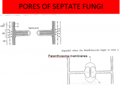
Pores of septate fungi |
|
|
|
How big is the nucleus in fungi walls? |
1-2um and up to 20-25um |
|
|
How many membranes does the nucleus have? |
double membrane |
|
|
Notable peculiarities: -Nuclear membrane / nucleolus remain intact during cell division (mitosis) -Prevents dispersal of nuclear contents in cytoplasm? -No metaphase plate; randomly disperse -All fungi still need microtubule organizing centre so chromosomes will separate properly. |
Vast majority of nucleus are haploid, all other plants, animals and pseudo fungi are 2N |
|
|
What does the phospholipid bilayer in fungi contain? |
-Transmembrane proteins(nutrient uptake) -Signal transduction -Anchor enzymes, chitinase and glucanase: integral membranes, that produce and deposit polysaccharides chains |
|
|
What membrane sterol is in fungi? |
Ergosterol, this is what fungicides go after |
|
|
What sterols do oomycota have in their plasma membrane? |
B-sitosterol and phytosterols |
|
|
What are the three main components in the secretory system? |
Endoplasmic reticulumn, golgi apparatus, and membrane bound vesicles |
|
|
46 |
46 |
|
|
What is defined as the peripheral growth zone? |
Point at which a cut on the hyphae does no affect growth or metabolism |
|
|
Growth thought to involve to two processes -Continuous extension of the plastic deformable tip -Rigidification of wall behind the tip |
Tips very sensitive to environmental or disturbance influences Tips undergo ‘stop-switch-branch’ reaction |
|
|
Chitin synthese |
one of the major enzymes involved with forming chitin |
|
|
Glucan synthase |
Other major ennzyme that catalyses the glucan chains. Composes bulk of fungal cell wall. Through to arrive in vesicles as well. Inserted into wall at tip of hypha. |
|
|
Negative autotropism: spores respond to touch of other spores |
s |
|
|
s |
s |