![]()
![]()
![]()
Use LEFT and RIGHT arrow keys to navigate between flashcards;
Use UP and DOWN arrow keys to flip the card;
H to show hint;
A reads text to speech;
57 Cards in this Set
- Front
- Back
- 3rd side (hint)
|
What does the Musculoskeletal System consist of?
|
2. Joints 3. Tendons 4. Ligaments 5. Muscles 6. Nerves 7. Blood Vessels |
Bite Just The Little Mangoes, Nuts and Bananas |
|
|
Define Bone. (Composition? )
|
D: A hard form of connective tissue that makes up most of the skeleton C: It is composed chiefly of calcium phosphate and calcium carbonate. |
|
|
|
What is the function of Bone? |
F: 1. Supports body and its vital cavities 2. Static attachment of muscle/tendon; 3. Organ and Vital Structure protection 4. Storage for Salts (e.g. calcium and phosphorus); 5. Haemopoiesis
|
1. Support 2. Protection 3. Storage 4. RBC production
|
|
|
The human skeleton can be divided into the...
|
Axial and Appendicular Skeleton
|
|
|
|
What does the Axial skeleton consist of? |
Bones of the: 1. Head (Skull/Cranium) 2. Neck (Cervical Vertebrae) 3. Trunk (Vertebrae, Sacrum, Ribs and Sternum) |
|
|
|
What is the function of the Axial Skeleton?
|
To protect viscera and act as a large area for muscle attachment
|
|
|
|
What does the Appendicular skeleton consist of? |
Bones of the: 1. Pectoral Girdle Bones 2. Upper and Lower Limb 3. Pelvic Girdle Bones |
Pectoral Girdle: Clavicle and Scapula Mostly Bones of the Limb Pelvic Girdle: 2 Hip Bones, Sacrum and Coccyx, but only hip bones are Appendicular |
|
|
What are the 2 types of Bone?
|
1. Compact: provides strength for weight bearing 2. Spongy (trabecular or cancellous) |
The difference depend on the relative amount of solid matter, the number and size of spaces they contain.
|
|
|
Bone is Classified according to its shape. What are the different kinds of bone? Give an example. |
1. Flat Bone 2. Irregular Bone 3. Short bone 4. Long Bone 5. Sesamoid Bone |
1. Sternum 2. Vertebra 3. Medial Cuneiform 4. Femur 5. Patella |
|
|
Define Joint/Articulation.
|
The point at which two or more bones are connected.
|
The place of union/junction between 2 or more rigid components (bones, cartilages or even parts of the same bone)
|
|
|
What are the 3 classes of Joints? Give examples.
|
1. Diarthrosis (freely moveable); shoulder 2. Amphiarthrosis (slightly movable/ semi-mobile joints); ribs and vertebra 3. Synarthrosis; sutures of the skull |
All Diarthrosis joints are synovial joints
|
|
|
The opposing surfaces of the two bones of a joint are... |
...lined with Cartilagenous, Fibrous or Soft/Synovial Tissue
|
|
|
|
What is Cartilage? Where? |
A type of connective tissue that forms parts of the skeleton where more flexibility is necessary (e.g. Costal Cartilage) |
|
|
|
Which type of joint is functionally the most common and important type of joint?
|
Synovial Joint: a freely movable joint
|
|
|
|
Synovial Joint: The 2 bones are separated by...
|
Synovial cavity, which contain synovial fluid to lubricate the joint surfaces and nourishes articular cartilage.
|
|
|
|
The bones of the Synovial Joint are joined by... What are the two parts of it? Functions? |
...the Joint capsule, consisting of: 1. Synovial membrane: secretes lubricant 2. Fibrous Capsule: supports and protects joints |
|
|
|
What are the different types of Synovial Joints?
|
1. Pivot 2. Plane 3. Ball and Socket 4. Hinge 5. Saddle 6. Condyloid |
1. P 2. P 3. B & S 4. H 5. S 6. C
|
|
|
The Atlanto-axial joint is an example of...
|
Pivot joint: permits uniaxial rotation, usually a rounded process of bone which fits into a ligamentous socket.
|
C1 (1st Cervical vertebra) is called 'Atlas' after the Titan C2 is called Axis |
|
|
The Hip joint is an example of...
|
Ball and Socket: Permits multiaxial movement, a rounded head fitted in a concavity.
|
|
|
|
The Acromioclavicular joint is an example of...
|
Plane: permits gliding/sliding movements, usually uniaxial
|
|
|
|
The Elbow joint is an example of...
|
Hinge: Permits uniaxial flexion and extension only
|
|
|
|
The Carpometacarpal joint of the thumb is an example of...
|
Saddle: Permits biaxial movement of saddle shaped heads
|
Allows abduction, adduction, flexion, extension and opposition (touch tip of 4 fingers!)
|
|
|
The Metacarpophalangeal joint is an example of...
|
Condyloid: Permits biaxial flexion, extension, abduction, adduction and circumduction
|
|
|
|
What are the 4 functions of Muscle Tissue?
|
1. Thermoregulation 2. Movement 3. Stabilise Position 4. Storing and Moving Substances |
1.Thermogenesis: Shivering Refl. 2.Contraction 3.Continual Contraction 4.Skeletal Muscle Pump: muscles squeeze deep veins in legs |
|
|
What are the 3 types of muscle?
|
1. Skeletal Muscle 2. Cardiac Muscle 3. Smooth Muscle |
1. Attached by tendon to bone 2. Lines the heart 3. Lines Hollow Organs (e.g. Oesophagus, Intestines) |
|
|
Skeletal muscle are under...
|
Voluntary control, supplied by nerves from the Somatic Nervous System
|
PNS
|
|
|
Cardiac and Smooth muscle are...
|
Under Involuntary control, supplied by nerves from the Autonomic Nervous System
|
PNS
|
|
|
Describe the structure/microscopic features of Skeletal Muscle.
|
1. Regular Cylindrical Fibres 2. Peripheral Nuclei 3. Striated |
Striated: Stripy appearance due to arrangement of Myosin and Actin.
|
|
|
Describe the structure/microscopic features of Cardiac Muscle.
|
1. Branched Cylindrical Fibres 2. Intercalated Disks 3. Single Central Nucleus 4. Striated |
Have Autorhythmicity: AP at constant rate
|
|
|
Describe the structure/microscopic features of Smooth Muscle.
|
1. Elliptical Shaped Fibres 2. Single Central Nucleus 3. Not Striated |
|
|
|
What are the 4 properties of Muscle?
|
1. Electrical Excitability 2. Extensibility 3. Elasticity 4. Contractile |
1. React to chemical, hormonal, local pH changes and stimuli 2. Can stretch without damage 3. Can recoil to original shape 4. Myosin and Actin |
|
|
APs that allow Voluntary muscle contraction begin...
|
In the motor cortex of the brain. Reflexes only reach the spinal cord. |
Where in the brain? |
|
|
Axon terminals/Synaptic End-Bulbs/Boutons attach to... Which allows... |
1. The centre of 1 skeletal muscle fibre, specifically to the Plasma Membrane (Sarcolemma). 2. The whole muscle fibre to contract simultaneously. |
1. Sarcolemma 2. Simultaneous |
|
|
What is a Neuromuscular Junction/Moor End Plate? Where?
|
The region of contact between a motor axon and a muscle fibre. L: Between Bouton and Sarcolemma |
|
|
|
Excitation Contraction Coupling: AP runs through a motor neurone and...
|
Reaches a Bouton, causing depolarisation.
|
|
|
|
The depolarisation causes V-Gated Calcium channels to...
|
Open, leading to an influx of Ca++ ions
|
|
|
|
This causes vesicles to fuse...
|
With the membrane and release the neurotransmitter via exocytosis into the synaptic cleft. e.g. Acetyl Choline
|
|
|
|
2 molecules of ACh bind to...
|
Nicotinic-ACh-Receptors on the postsynaptic membrane, causing a conformational change in structure.
|
|
|
|
Non-selective cation channels open, which allows a flow of...
|
Na+ in, which causes a change in membrane potential; Depolarisation of Sarcolemma=End Plate Potential
|
|
|
|
The AP continues down the Sarcolemma...
|
Down through T-tubules and up the Triad zone.
|
|
|
|
The Sarcoplasmic Reticulum...
|
Releases Ca++, which activates Troponin C on the Tropomyosin-Troponin complex. This causes Muscle Contraction.
|
|
|
|
What do the Thin filaments consist of?
|
Actin molecules, blocked by Tropomyosin and governed by Troponin C |
Actin has binding sites for the Myosin Head |
|
|
What do the Thick filaments consist of?
|
Myosin Head and Myosin Tail
|
|
|
|
How is Acetylcholine recycled?
|
ACh is broken into Acetic Acid and Choline by Actylcholinesterase. Choline Acetyltransferase converts them into Acetylcholine. |
AChE and ChAT
|
|
|
Contraction Cycle: Calcium binds to Troponin C causing a conformational change...
|
So Troponin slides Tropomyosin off of binding sites on Actin
|
|
|
|
Myosin binding sites are exposed on the Actin, outline Cross Bridge Formation.
|
1. Myosin has ADP+Pi from previous contraction cycle 2. Pi phosphorylates the head 3. The conformational change allows the head to attach to the Myosin binding site. |
|
|
|
Movement is caused by the Power Stroke. What causes the Power Stroke?
|
1. Another conformational change is caused by the release of ADP+Pi from the Myosin Head. 2. The head bends 45 degrees and pulls the Actin molecule to the Sarcomere/M-Line |
|
|
|
What causes the Myosin head to release the Actin?
|
ATP binds on the Myosin head's Actin Binding site
|
|
|
|
The Myosin head's ATPase enzyme ATP to...
|
ADP+Pi, returns to 'Cocked position' ready for use.
|
Troponinc C goes back to its original position
|
|
|
How does Ca++ leave?
|
1. Active Transport pumps calcium back (out of sarcoplasm into the Sarcoplasmic Reticulum) 2. Calsequestrin binds 20 Ca++, helps hold calcium in the cisterna of the SR |
Calsequestrin is a calcium-binding protein of the sarcoplasmic reticulum
|
|
|
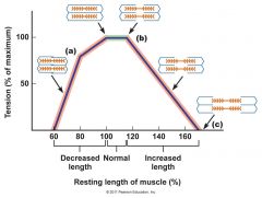
What does the Length-Tension Relationship state? (Tension% against Striation Spacing micrometres)
|
Forcefulness of muscle contraction depends on the Sarcomere's length within a muscle before contraction occurs.
|
|
|
|
Maximal Tension is lead to by...
|
Optimal overlap (2.0-2.4 micrometres) between the thick and thin filaments, without compressing the A band. (Overlap allows Cross-Bridge formation)
|
A-Band: Dark/Thick I-Band: Light/Thin Z-disc: End of Thin F. (crumples Myosin heads if overlap is too small) |
|
|
Ca++ and ATP is needed for muscle contraction. As ATP is used up...
|
It is rapidly replenished by Creatine Phosphate/Phosphocreatine. Action of Creatine Kinase |
Note: Its phosphate group is readily transferred to ATP
|
|
|
What are the roles of ATP in Muscle contraction?
|
1. Power Stroke (Dissociation) 2. 'Recocking' in ATPase of MH |
Muscle synthesises ATP rapidly, 1 ATP is split in each cycle
|
|
|
What is Rigor Mortis?
|
A sign of death, the limbs of corpse stiffen as chemical changes occur in muscle.
|
|
|
|
What is the physiological cause of Rigor Mortis?
|
1. The Myosin head continues binding with Actin binding sites. 2. No ATP means no Cross-bridge separation, therefore no muscle relaxation. |
|
|
|
Define Motor Unit.
|
The number of muscle fibres innervated by 1 motor neurone
|
|