![]()
![]()
![]()
Use LEFT and RIGHT arrow keys to navigate between flashcards;
Use UP and DOWN arrow keys to flip the card;
H to show hint;
A reads text to speech;
40 Cards in this Set
- Front
- Back
|
Muscle Fibers |
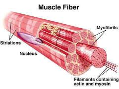
AKA- Skeletal muscle cells. Composes the skeletal muscle. |
|
|
Fascicles |
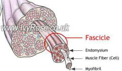
Groups of muscle fibers. Covered by epimysium. |
|
|
Epimysium |
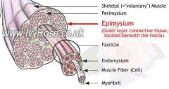
a sheath of fibrous elastic tissue surrounding a muscle. (Epi- (means upon) (entire, whole muscle) |
|
|
Root mysium |
muscle |
|
|
Endomysium |
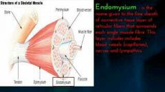
the delicate connective tissue surrounding the individual muscular fibers within the smallest bundles (a muscle fiber/cell) |
|
|
Fascia |
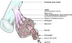
a thin sheath of fibrous tissue enclosing a muscle or other organ. |
|
|
Sarcolemma |
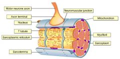
Plasma membrane wrapped around skeletal muscle fibers. |
|
|
Sarcoplasm |
the cytoplasm of striated muscle cells |
|
|
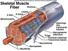
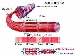
Myofibrils |
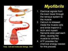
any of the elongated contractile threads found in striated muscle cells |
|
|
Sarcoplasmic Reticulum |
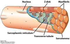
(SR) the endoplasmic reticulum (ER) of cardiac muscle and skeletal striated muscle that functions especially as a storage and release area for calcium. |
|
|
Tendon |
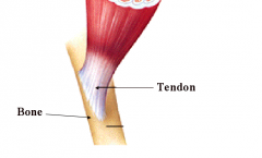
a flexible but inelastic cord of strong fibrous collagen tissue attaching a muscle to a bone. |
|
|
A band |

Any of various dark-staining anisotropic cross striations in the myofibrils of muscle fibers. |
|
|
I band |

A pale band of actin on each side of the Z line of a striated muscle fiber.
|
|
|
Z Disk/Z line |
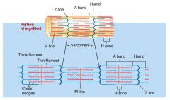
A dark thin protein band to which actin filaments are attached in a striated muscle fiber, marking the boundaries between adjacent sarcomeres.
|
|
|
Triad |
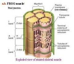
The transverse tubule, and the terminal cisternae on each side of it, in a skeletal muscle fiber. |
|
|
Sphincters |
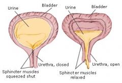
(circular muscles) a ring of muscle surrounding and serving to guard or close an opening or tube, such as the anus or the openings of the stomach.
|
|
|
Terminal cisternae |
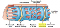
enlarged areas of |
|
|
T-tubule |
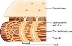
A T-tubule (or transverse tubule) |
|
|
Myofilaments |
the filaments of |
|
|
Thick filament |
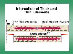
consist primarily of |
|
|
Thin filament |

Thin filaments, 7 nm in
|
|
|
Skeletal Muscle |
Striated, voluntary Skeletal muscle fibers are long and thin. Each skeletal muscle fiber has multiple nuclei located next to the sarcolemma. |
|
|
Cardiac Muscle |
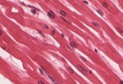
Striated, non-voluntary Cardiac myoctes are short, wide, and branching. Cardiac myocytes usually have a single, large nucleus located among the myofibrils. The intercalated disk of cardiac muscle tissue appear as black lines oriented parallel to the striations. |
|
|
Smooth Muscle |
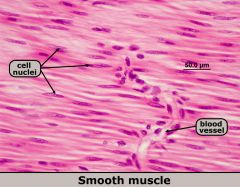
Non-striated, non-voluntary. (lack T-tubules) Smooth muscle cells are thin and flat. Nucleus is usually in the middle of the cell. Smooth muscle tissue is typically found on a slide with other types of tissue because it lines hollow organs.
|
|
|
Perimysium |
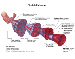
sheath of |
|
|
Striations |
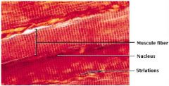
refers to the stripe-like visual features found in skeletal and cardiac muscle. |
|
|
Titin
|
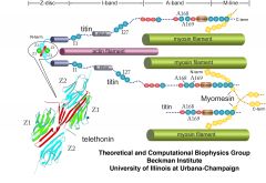
A very large fibrous protein that connects thick myosin filaments to Z discs in the sarcomere. The elasticity of skeletal muscle fibers is primarily due to the presence of this protein |
|
|
Neuromuscular Junction |
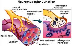
A neuromuscular junction is a synapse between a motor neuron and skeletal muscle. This lesson describes the events of synaptic transmission leading to contraction of skeletal muscle. |
|
|
After the power stroke has occurred during skeletal muscle contraction, what must occur in order for the acto-myosin cross-bridge to be released? |
ATP must bind with the myosin head
|
|
|
Smooth muscle is capable of mitosis and hyperplasia |
True |
|
|
The ability of the muscle cell membrane to respond to stimuli with electric changes is known as
|
excitability
|
|
|
The ability of the muscle cell to stretch under tension is known as
|
extensibility |
|
|
The ability of the muscle cell to return to its initial length after being stretched is called
|
elasticity |
|
|
Which of the following is a regulatory protein of muscle?
|
tropomyosin
|
|
|
Elastic filaments are composed of
|
titin |
|
|
The segment from one Z disc to the next is called a(n)
|
sarcomere |
|
|
The resting membrane potential (RMP) for a skeletal muscle cell is
|
-90mV |
|
|
Neuromuscular junction steps |
1) action potential comes down and causes vesicles to release ACh into synaptic cleft 2) ACh binds to Na channels 3) Na goes through Na channels 4) Na causes an action potential to form in the muscle 5) AChE recycles ACh and the process starts again with a new action potential |
|
|
One nerve fiber and all the muscle fibers it innervates are collectively called a:
|
motor unit
|
|
|
synaptic vesicles
|
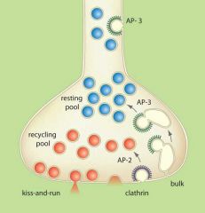
store
various neurotransmitters that are released at the synapse. The release is regulated by a voltage-dependent calcium channel. Vesicles are essential for propagating nerve impulses between neurons and are constantly recreated by the cell. |