Reading...
![]()
Play button
![]()
Play button
![]()
Use LEFT and RIGHT arrow keys to navigate between flashcards;
Use UP and DOWN arrow keys to flip the card;
H to show hint;
A reads text to speech;
115 Cards in this Set
- Front
- Back
|
What are the functions of bone?
|
1) Support of soft tissue
2) Protection of internal organs 3) Movement 4) Blood cell production 5) Mineral storage 6) Energy storage (adipose cells) |
|
|
What are the 4 types of cells found in bone?
|
Osteoprogenitor (osteogenic) cells
Osteoblasts Osteoclasts Osteocytes |
|
|
What are osteoprogenitor cells?
|
They divide and differentiate into osteoblasts
|
|
|
What do osteoblasts do?
|
Build bone (secrete collagen and organic cpds around themselves and differentiate into osteocytes)
|
|
|
What are osteocytes?
|
Mature bone cells that occupy lacunae of compact bone, exchange nutrients & wastes with blood via canaliculi
|
|
|
What function do osteoclasts perform?
|
Resorb bone matrix, releasing minerals back into blood. Believed to develop from monocytes
|
|
|
Diaphysis
|
Long shaft of typical long bone
|
|
|
Metaphysis
|
ends of long bone, contain epiphyseal plate
|
|
|
Epiphysis
|
ends of long bone, covered in articular cartilage
|
|
|
Epiphyseal plate
|
Sheet of cartilage in metaphysis where long bones grow in length
|
|
|
Spongy bone
|
Low density, high SA osseous tissue that fills inner cavity of long bones. Site of hemopoiesis
|
|
|
What is hemopoiesis and where does it occur?
|
Production of red bloods cells, occurs in spongy bone
|
|
|
Compact bone
|
Surrounds medullary cavity (contains yellow bone marrow) in long bone. Dense, highly organized, composed of units of osteons, constantly being remodeled
|
|
|
Haversian (central) canals
|
Channels in osteons (of compact bone) that contain blood & lymph vessels, connected by Volkmann's canals. New matrix constantly being laid down on tunnel walls (forming lamellae)
|
|
|
Lamallae
|
hardened concentric layers of matrix formed by osteoblasts in an osteon, separate rings of trapped osteocytes
|
|
|
Canaliculi
|
small canals that connect osteocytes to each other and to blood vessels
|
|
|
Volkmann's canals
|
Channels in osteons that run perpendicular, contain blood & lymph vessels, and connect osteons to each other and to periosteum
|
|
|
Lacunae
|
Small chambers occupied by osteocytes
|
|
|
Osteon (Haversian system)
|
Functional unit of compact bone
|
|
|
What is bone matrix composed of?
|
Hydroxyapatite crystals and collagen
|
|
|
How do collagen fibers affect bone strength?
|
Give bone tensile strength, elasticity
|
|
|
How does hydroxyapatite affect bone strength?
|
Gives bone great compressive strength (greater than best reinforced concrete), rigidity
|
|
|
In what form does ca2+ exist in bone?
|
Mostly in form of Hydroxyapatite crystals; some in form of slightly soluble calciums salts (i.e. CaHPO4)
|
|
|
What minerals does bone store?
|
Ca2+ and (HPO4)2-
|
|
|
What buffers plasma Ca2+ levels?
|
Slightly soluble calcium salts such as CaHPO4
|
|
|
What are the 4 types of bone?
|
1) Long bone
2) Short bone 3) Flat bone 4) Irregular bone |
|
|
Name some long bones
|
Leg, arm, finger, and toe bones
|
|
|
Define a short bone and give an example
|
ex. wrist, ankle; intended for limited movement, many break more easily due to lack of support and extensive bone marrow
|
|
|
What are the primary functions of flat bones? Name some flat bones
|
ex. skull, sternum, ribs, shoulder blades; large areas for muscle attachment and organ protection
|
|
|
In what form does calcium exist in the blood?
|
Most calcium is bound to proteins (calcium salts are only slightly soluble); some is bound to phosphates (HPO4)2- and other anions; a fraction is in form of free ions
|
|
|
What are the effects of having too high a concentration of calcium ions in the blood? What are the effects of too low a concentration?
|
too much Ca2+ causes membranes to become hypo-excitable, producing lethargy, fatigue, and memory loss. Too little produces cramps and convulsions.
|
|
|
What type of tissue is cartilage?
What is it composed of? |
a connective tissue, primarily collagen (a protein)
|
|
|
Are there blood vessels or nerves in cartilage?
|
no, except in its very outer membrane, the perichondrium
|
|
|
What are the three types of cartilage?
Which one is the most common and what it its function? |
1. Hyaline
2. Fibrocartilage 3. Hyaline Hyaline cartilage is the most common, it reduces friction and absorbs shock in joints |
|
|
What are the three types of joints?
|
1. Fibrous joints
2. Cartilaginous joints 3. Synovial joints |
|
|
What are fibrous joints and where in the body can some of them be found?
|
they occur between two bones held tightly together with little or no movement; skull bones form fibrous joints with each other, and the teeth form fibrous joints with the mandible
|
|
|
What are cartilaginous joints and what are some examples?
|
they allow little or no movement (like fibrous joints) and occur between bones tightly connected by cartilage; the ribs and the sternum, and the pubic symphysis in the hip bone are examples of cartilaginous joints
|
|
|
What are synovial joints?
|
occur between bones that are not bound by cartilage but rather are separated by a capsule filled with synovial fluid; synovial fluid lubricates and nourishes the cartilage, contains phagocytotic cells that eat microbes and particles from normal wear and tear, and allow a very wide range of movement
|
|
|
Is skin a tissue or an organ?
|
an organ
|
|
|
What are some important functions of skin?
|
thermoregulation, protection, sensation, excretion, immunity, blood reservoir, and vitamin D synthesis
|
|
|
How does skin aid in the regulation of body temperature?
|
dissipates heat by evaporative cooling and radiation, shunting of blood away from skin, piloerection, has sensory receptors for both heat and cold, fatty subcutaneous tissue (superficial fascia or hypodermis) insulates
|
|
|
How does skin function in sensory input?
|
senses temperature, pain, pressure, touch
|
|
|
What role does skin have in excretion?
|
excretes water (insensible fluid loss by diffusion) and salts
**burning of skin increases insensible fluid loss |
|
|
How is skin involved in vitamin D synthesis?
|
UV radiation activates molecule in skin that is modified by enzymes in liver and kidneys to produce vitamin D
|
|
|
What are the two principle parts of the skin?
|
epidermis and dermis
|
|
|
What is the hypodermis or superficial fascia?
|
subcutaneous layer of fat that aids in insulation
|
|
|
What part of the skin is avascular?
|
the epidermis
|
|
|
What are the four major cell types of the epidermis and their functions?
|
1. Keratinocytes- produce keratin which waterproofs skin
2. Melanocytes- transfer the skin pigment melanin to keratinocytes 3. Langerhans cells- interact with helper T cells 4. Merkel cells- attach to sensory neurons and function in sensation of touch |
|
|
How many layers of strata are in the epidermis?
|
5
|
|
|
Which stratum of the epidermis contains merkel and stem cells?
|
the deepest
|
|
|
Describe the process of keratinization
|
stem cells continuously divide to make keratinocytes and other cells; keratinocytes are pushed to top layer and on the way accumulate keratin and die, losing cytoplasm, nucleus, and other organelles; at top they eventually slough off body. process takes 2-4 weeks
|
|
|
How many layers of flat, dead cells comprise the outermost layer of the epidermis?
|
25-30 layers
|
|
|
How do calluses form?
|
friction or pressure stimulates the epidermis to thicken
|
|
|
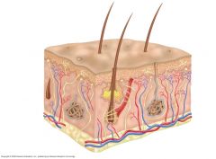
Hair shaft, sweat pore, Corpuscle of touch (Meissner's corpuscle), sebaceous gland, arrector pili muscle, sensory nerve, hair follicle, Lameliated (Pacinian) corpuscle, sudoriferous gland, vein, artery, adipose tissue, epidermis, dermis, subcutaneous layer
|
|
|
What type of tissue is the dermis?
|
connective tissue
|
|
|
What type of tissue is the epidermis?
|
epithelial tissue
|
|
|
What is the dermis derived from?
|
mesodermal cells
|
|
|
What is in the dermis?
|
blood vessels, nerves, glands, hair follicles, collagen and elastic fibers
|
|
|
What gives the dermis strength, extensibility, and elasticity?
|
collagen and elastic fibers
|
|
|
What is the integumentary system comprised of?
|
hair, nails, skin, glands, and some nerve endings
|
|
|
Where on the body is the dermis very thick?
|
palms and soles
|
|
|
Where do hair, nails, and some glands of the integumentary system derive from?
|
embryonic epidermis
|
|
|
What is hair, and what glands are most hairs associated with?
|
hair is keratinized cells tightly bound together; most hairs are associated with sebaceous glands that empty oil directly into follicle and onto skin
|
|
|
What smooth muscle are hairs closely associated with?
|
arrector pili
|
|
|
What type of cells are nails?
|
keratinized cells
|
|
|
What types of glands does the skin contain?
|
sebaceous, sudoriferous, and ceruminous (ears)
|
|
|
What are sudoriferous glands?
|
sweat glands
|
|
|
What are ceruminous glands?
|
produce ear wax
|
|
|
What are the three types of muscle tissue?
|
skeletal, cardiac, and smooth
|
|
|
How does any type of muscle tissue generate force?
|
by contracting its cells
|
|
|
Are the mechanisms by which muscle cells contract the same among the three types?
|
no
|
|
|
List the four possible functions of muscle contraction
|
1. body movement
2. stabilization of body position 3. movement of substances thru the body 4. generating heat to maintain body temp |
|
|
What connects muscle to bone?
|
a tendon
|
|
|
What connects bone to bone?
|
a ligament
|
|
|
What are the origin and insertion of a muscle?
|
Of a muscle that stretches across a joint, the origin is on the larger, more stationary bone and the insertion is on the smaller bone, which moves relative to the larger bone upon contraction of the muscle
|
|
|
Define agonist/antagonist
|
Muscles work in groups; the agonist is the muscle responsible for movement, the antagonist acts in opposition to the agonist and is responsible for returning a limb to its initial position (ex. biceps/triceps)
|
|
|
Define synergistic muscles
|
they assist the agonist by stabilizing the origin bone or by positioning the insertion bone during movement
|
|
|
What is the relationship between skeletal muscle and circulation?
|
contraction of skeletal muscle can squeeze blood and lymph vessels aiding in circulation
|
|
|
What control is skeletal muscle under?
|
it is voluntary muscle tissue and is innervated by the somatic nervous system
|
|
|
Contraction of skeletal muscle produces massive amounts of heat. What mechanism controlled by the hypothalamus takes advantage of this?
|
shivering to warm the body (rapid contraction of skeletal muscle)
|
|
|
What is the smallest functional unit of skeletal muscle?
|
the sarcomere
|
|
|
Describe the microanatomy of skeletal muscle
|
a skeletal muscle is a bundle of fasciclae, each fasciculus containing many muscle cells (fibers); A skeletal muscle fiber is many myofibrils wrapped together in a sarcolemma; each myofibril is itself wrapped in the sarcoplasmic reticulum and mitochondria and many nuclei are lodged between the myofibrils; Thick (myosin) and thin (actin) filaments comprise a myofibril, which is divided into the functional unit of a sarcomere
|
|
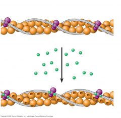
In the figure Identify actin, tropomyosin, troponin complex, the myosin-binding site, and ca2+. Describe the events depicted as Ca2+ are released from the sarcoplasmic reticulum
|
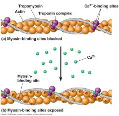
calcium ions bind to troponin which then pulls tropomyosin back, exposing the myosin binding site and allowing the myosin head to bind to actin
|
|
|
What is the A band in a skeletal muscle sarcomere?
|

The A band is the dark band and corresponds to the length of a bundle of myosin filaments- the width of the A band does not change upon contraction
|
|
|
What gives skeletal (and cardiac) muscle their characteristic light and dark bands?
|
the orderly overlapping of the actin and myosin filaments give cardiac and skeletal muscle their striated appearance (light and dark bands)
|
|
|
What is the I band in a skeletal muscle sarcomere?
|

The I bands are the light bands and are composed mainly of actin filaments- the I band shortens during contraction
|
|
|
What do the Z lines denote in a skeletal muscle sarcomere?
|

The area between two Z lines is known as a sarcomere, The Z lines bisect the I band and are the point of attachment of actin filaments
|
|
|
What is the H zone in a skeletal muscle sarcomere?
|
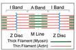
The myosin filaments that do not overlap with actin- the H zone gets smaller upon contraction
|
|
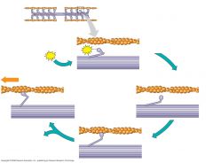
Describe the 5 stages of the myosin-actin interaction underlying muscle fiber contraction
|
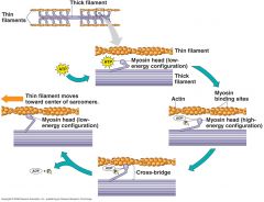
1. myosin head is bound to ATP and is in its low-energy configuration
2. myosin head hydrolyzes ATP to ADP and inorganic phosphate which causes head to extend (high-energy configuration) 3. myosin head binds to actin forming cross-bridge 4. myosin releases ADP and inorganic phosphate, bending head which pulls actin (power stroke) 5. binding of new ATP molecule releases myosin head from actin returning it to low-energy configuration |
|
|
How is a muscle cell excited into contraction?
|
a neuron attaches to muscle fiber at neuromuscular synapse, releasing acetylcholine into synaptic cleft which activates ion channels in sarcolemma and creates an action potential
|
|
|
What role do the T-tubules play in contraction of the muscle cell?
|
they transmit the action potential deep into the cell thereby spreading the AP rapidly and allowing for uniform contraction
|
|
|
What is the function of the sarcoplasmic reticulum in muscle contraction?
|
when the action potential reaches the SR, it releases Ca2+ into the cytosol, so it can then bind to troponin and expose the myosin binding site. The Ca2+ begins the 5 stage cycle of myosin-actin binding
|
|
|
How is the Ca2+ concentration in the cytosol maintained during muscle contraction?
|
at the end of each cycle (1 action potential) the Ca2+ is actively transported back into the SR
|
|
|
What is a motor unit and why is it important?
|
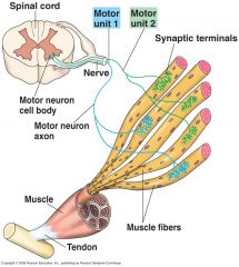
a motor unit consists of the muscle fibers served by a single neuron (1 neuron can innervate between 2 and 2000 muscle fibers). In a single muscle, not all motor units are recruited (graded response). In this way, motor units allow for selective contraction of muscle fibers so that we may control the strength and extent of muscle contraction
|
|
|
What is the force of a muscle contraction dependent on?
|
1. The number and size of motor units
2. The frequency of action potentials in each neuron |
|
|
What sorts of motor units are likely to be recruited first and why?
|
small motor units, makes for a smooth increase in the force
|
|
|
What type of motor units are involved in intricate movements such as the muscle contractions of the fingers?
|
small motor units
|
|
|
what is the most fatigue-resistant type of skeletal muscle?
|
slow oxidative
|
|
|
what type of skeletal muscle has the greatest amount of myoglobin? The least?
|
slow oxidative; fast glycolytic
|
|
|
What types of activities are best suited for slow-twitch (Type I) fibers?
|
endurance-type activities; running a marathon, maintaining posture
|
|
|
What types of activities are ideal for fast-twitch A (Type II A) fibers?
|
sprinting, walking
|
|
|
Fast-twitch fibers (Type II B) are best used for what sorts of activities?
|
short-term intense or powerful movements, such as hitting a baseball
|
|
|
What types of skeletal muscle contract rapidly?
|
Fast oxidative and fast glycolytic fibers
|
|
|
What skeletal muscle fiber type appears white under a light microscope?
|
fast glycolytic
|
|
|
What muscles would have great numbers of slow-twitch fibers?
|
postural muscles
|
|
|
What muscles would have larger amounts of fast-twitch A fibers?
|
upper leg muscles
|
|
|
What muscles would have larger numbers of fast-twitch B fibers?
|
upper arm muscles
|
|
|
What does the ratio of fiber types in a muscle depend on?
|
1. the contraction requirements of the muscle
2. genetics |
|
|
What happens when muscles strengthen?
|
The diameter of the muscle fibers increases, the number of sarcomeres and mitochondria in the fibers increase, and the sarcomeres lengthen
|
|
|
What do "slow" and "fast" refer to in describing skeletal muscle?
|
The rate at which myosin heads hydrolyze ATP
|
|
|
How does cardiac muscle differ from skeletal muscle?
|
only 1 nucleus
intercalated discs larger mitochondria contracts in upon self (not bone) involuntary muscle (myogenic) AP plateaus after depolarization |
|
|
What are the characteristics of smooth muscle?
|
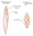
spindle-shaped, involuntary muscle (autonomic nervous system), 1 nucleus, no sarcomeres, intermediate filaments that pull dense bodies together to contract
|
|
|
What are the two types of smooth muscle?
|
1. single-unit (visceral): cells connected by gap junctions and contract as single unit
2. multi-unit: each smooth muscle fiber is attached directly to a neuron; group of multi-unit fibers can be in same location as single-unit while maintaining ability to contract independently |
|
|
Where in the body is single-unit smooth muscle found?
|
small arteries and veins, stomach, intestines, uterus, and urinary bladder
|
|
|
Where is multi-unit smooth muscle found?
|
large arteries, bronchioles, pili muscles attached to hair follicles, and the iris
|