Reading...
![]()
Play button
![]()
Play button
![]()
Use LEFT and RIGHT arrow keys to navigate between flashcards;
Use UP and DOWN arrow keys to flip the card;
H to show hint;
A reads text to speech;
27 Cards in this Set
- Front
- Back
|
What are the 3 types of muscle?
|
Visceral muscle, cardiac muscle, skeletal muscle
|
|
|
Describe how skeletal muscle tissue is formed
|
Mesenchymal cells in myotome differentiate into myoblasts
Myoblasts fuse and form multinucleated myotubes Contractile proteins laid down pushing nucleus to side and producing myofibres Some precursor cells (myoblasts) remain as satellite cells |
|
|
What are muscle antagonists?
|
Muscles that work opposite each other
|
|
|
What are muscle synergists?
|
Muscles that work together
|
|
|
What are the origin and insertion of a muscle?
|
Origin - proximal or central attachment of muscle tendon to bone (less movement)
Insertion - distal or peripheral attachment of muscle tendon to bone (more movement) |
|
|
What are the head and belly of a muscle?
|
Head - belly part closest to origin
Belly - main part |
|
|
Compare fat muscles to thin muscles
|
Fat muscles have more force generating capacity because they have more parallel contractile units
|
|
|
Compare long muscles to short muscles
|
Long muscles contract faster because they have more contractile units in series
|
|
|
What are the 5 muscle patterns?
|
Strap
Spindle Unipennate Bipennate Multipennate |
|
|
What do myofibrils divide into?
|
Sarcomeres - repetitive units
|
|
|
What myofilament proteins do sarcomeres contain?
|
Myosin, actin
|
|
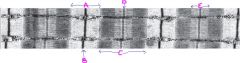
Name A, B, C, D & E
|
A= I band
B= Z line C= A band D= M band E= H zone |
|
|
What are the filaments found in the I band?
|
Actin filaments
|
|
|
What filaments provide for the dark banding?
|
Myosin
|
|
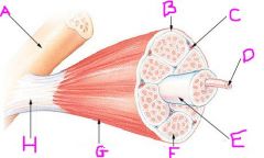
Name A to H
|
A= bone
B= Perimysium C= Blood vessel D= Muscle fibre E= Fascicle F= Endomysium G= Epimysium H= Tendon |
|
|
What are the tubules that are arranged parallel to the myofibrils called?
|
Sarcoplasmic reticulum
|
|
|
What are the tubules arranged at right angles (transversely) to the myofibrils called?
|
T-tubules
|
|
|
What do epimysium, perimysium and endomysium surround?
|
Epimysium - whole muscle
Perimysium - bundles of muscle fibres Endomysium - individual muscle fibres |
|
|
What is a sarcolemma?
|
Cell membrane of myofibril
|
|
|
What is a triad?
|
T-tubule and ends of two adjoining sarcoplasmic reticula
|
|
|
What is the function of the sarcoplasmic reticulum?
|
To provide a means of conduction of an impulse from the surface of a muscle fibre to the innermost parts
|
|
|
What are the 3 types of muscle fibre, what type of twitch do they give and what fibres are contained within them?
|
Type I - slow twitch, red fibres, aerobic
Type IIa - fast twitch, intermediate fibres, aerobic and anaerobic Type IIb - fast twitch, white fibres, anaerobic |
|
|
How is the plasticity of muscle shown?
|
Innervation
Growth Injury Exercise |
|
|
How does innervation affect elasticity?
|
If innervation is lost muscle fibres go into atrophy (loss and decrease in size)
Muscle fibres develop in accordance to size of motor neuron |
|
|
How does growth affect plasticity?
|
When growth occurs, muscle fibres lengthen by addition of more sarcomeres
Or can decrease by inappropriately set fracture shortens the limb |
|
|
How does exercise affect plasticity?
|
Exercise will increase oxidative metabolic capacity of motor units
Maximal strength training will add more parallel sarcomeres (hypertrophy of muscle fibres) |
|
|
How does injury affect plasticity?
|
Fibrosis - scar tissue laid down instead of muscle fibres
Evidence that satellite cells can lay down new muscle fibres or repair old ones |