Reading...
![]()
Play button
![]()
Play button
![]()
Use LEFT and RIGHT arrow keys to navigate between flashcards;
Use UP and DOWN arrow keys to flip the card;
H to show hint;
A reads text to speech;
54 Cards in this Set
- Front
- Back
|
Drawing a flow chart, how would you classify inflammatory arthritis.
|
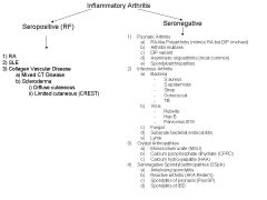
Seropositive and Seronegative based on the absence of presence of RF antibodies.
|
|
|
What type of nodules/deformities are found in someone with OA? RA?
|
OA = Heberden's and Bouchard's nodes
RA = Swan neck and Boutonerrie deformities |
|
|
What are Heberden's nodes? Bouchard's nodes?
|
They are bony prominences found on the DIP joints of someone with OA.
Bony prominences found on the PIP joints of someone with OA. |
|
|
What gold standard protocol should be performed on someone with a monoarthritis?
|
Fluid aspiration to investigate the synovial fluid.
|
|
|
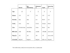
What does normal synovial fluid look like?
|
Colorless/straw color, viscous (due to HA) and has a WBC <200/mm3.
|
|
|
What are the various types of synovial fluid, what disease is associated and how many WBC counts?
|
Type 1 (Non-inflammatory)(OA) = <2000 WBC/mm3
Type 2 (Inflammatory)(RA) = >25,000 WBC/mm3 Type 3 (Septic) = >100,000 WBC/mm3 Type 4 (Hemorhagic) = RBC |
|
|
Compare and contrast the synovial fluid differences between Types 0-4?
|
|
|
|
What type of immune cells are found in Type 1- 4 synovial fluids?
|
Type 1 = mononuclear (lymphocytes)
Type 2 & 3 = PMNs Type 4 = RBC |
|
|
What sort of investigations should be performed on a synovial fluid?
|
Naked eye inspection of color, viscosity and turbidity.
Gram stain, C&S, AFB and fungal Microscopy looking for crystals WBC count and differential. Albumin and glucose concentration. |
|
|
Compare and contrast stiffness in RA and OA?
|
RA stiffness has prolonged (>45min) in the morning that gets better with activity and worse with inactivity. Can cause night pain.
OA stiffness is also in the AM but only lasts for 10 minutes. Activity makes it worse. |
|
|
What type of diseases are expected with Type 2 synovial fluid?
|
Inflammatory including RA, psoriatic arthritis, SLE.
|
|
|
Why is Type 2 synovial fluid watery and turbid?
|
Watery because the HA is being broken down by enzymes
Turbid because of the increased proteins and WBC |
|
|
What happens to the glucose and albumin (protein) levels in Type 2 & 3 synovial fluid?
|
Protein levels increase, glucose levels decrease in both inflammatory and septic causes.
|
|
|
What are the two classifications of arthritis?
|
Inflammatory or Non-inflammatory
|
|
|
How do you classify inflammatory arthritis?
|
Seropositive (for RF) or Seronegative
|
|
|
Give examples of non-inflammatory arthritis, seropositive and seronegative inflammatory arthritis?
|
Non-inflammatory = OA
Inflammatory seropositive = RA, SLE, Collagen vascular diseases Inflammatory seronegative - 15% of RA, psoriatic arthritis, ankylosing spondylitis, infection, crystal arthropathies |
|
|
What are some seronegative inflammatory arthritis?
|
1) RA (15%)
2) Psoriatic 3) Ankylosing spondylitis 4) Infection 5) Crystal arthropathies |
|
|
What are some infectious agents that cause seronegative inflammatory arthritis?
|
1) Bacteria - S.aureus, S.epidermidis, Strep and N.gonorrhea
2) Viral - rubella, parvovirus B19, HBV 3) Fungal 4) TB 5) Lyme |
|
|
How does subacute bacterial endocarditis cause inflammatory arthritis?
|
Mimicry - infectious Ag must look like a self-Ag in the joints therefore creating antibodies to the infectious agent also causes you to attack your own joints.
|
|
|
What are the three types of crystal arthropathies?
|
1) Monosodium urate (MSU)
2) Calcium pyrophosphate dihydrate (CPPD) 3) Calcium hydroxyapatite (HAA) |
|
|
How can you classify inflammatory arthritis based on # of joints?
|
1) Monoarthritis - one joint involved
2) Oligoarthrits = <5 jts involved 3) Polyarthritis = >4 jts involved Oligo and poly can be symmetrical or asymmetrical. |
|
|
What joints does RA usually affect?
|
The small joints of the hand including PIP and MCP
The wrist, elbow, shoulder, hips knee, ankles and feet (MTP). The DIP joints are NEVER involved in RA. |
|
|
What are some joint features of RA?
|
Seropositive (85%) inflammatory arthritis
Symmetrical Polyarthritis - can present as mono Causes nodules, soft to palpate. Causes joint erosion and deformity |
|
|
Define pannus?
|
Pannus is the hypertrophied synovium (Type A and B cells) of RA that occur at the double reflection of the synovial membrane.
The pannus invades the cartilage and juxta-articular margins causing erosion = pocket erosions |
|
|
What changes does the pannus caused by RA cause on X-ray?
|
Joint space loss (equally throughout the join unlike OA)
Joint erosion Osteopenia or bite markings Joints can b/c subluxed/ankylosed |
|
|
What type of deformities in the hand can be observed in RA?
|
1) Swan neck deformities
2) Boutienneir deformities 3) Ulnar deviation 4) Z or duckbill deformity of the thumb |
|
|
What is Felty's syndrome?
|
Triple syndromes
1) RA 2) Splenomegaly 3) leukopenia |
|
|
What is Sjogren's syndrome?
|
Endocrine system attacks the salivary and tear glands therefore your eyes (xerophthalmia) and mouth (xerostomia) get very dry.
This can cause a huge issue with dental caries. |
|
|
How does lupus arthritis look?
|
Lupus arthritis is associated with other features, never on its own.
Symmetrical and affects the small joints of the hands and knee - it doesn't affected the DIP joints. It does NOT cause erosion of the joints (unlike RA) |
|
|
What is the Sicca syndrome?
|
Sjogern's features of dry eyes (xerophthalmia) and dry mouth (xerostomia)
Is associated with RA, SLE and scleroderma |
|
|
What are the 11 criteria to diagnosis SLE? How many total do you need to diagnosis?
Need 4/11 to be +ve for SLE |
1) Malor rash (butterfly rash)
2) Photosensitivity rash 3) Maculopapular rash 4) Mouth ulcers 5) Inflammatory arthritis 6) Pleuritis or pericarditis 7) Nephrosis or Nephritis 8) CNS manifestations 9) +ve ANA 10) +ve Smith Ab (Sm) or DNA Ab or anti-phospholipid antibody 11) Lymphocytopenia or Coomb's positive hemolytic anemia or thrombocytopenia |
|
|
What is scleroderma?
|
Idiopathic sclerosis (thickening/hardening) of the skin.
|
|
|
What are the 2 types of scleroderma?
|
1) Diffuse systemic
2) Limited cutaneous (CREST) |
|
|
What systemic diseases are found in scleroderma?
|
1) Severe Raynauds
2) Hypertensive crisis 3) Renal failure 4) Interstitial lung disease |
|
|
What does CREST stand for in Limited cutaneous scleroderma?
|
C - Calcinosis (Ca+2 deposits in soft tissue)
R - Raynaud's syndrome E - Esophageal dilatation S - sclerodactyly (binding at the knuckles) T - Telangiectasia (small dilated blood vessels near the surface of skin or mucosa) |
|
|
What is mixed connective tissue disease (MCTD)?
|
Mix of lupus, RA, scleroderma and polymyositis.
|
|
|
What is psoriatic arthritis?
|
Arthritis with psoriasis rash/nail changes (onycholysis)
|
|
|
How does psoriatic arthritis affect the joints?
|
1) Asymmetrical
2) Oligoarthritis (<5 jts) 3) Sausage digits 4) Involved the DIP joints 5) Sclerosis (thickening) and periostitis (inflammation of the periosteum) instead of osteopenia as seen in RA. |
|
|
What type of inflammatory arthritis causes the cup appearance or the "opera glass hand"
|
Arthritis mutilans - a subset of psoriatic arthritis.
|
|
|
What are the 5 subsets of psoriatic arthritis?
|
1) RA-like Polyarthris (mimics RA but DIP involved)
2) Arthritis mutilans 3) DIP variant 4) Asymmetric oligoarthritis (most common) 5) Spondyloarthropathy |
|
|
What joints are inflammaed in seronegative spondylarthropathies?
|
1) Inflammatory axial spine involvement
2) Peripheral joint involvement, oligoarthritis, large joints and asymmetric 3) Enthesisi is involved (where the tendon inserts into the bone) |
|
|
What mutation is observed in seronegative spondylarthropathies?
|
HLA-B27
|
|
|
What are the four subsets of seronegative spondylarthropathies (SSpA)
|
1) Reactive Arthritis (AKA Reiters)
2) Ankylosing spondylitis (AS) 3) Spondylitis of psoriasis (PsorSp) 4) Spondylitis of IBD (IBD) |
|
|
What are some features of inflammatory back pain?
|
1) Insidious onset before 40yo
2) Gets worse with inactivity and better with exercise 3) Night pain* 4) Morning stiffness >30min |
|
|
What is a defining feature to distinguish between seronegative spondylarthropies (SSpA) and anklyosing spondylitis (AS)?
|
AS will have sacroilitiis, SSpA will not.
|
|
|
What are some common features seen in anklyosing spondylitis (AS)?
|
1) Sacroilitis
2) Syndesmophytes (new bone) bridging vertebral bodies 3) Enthesopathy (heel spurs) 4) Can get bamboo spine (fuse) |
|
|
What review of system questions should you ask if you suspect seronegative spondylopathies (SSpA)?
|
1) Iritis
2) Conjuctivitis 3) Psoriasis 4) Inflammatory bowel diseases 5) Recent travel and dysentry 6) Urethritis, cervicitis |
|
|
What type of bugs commonly trigger dysentry and reactive (reiter's) arthritis?
|
1) Salmonella
2) Yersinia 3) Shigella 4) Campylobacter But any microorganism can trigger reactive arthritis |
|
|
What is the triad found in reactive (reiter's) arthritis?
|
1) Arthritis
2) Conjuctivitis 3) Urethritis - chlamydia (can be associated with dysentry instead) |
|
|
Explain the concept of molecular mimicry and its role in inflammatory arthritis?
|
Some bacterium express immune epitopes that are similar to self HLA-B27. If this occurs, your body will produce antibodies that will X-react with self auto-antibodies and cause a reactive arthritis.
|
|
|
Compare the polymerized microscope results in someone with gout/pseudogout caused by MSU crystals and CPPD?
|
Gout (MSU) will cause a -ve birefringement which produces yellow crystals that are parallel with the axis and blue that are perpendicular.
Pseudgout (CPPD) produces a +ve birefringment with yellow crystals perpendicular and blue parallel. |
|
|
What is a classic case of gout?
|
Some has cardiac bypass surgery and they wake up with a inflammed toe - sheets can't even touch it.
|
|
|
What does CPPD look like on X-ray?
|
CPPD crystals cause chondrocalcinosis (cartliage becomes calcified)
CPPD can cause OA of a severe and inflammatory nature in atypical joints like the true wrist and shoulder. |
|
|
What causes calcified tendons to show up on X-rays and peri-arthritis?
|
Calcium hydroxyapatite crystals (HAA)
|