Reading...
![]()
Play button
![]()
Play button
![]()
Use LEFT and RIGHT arrow keys to navigate between flashcards;
Use UP and DOWN arrow keys to flip the card;
H to show hint;
A reads text to speech;
10 Cards in this Set
- Front
- Back
|
What are the 5 key characteristics of synovial joints?
|
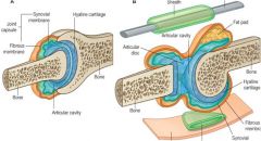
Articular Cartilage
Joint Space Synovial Fluid Synovial Membrane Joint Capsule |
|
|
Why is the tibiofibular joint important?
|
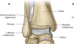
- Composed to 2 joints = superiorly a planar synovial joint, and inferiorly a syndesmosis (compound fibrous joint)
- The 2 bones are held together by the inter-osseous membrane - Holds ankle in alignment and stregthens it by keeping the lateral malleolus firmly against the lateral surface of the talus, helping to add stregth to a weak structure |
|
|
Describe the effects of the differences in planes of the tibia and fibula
|
- Tibia and fibula are not completely in the same plane = the fibula is more posterior
- This alignment means that the foot turns out 15-20 degrees when walking - The medial malleolus is also smaller than the lateral one - so stability increases on the lateral rather than the medial side |
|
|
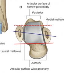
Describe the anatomy of the talus
|
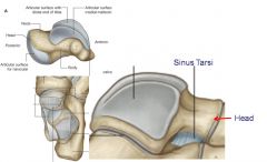
- 3 parts = head, neck and body
- Head articulates with the navicular - Neck receives the blood supply from the sinus tarsi, so that if there is a fracture here, there is a high chance of avascular necrosis as the rest of the bone will no longer be fed - Body has the main articular surfaces for the tibia and fibula, and inferiorly for the calcaneus. The facets on the lateral surface are larger than the facet on the medial surfaces as there is more contact with the fibula = more stable |
|
|
Describe the ligaments of the ankle
|
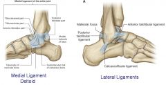
- Medial ligament is larger than lateral and stronger
• Runs between the medial malleolus and talus, navicular and calcaneus • Described as having 4 continuous parts = tibionavicular, tibiocalcaneal, anterior and posterior tibiotalar parts • Stabilises the ankle joint during eversion and prevents subluxation of the joint - Lateral ligament reinforces the ankle laterally • Compound structure formed from 3 separate ligaments = anterior talofibular, posterior talofibular and calcaneofibular ligaments |
|
|
What movements are possible at the ankle?
|
- Ankle is purely a hinge joint so can only dorsiflex and plantarflex
- Other movements, that appear to occur at the ankle, actually occur at the tarsal joints |
|
|
Summarise the ankle joint:
1. Type 2. Articular surfaces 3. Capsule 4. Ligaments 5. Special features 6. Movements |
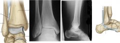
1. Type: Synovial hinge joint
2. Articular surfaces: distal ends of the tibia and fibula and superior part of the talus 3. Capsule: Think anteriorly and posteriorly but supported on either side by strong collateral ligaments. Fibrous layer attached superioly to the borders of the tibia, the malleoli and inferiorly to the talus. Synovial membrane is loose and lines the fibrous layer of the capsule 4. Ligaments: - Lateral ligament: 3 ligaments = anterior and posterior talofibular, calcaneofibular ligaments - Medial ligament (deltoid): very large and strong and attaches to the talus, navicular, and calcaneus 5. Special features: flextor retinaculum attaches from the tip of the medial malleolus to the medial calcaneal process and plantar aponeurosis 6. Dorsiflexion and planterflexion |
|
|
Describe the subtalar joint:
1. Type 2. Articular surfaces 3. Capsule 4. Ligaments 5. Movements |
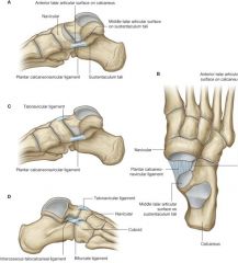
1. Type: plane synovial joint
2. Articulating surfaces: inferior surface of body of talus with superior surface (posterior facet) of calcaneus 3. Capsule: fibrous later of joint capsule is attached to margins of the articular surface 4. Ligaments: Medial, lateral and posterior talocalcaneal ligaments support capsule; interosseous talocalcaneal ligament binds bones together 5. Movements: inversion (always with some adduction of toes) and eversion of the foot (always with some abduction of toes) |
|
|
Describe the 3 parts of the foot
|
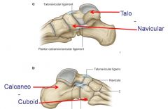
1. Hindfoot = talus and calcaneus joints
2. Midfoot = Navicular, cuneiforms and cuboid joints 3. Forefoot = metatarsals and phalanges |
|
|
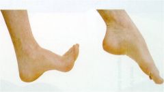
What are the arches of the foot and why are they significant?
|
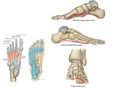
- If feet were rigid strcutres each impact with the ground would generate extremely large forces to the skeletal system
- Because the foot is composed of numerous bones connected by ligaments it has flexibility, allowing it to deform with ground contact, therefore absorbing most of the shock - The tarsal and metatarsal bones are also arranged in longitudinal and transverse arches that add to the weight-bearing capability and resilience of the foot - The arches distribute weight over the foot, acting as shock absorber and springboards for walking etc and also add to the foots ability to change its surface contour - When walking the weight is transfered to the talus from the tibia, posterioly to the calcaneus, then down the lateral margins of the foot to the base of the 5th metatarsal, then on to the big toe - Between these weight bearing points are the arches - Factors influencing the formation of the arches 1. shape of the bones 2. layers of fibrous tissue that bowstring the longitudinal arch (plantar aponeurosis etc) 3. active bracing of the intrinsic muscles of the foot 4. active and tonic contraction of muscles with long tendons entering the foot |