![]()
![]()
![]()
Use LEFT and RIGHT arrow keys to navigate between flashcards;
Use UP and DOWN arrow keys to flip the card;
H to show hint;
A reads text to speech;
107 Cards in this Set
- Front
- Back
|
Name the 3 general functions of the nervous system. |
1. Sensory 2. Integration 3. Motor |
|
|
Describe the sensory function of the nervous system. |
Monitors/receives information about the external and internal environments and communicates this information to integrative areas. |
|
|
Describe the integration function of the nervous system. |
Processing, distribution and interpretation of input signals and processing and distribution of output signals to initiate body responses/activities. |
|
|
Describe the motor function of the nervous system. |
Activation of effector organs/cells (muscles and glands) to cause an appropriate action/response in relation to existing sensory information. |
|
|
What are the 2 main divisions of the nervous system? |
1. Central Nervous System (CNS) 2. Peripheral Nervous System (PNS) |
|
|
What does the CNS consist of? |
Brain and spinal cord |
|
|
What does the PNS consist of? |
Nerves |
|
|
What are receptors? |
Receptors are sensory structures that detect changes in the internal or external environment. |
|
|
Name the 3 types of sensory receptors. |
1. Somatic sensory receptors 2. Special sensory receptors 3. Visceral sensory receptors |
|
|
What do somatic receptors sense? |
Position, touch, pressure, pain and temperature sensations. |
|
|
What do special receptors sense? |
Smell, taste, vision, balance and hearing. |
|
|
What is the function of the sensory division of the PNS? |
Brings information to the CNS from receptors in peripheral tissues and organs. |
|
|
What is the function of the CNS? |
Information processing which includes the integration and distribution information in the CNS. |
|
|
What is the function of the motor division of the PNS? |
Carries motor commands from the CNS to peripheral tissues and systems. |
|
|
Name the 2 branches of the motor division of the PNS. |
1. Somatic nervous system 2. Autonomic nervous system |
|
|
What is the function of the somatic nervous system? |
Controls skeletal muscle contractions. |
|
|
What is the function of the autonomic nervous system? |
Provides autonomic regulation of smooth muscle, cardiac muscle, glands, and adipose tissue. |
|
|
What are effectors? |
Effectors are target organs whose activities change in response to neural commands. |
|
|
Name the only effector under somatic nervous system control. |
Skeletal muscle |
|
|
Name the 4 effectors under autonomic system control. |
Smooth muscle Cardiac muscle Glands Adipose tissue |
|
|
What are neurons? |
Neurons are cells which communicate by receiving chemical signals, conducting electrical impulses and releasing neurotransmitters |
|
|
What is the function of a neuron? |
Their function is to receive, transmit (conduct) and send signals between each other and other cells of the body. |
|
|
Name the 3 interesting feature of neurons. |
1. Longevity 2. Amitotic 3. High metabolic rate |
|
|
Name the 3 functional regions of a neuron. |
1. Receptive region 2. Conducting region 3. Secretory region |
|
|
What does the receptive region of a neuron consist of? |
Dendrites and cell body (except unipolar neurons) which receive/respond to incoming chemical signals. |
|
|
What does the conducting region of a neuron consist of? |
Axon which transmits electrical impulses (action potentials). |
|
|
What does the secretory region of a neutron consist of? |
Axon (synaptic) terminals which secrete neurotransmitters (chemical messengers) that stimulate or inhibit other neurons or body cells. |
|
|
Name the 4 anatomical (structural) classifications of neurons. |
1. Multipolar neurons 2. Unipolar neurons 3. Anaxonic neurons 4. Bipolar neurons |
|
|
Describe a multipolar neuron. |
Have two or more dendrites and a single axon. Most common neuron type in the nervous system. |
|
|
Describe a unipolar neuron. |
Dendrites are continuous with the axon and cell body lies off to one side of axon. Most sensory neurons are unipolar neurons. |
|
|
Describe a anaxonic neuron. |
All cell process look alike (no distinct axon). Located in brain and some special sense organs, but functions are poorly understood. |
|
|
Describe a bipolar neuron. |
Have two distinct processes- a single dendrite with distal branches and a single axon. Rare, found only in some special sense organs. |
|
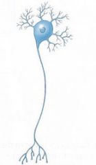
Name this type of neuron. |
Multipolar |
|
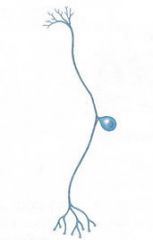
Name this type of neuron. |
Unipolar
|
|
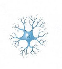
Name this type of neuron. |
Anaxonic
|
|
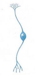
Name this type of neuron. |
Bipolar |
|
|
What are neuroglia? |
Neuroglia (glia or glial cells) are cells which support and protect neurons in the CNS and PNS. They are abundant and diverse, making up nearly half the volume of the nervous system. |
|
|
Name the 4 types of neuroglia found in the CNS. |
1. Astrocytes 2. Ependymal cells 3. Microglia 4. Oligodendrocytes |
|
|
Name the 2 types of neuroglia found in the PNS. |
1. Schwann cells 2. Satellite cells |
|
|
Name the 5 functions of astrocytes. |
1. Provide structural and metabolic support for neurons 2. Regulate ion, nutrient and gas concentrations in the ECF 3. Maintain the BBB 4. Modulate synaptic transmission 5. Form scar tissue after CNS injury |
|
|
Name the 2 main functions of ependymal cells. |
1. Form an epithelium called the ependyma which lines fluid filled spaces in the brain and spinal cord. 2. These cells produce monitor and circulate CSF. |
|
|
Name the main function of microglia. |
Moving through nervous tissue and removing cellular debris, waste products and pathogens by phagocytosis. |
|
|
Name the 2 main function of oligodendrocytes. |
1. Provide structural framework by stabilizing the position of neuronal axons 2. Produce myelin which wraps neuronal axons in myelin sheaths. |
|
|
Describe the function of schwann cells. |
Schwann cells form a sheath around segments of axons of peripheral neurons. This isolates the neurons from contact with the ECF. In most cases, the schwann cells form multi-layered myelin sheaths around the axon segments, just like oligodendrocytes in the CNS. |
|
|
Describe the function of satellite cells. |
These cells surround neuron cell bodies within ganglia and regulate the environment around the neurons (much like astrocytes in the CNS). |
|
|
What is the grey and white matter located in the brain? |
White matter - central Grey matter - superficial |
|
|
Where is the grey and white matter located in the spinal cord. |
White matter - superficial Grey matter - central |
|
|
What allows neurons to spread electrical signals? |
Changes in membrane voltage. |
|
|
What is the voltage of the membrane determined by? |
The concentration of ions inside and outside the cell. |
|
|
Movement of what ions changes the voltage of the cell membrane? |
Sodium Potassium Chloride |
|
|
What is the cells resting membrane potential? |
The resting neutron is negative inside the cell compared to outside the cell. The value of the RMP is around -70mV. |
|
|
What happens during depolarisation? |
The cell membrane becomes more positively charged. The RMP moves towards 0mV. |
|
|
What causes depolarisation? |
Excitatory stimuli. |
|
|
What happens during hyperpolarisation? |
The cell membrane becomes more negatively charged. The RMP moves further from 0mV. |
|
|
What causes hyperpolarisation? |
Inhibitory stimuli. |
|
|
What are the 2 types of changes that can occur to the RMP of a neutron due to depolarisation and hyperpolarisation? |
Graded (local) membrane potentials Action potentials |
|
|
Describe 4 distinguishing features of graded membrane potentials. |
1. Occur in the receptive region 2. Includes small depolarisations 3. Includes small hyperpolarisations 4. Can excite or inhibit the neuron |
|
|
Describe 4 distinguishing features of action potentials. |
1. Voltage change between -59mV and +30mV 2. Is a large depolarisation 3. Conducted along the length of the axon 4. Will cause the release of neurotransmitters |
|
|
What are EPSPs? |
Excitatory post-synaptic potentials (EPSPs): these are graded potentials caused by excitatory stimuli which lead to depolarization. |
|
|
What are IPSPs? |
Inhibitory post-synaptic potentials (IPSPs): these are graded potentials caused by inhibitory stimuli which lead to hyperpolarization. |
|
|
What happens when potentials summate? |
'Adding up' of multiple graded potentials (across time and/or space) to make larger changes in the RMP. However, the addition of EPSPs and IPSPs of equal value can cancel each other out. |
|
|
Define summation. |
TOTAL change of voltage within the entire receptive region of a neuron within a certain period of time. |
|
|
What is the threshold value? |
The particular voltage value that must be met for an action potential to fire. |
|
|
Where does summation occur? |
At the axon hillock where all the graded potentials accumulate. |
|
|
What voltage gated ion channels are utilised in action potentials? |
Na+ ion channels. |
|
|
When are Na+ voltage gated ion channels triggered? |
When depolarisation reaches threshold. |
|
|
What is depolarisation caused by? |
Na+ flowing into the cell. |
|
|
What is repolarisation caused by? |
K+ flowing out of the cell. |
|
|
What is hyperpolarisation caused by? |
K+ continuing to exit the cell. |
|
|
What does the initial large depolarisation at the axon hillock trigger? |
The voltage gated ion channels in the neighbouring region of the axon to open, which allows for the influx of ions. |
|
|
What is ion channel inactivation? And what does it achieve? |
Inactivating Na+ channels after they close at the end of depolarisation, which ensures the conduction of the AP is in one direction only. |
|
|
Name the 2 refractory periods. |
1. Absolute refractory period. 2. Relative refractory period. |
|
|
When do absolute refractory periods occur? |
While Na+ channels on an axon segment are open, the neuron cannot respond to any incoming stimuli (i.e. the cell is incapable of generating a new AP). |
|
|
When do relative refractory periods occur? |
When the Na+ channels on an axon segment become inactive it causes a brief increase in the cell's threshold value. During this period only very strong stimuli can cause the generation of a new AP. |
|
|
What 2 main factors does the velocity of an action potential depend on? |
1. Axon diameter 2. Myelination |
|
|
How does axon diameter affect AP speed? |
The wider the axon diameter, the faster the AP travels. This is because there is less resistance to the flow of the local currents due to the increased space. |
|
|
How does myelin affect AP speed? |
As myelin sheaths wrap only segments of axon, they leave gaps of exposed axon called nodes of Ranvier. It is at these nodes that the movement of ions can take place (not across the areas of membrane covered by myelin sheaths). Therefore the AP "skips' from node to node, getting to the axon terminals more quickly. |
|
|
Name the 3 main classifications of nerve fibres based on velocity. |
1. Group A fibres 2. Group B fibres 3. Group C fibres |
|
|
Describe Group A fibres. |
Large diameter axons with thick myelin sheaths; capable of conducting APs up to 150 meters/second (m/s). |
|
|
Describe Group B fibres. |
Medium diameter axons which are lightly myelinated; transmit APs at an average of 15 m/s. |
|
|
Describe Group C fibres. |
Small diameter, unmyelinated axons; conduct APs at around 1m/s or less. |
|
|
How is the strength of a stimulus coded? |
The frequency of the APs generated. |
|
|
What is a synapse? |
A synapse is the functional junction between a neuron and another neuron or between a neuron and an effector cell (muscles and glands). |
|
|
Name the 2 types of synapses. |
1. Electrical - rare 2. Chemical - majority |
|
|
What triggers the release or neurotransmitter at the ends of an AP? |
The influx of Ca+2 into the cell. |
|
|
Describe synaptic potentiation. |
Repeated or continuous use of a synapse enhances the presynaptic neuron's ability to excite a postsynaptic neuron. This increased excitability increases the efficiency of neurotransmission between these neurons and strengthens the neural pathway.Synaptic potentiation can therefore be considered a learning processes- whereby neural connections/synapses that are used are strengthened, whereas ones that are not are weakened and may even disappear. |
|
|
What is the type and amount of neurotransmitter likely to be linked to? |
Frequency of neuronal stimulation. |
|
|
What affect does the amount of neurotransmitter released have on the postsynaptic neuron? |
The amount of neurotransmitter released into the synaptic cleft influences the strength of the graded potentials at the postsynaptic cell |
|
|
What determines the excitatory or inhibitory response to a neurotransmitter? |
The type of neurotransmitter AND the type of receptor for that neurotransmitter. |
|
|
Describe direct action by a neurotransmitter. |
Direct action occurs when neurotransmitter binding directly stimulates the opening of ion channels in the postsynaptic cell. |
|
|
Describe indirect action by a neurotransmitter. |
Indirect action occurs when neurotransmitter binding stimulates a second messenger molecule (e.g. G proteins) to stimulate the opening of ion channels in the postsynaptic cell. |
|
|
Is Acetylcholine excitatory, inhibitory or both? Where would you find it? General functions? |
Both CNS - brain and spinal cord PNS - NMJ and ANS Learning and memory/brain excitability. Activation of skeletal muscles and autonomic regulation. |
|
|
Is Norepinephrine excitatory, inhibitory or both? Where would you find it? General functions? |
Both CNS PNS - ANS Attention, mood, consciousness, emotions, body temp. Regulation of sympathetic effectors. |
|
|
Is Epinephrine excitatory, inhibitory or both? Where would you find it? General functions? |
Both CNS - diencephalon, brainstem, spinal cord PNS - ANS Motor control Enhances sympathetic activity as a hormone |
|
|
Is Dopamine excitatory, inhibitory or both? Where would you find it? General functions? |
Mostly inhibitory
CNS regulation of mood, sleep, temperature, emotions, food |
|
|
Is Serotonin excitatory, inhibitory or both? Where would you find it? General functions? |
Mostly inhibitory
CNS Regulation of mood, sleep, temperature, emotions, food. |
|
|
Is Histamine excitatory, inhibitory or both? Where would you find it? General functions? |
Mostly excitatory
CNS - hypothalamus Wakefulness, appetite, learning and memory, body temp, fluid balance. |
|
|
Is Glutamate excitatory, inhibitory or both? Where would you find it? General functions? |
Excitatory CNS Major excitatory NT in brain - learning and memory |
|
|
Is GABA excitatory, inhibitory or both? Where would you find it? General functions? |
Inhibitory CNS - brain Major inhibitory NT in brain |
|
|
Is Glycine excitatory, inhibitory or both? Where would you find it? General functions? |
Inhibitory PNS - spinal cord Major inhibitory NT in spinal cord |
|
|
Is Endorphins excitatory, inhibitory or both? Where would you find it? General functions? |
Inhibitory CNS and lymphatics Inhibits pain signals |
|
|
Is Substance P excitatory, inhibitory or both? Where would you find it? General functions? |
Excitatory CNS Excites and stimulates pain pathways |
|
|
Is Adenosine excitatory, inhibitory or both? Where would you find it? General functions? |
Inhibitory CNS and PNS Vasodilation |
|
|
Is Cholecystokinin (CKK) excitatory, inhibitory or both? |
Excitatory CNS and gut Gut-brain communication, appetite regulation, pain and anxiety |
|
|
Is Nitric Oxide excitatory, inhibitory or both? Where would you find it? General functions? |
Both CNS PNS - adrenal glands, penis, smooth muscle Memory via synaptic potentiation Smooth muscle relaxation. |
|
|
Is Endocannabinoids excitatory, inhibitory or both? Where would you find it? General functions? |
Inhibitory CNS Memory, petite regulation, nausea, neuronal development |
|
|
Name the 4 ways of affecting neurotransmitter cellular communication. |
1. Enhancing neurotransmitter release 2. Inhibiting neurotransmitter release 3. Inhibiting removal of the neurotransmitter from the synapse 4. Blocking neurotransmitter receptors |