![]()
![]()
![]()
Use LEFT and RIGHT arrow keys to navigate between flashcards;
Use UP and DOWN arrow keys to flip the card;
H to show hint;
A reads text to speech;
138 Cards in this Set
- Front
- Back
|
Avian Influenza Virus |
(Orthomyxoviridae)
Infects poultry. Highly pathogenic (H5,H7) and mild pathogenic strain, due to differences in the cleavability of HA into HA1 and HA2.
Low to mild strain has a single arginine site, limited to cleavage by trypsin proteases in respiratory and intestinal tract. - Anorexia, lethargy, and variable dyspnea - Sinusitus and infraorbital swelling due to secondary infection
Highly pathogenic strains have multiple basic amino acids at cleavage sites, furin-like proteases ubiquitous distribution. Severe systemic disease. - Facial edema, with cyanosis of comb and wattles - Cutaneous petechia - Perivascular cuffing and necrosis of splenic lymphoid follicles and pancreatic acinar cells
Diagnosis: Serology and virus neutralization tests. Ddx: Fowl plague and Newcastle disease virus (more virulent strain) |
|
|
Avian Influenza Virus (picture) |

Respiratory and gastrointestinal tract (systemic). |
|
|
Canine Influenza Virus |
(Orthomyxoviridae) Infects dogs
H3N8 equine influenza virus (threonine and tryptophan substitution, loss of glycosylation site in HA2 subunit) |
|
|
Canine Influenza Virus (picture) |

Respiratory disease |
|
|
Equine Influenza Virus |
(Orthomyxoviridae)
Infects horses. Fast-spreading disease. Attacks lower respiratory epithelium. - Fever, muscle stiffness, cough, dyspnea, and nasal discharge - Edematous swelling of the limbs and abdomen - Chronic cough, 2-3 weeks sequela - Bronchopneumonia and secondary bacterial infection
Ddx: Initial clinical presentation, equine rhinopneumonitis (EHV) and equine viral arteritis virus |
|
|
Equine Influenza Virus (picture) |

Respiratory disease |
|
|
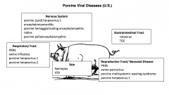
Swine Influenza Virus |
(Orthomyxoviridae) Infects swine
Stress is an important predisposing factor -Fever, anorexia, muscular weakness, paroxysmal coughing, and dyspnea - Mucopurulent tracheitis and bronchitis are principle lesions, alveolar necrosis and hyaline membranes - Secondary bacterial pneumonia |
|
|
Swine Influenza Virus (picture) |

Respiratory tract |
|
|
Hantaviruses |
(Bunyaviridae)
Rodents are carriers, species have different serotypes. Deer mouse is primary reservoir (pinyon mouse, brush mouse, and western chipmunk are adaptable), but common rat can harbor virus. Shed virus in saliva, urine, feces for many weeks. Infect humans (zoonotic).
Can develop hemorrhagic fever with a renal syndrome. Another pattern in Hantavirus pulmonary syndrome. - Fever, rapid onset of non-cardiogenic pulmonary edema, hypotension, shock - < 50% is fatal |
|
|
Rift Valley Fever Virus |
"Exotic" (Bunyaviridae)
Transmitted by Culex and Aedes mosquitos, or mechanically by biting flies.
Young sheep, goats, and cattle are susceptible (high mortality), can die without clinical signs. - Abortion is common in cattle and sheep
Humans are susceptible. - Liver injury - Anorexia and epigastric pain |
|
|
Lymphocytic choriomeningitis virus |
(Arenaviridae)
Rodent-borne disease that causes nonsuppurative meningioencephalitis (CNS) in humans.
Wild mice are natural host, usually inapparent infection. Lab rodents are a source of most infections (infected in utero shed for life). Split tolerance (antibody production by failure to generate cytotoxic T cells), cause lifelong viremia. Immune complex glomerulonephritis. If infected within first few days of life, develop lethal choriomeningioencephalitis. Transmitted in urine as aerosols or fomites.
Infects humans. - asymptomatic or mild febrile illness, headache, and/or myalgia - if pregnant, abortion, fetal hydrocephalus, chorioretinitis, and mental retardation
Lesions associated with viral infection can be prevented by immunosuppression of the host. |
|
|
Bovine Parainfluenza Virus 3 |
(Paramyxoviridae)
Experimental calves develop airway associated respiratory disease and interstitial pneumonia. - Syncytial giant cells with intracytoplasmic inclusions in lung tissue
Can be involved in shipping fever. Virus-induced damage to respiratory tract, secondary bacteria infections, plus stress. |
|
|
Bovine Parainfluenza Virus 3 (picture) |

Respiratory tract |
|
|
Bovine Respiratory Syncytial Virus |
(Paramyxoviridae)
Characteristic syncytia in infected cells. Experimental cattle develop rhinitis, tracheitis, bronchitis/bronchiolitis.
Also thought to play a role in shipping fever and enzootic pneumonia of calves (calves <4 months).
In sheep and goats, predisposed to chronic pasteurellosis. In humans, lower respiratory tract disease in infants and mild upper respiratory tract disease in adults. |
|
|
Bovine Respiratory Syncytial Virus (picture) |

Respiratory tract |
|
|
Canine Distemper Virus |
(Paramyxoviridae)
Dogs, wolves, coyotes, mink, ferrets, and otters, raccoons, civets, and mongoose also susceptible. Wild species are reservoir.
First phase, aerosolized virus gains access to epithelial cells of the upper respiratory tract, rapidly spreads to macrophages in tonsils and bronchial lymph nodes. Next 7 days, spread to bronchial and mesenteric lymph nodes, bone marrow, spleen, and thymus. - Asymptomatic
Second phase, colonization and destruction of epithelial cells of respiratory and GI tract. CNS is last area to be affected. Also infects uroepithelium of bladder and renal pelvis. - Fever, nasal discharge, conjunctivitis, coughing, and secondary bacterial infection of respiratory tract (Bordetella) which can cause bronchopneumonia - Diarrhea and dehydration are common complications Virus has an affinity for myelinated areas of CNS. - Seizures and neuromuscular twitching with excessive salivation, "gum-chewing fits" - Areas of malacia and gliosis in the cerbellar white matter and myelinated tracts of the cerebrum and spinal cord
Old dog encephalitis, in middle aged and old dogs from persistence of virus in the brain. |
|
|
Canine Distemper Virus (picture) |

Respiratory, digestive and CNS |
|
|
Canine Parainfluenza Virus |
(Paramyxoviridae)
Causes mild upper respiratory disease.
Vaccine available. Allegedly prevents canine tracheobronchitis (kennel cough), but significance is uncertain. |
|
|
Canine Parainfluenza Virus (picture) |
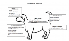
Respiratory disease |
|
|
Newcastle Disease Virus |
(Paramyxoviridae)
Infects domestic and wild bird populations (chickens)
Lentogenic strains, asymptomatic disease. Mesogenic strains, most commonly encountered strains. - Catarrhal nasal exudate, mild upper airway infection, air sacculitis - Cheesy exudate in air sacs of chronically infected birds - <4 weeks of age, respiratory distress, diarrhea, and CNS signs - Nonsuppurative encephalitis and bronchitis
Modified live vaccine.
Zoonotic, cause conjunctivitis in humans, contact transmission. |
|
|
Newcastle Disease Virus (picture) |

Respiratory tract, nervous system, GI tract |
|
|
Ebola Virus |
(Filoviridae)
Cause severe hemorrhagic disease in humans, can also be a threat to other primates - extensive subcutaneous and intestinal hemorrhage, intractable diarrhea, necrotizing hepatitis, prostration, and shock
Hemorrhagic fever from viral injury to hepatocytes and endothelial cells, leads to disseminated intravascular coagulopathy.
Spread via contact with fluids |
|
|
Rabies Virus |
(Rhabdoviridae)
All mammals are susceptible, with almost 100% morbidity (no birds, but can be carriers in saliva).
Reaches the CNS by antegrade spread from peripheral nerves (initial site of infection is muscle), intracellular route. Does not trigger an immune response. - CNS signs - Concentrates in salivary glands
Intracytoplasmic inclusions in brain tissue, can also detect with antibody test on fresh brain tissue (should not be frozen or submitted in formalin). |
|
|
Rabies Virus (picture) |

CNS disease |
|
|
Vesicular Stomatitis Virus |
(Rhabdoviridae)
Relatively harmless disease, but lesions are indistinguishable from foot and moth disease.
Outbreaks in cattle, swine, and horses (but deer, mountain goats, camelids, raccoons, bobcats, and monkeys) Humans can also be infected.
Insect vectors and mechanical transmission are most common. Once introduced, contact with saliva or fluid from rupture lesions.
"Influenza like" clinical signs. - Vesicular and erosive lesions on the tongue, gingiva, lips, teats, coronary band, and interdigital space |
|
|
Vesicular Stomatitis Virus |

Stomatitis |
|
|
Bluetongue Virus |
(Reoviridae)
Non-contagious disease isolated in domestic sheep. Transmitted by biting insects (primarily Culicoides insects, midges, gnats, and sand flies). Inapparent viremia in free-ranging white tailed deer, possible reservoir. Subclinical in cattle and goats, likely reservoir. Similar virus that causes epizootic hemorrhage in deer.
25 strains, wide variation of clinical disease. - fever, petchiae, cyanosis of nasal and oral mucosa - Swelling and ulceration of lips, tongue with erosions of dental pads, tongue, and gums - Lameness, ulceration and hyperemia of coronary band and laminitis - Hyperemia of skin damaged wool growth
Virus replication in endothelial cells, damage to pulmonary vessels, results in edema and cyanosis. Microscopic skeletal myocytis and arteritis of vessels in oral mucosa.
Vaccine, but has complications. |
|
|
Bluetongue Virus (picture) |

Nervous system, stomatitis, reproductive/neonatal |
|
|
Epizootic Hemorrhagic Disease Virus |
(Reoviridae)
Closely related to bluetongue virus. One of the most important disease of deer (white tailed and mule). Cattle can serve as reservoir host. Vector is Culicoides mosquito.
Lesions are similar to sheep with bluetongue. Severe disease with gastrointestinal hemorrhage and shock.
Viral injury to endothelial cells and subsequent disseminated intravascular coagulation.
Diagnosis thru serological confirmation. Ddx: Bovine viral diarrhea, malignant catarrhal fever, bluetongue.
|
|
|
Rotaviruses |
(Reoviridae)
Neonatal sheep, pigs, horses, mice, primates, and humans.
Target epithelial cells of distal half of intestinal villi. Undergo necrosis and are shed, shortened and blunted villi, reduce absorptive efficiency. - Diarrhea
Less pathogenic than coronaviruses, most recover. - Dehydration and secondary bacterial infection complications
Virus shed in feces.
Maternal antibody is protective, colostrum. Vaccination of dam is a major preventative strategy. |
|
|
Rotavirus (picture) |
GI tract |
|
|
Infectious Bursal Disease Virus |
(Birnaviridae)
Chickens 3-6 weeks are most susceptible. Target pre-B lymphocytes in bursa of fabricus primarily, when organ is developing. Results in B cell deficiency, "viral bursectomy" - Low levels of antibodies - Increased risk of opportunistic infection, immunosuppression - Gangrenous dermatitis from Clostridial infection - Aplastic anemia
Ingested, replicates in GALT, reach and localize in bursa of fabricus. - Swollen, subserosal edematous yellow fluid - Necrosis of lymphocytes |
|
|
Infectious Bursal Disease Virus (picture) |

GI and hemolymphatic system. |
|
|
Avian Leukosis and Sarcoma Virus |
(Retroviridae)
Causes lymphoid neoplasms thru cis-activation of c-myc gene of host. Vertical transmission, lifelong viremia, and tumor development at 3-7 months of age. - Enlarged bursa of Fabricus - Transformed B lymphocytes metastasize to liver, spleen, and other organs.
Can also cause nephroblastomas and renal cell carcinomas
Transduced variants of virus can cause myeloblastosis, fibrosarcoma, eythroblastosis (myb, src, erb, and myc oncogenes)
Rous sarcoma virus (src-transducing variant of ALV)
Can cause non-cancerous osteopetrosis from aberrant bone remodeling of infected osteoblasts. |
|
|
Bovine Immunodeficiency Virus |
(Retroviridae)
Infects a wide spectrum of cells of immune system, moderate immunosuppression. - Weight loss, secondary infections, lymphadenopathy, lymphocytosis, and CNS lesions
Jembrana disease virus is related, panleukopenia and widespread hemorrhages. |
|
|
Bovine Immunodeficiency Virus (picture) |

Hemolymphatic system |
|
|
Bovine Leukemia Virus |
(Retroviridae)
Cattle are infected at a young age (vertical transmission, ingestion thru milk, exposure to infected lymphocytes following trauma, iatrogenic transmission)
Mostly asymptomatic, some may develop clinical disease. - persistent lymphocytosis, lymphoid tumors develop in lymph nodes and various organs, leukemia typically is not present
Experimental in sheep causes lymphosarcoma, but does not spread from cattle to sheep normally. |
|
|
Bovine Leukemia Virus (picture) |

Hemolymphatic system |
|
|
Caprine Arthritis-Encephalitis Virus |
(Retroviridae)
Most important disease of goats, 80% infection rates, cause substantial economic loss.
Encephalomyelitis rarely occurs in kids 2-4 months. Progressive leukoencephalomyelitits and demyelination, ascending paralysis. Joints become swollen and painful, proliferative synovitits.
More commonly, disease manifests as arthritis in goats over 1 year.
Can be transmitted to sheep experimentally, but does not occur naturally.
Transmitted via colostrum and milk, can heat treat, but loss of antibodies and immune transfer from mother. |
|
|
Caprine Arthritis- Encephalitis Virus (picture) |
Musculoskeletal system. Nervous system (kids 2-4 months, rare form) |
|
|
Equine Infectious Anemia Virus |
(Retroviridae) Can manifest as either an acute syndrome causing death within one month, a chronic relapsing disease, or a silent infection.
Acute EIA - Severe anemia, petechial hemorrhages of the mucosae, icterus, edema - 80% mortality
Some horses may be asymptomatic or have recurrent episodes of varying severit (antigenic drift of viral envelope glycoprotein).
Anemia from clearance of erythocytes from circulation, bone marrow suppression, and autoimmune destruction of erythrocytes. - Vasculitis and glomerulonephritis rom immune complexes
Transmitted mechanically by flies and mosquitoes, or iatrogenic thru contaminated needs or tack. |
|
|
Equine Infectious Anemia Virus (picture) |

Hematopoetic system |
|
|
Feline Immunodeficiency Virus |
(Retroviridae) Domestic cats, and other cat species such as lions, pumas, and bobcats.
Incubation period may last several years, disease progresses from a decline in CD4+ T lymphocytes - Diarrhea, chronic wasting, upper respiratory infection, or stomatitis and periodontitis - Secondary infections with numerous opportunistic organisms leading to terminal stages
Shed mainly in saliva, or thru bites.
Diagnosis: Virus in peripheral blood leukocytes (ELISA or PCR), but false positive ELISA occur frequently. |
|
|
Feline Immunodeficiency Virus (picture) |

Immune system and hemolymphatic system. |
|
|
Feline Leukemia and Sarcoma Viruses |
(Retroviridae)
FeLV associated with lymphosarcoma, myeloproliferative diseases, and fibrosarcomes, anemia and immunopathologic diseases. - 4 forms of lymphosarcoma (muticentric T cell, thymic T cell, alimentrary B cell, and unclassified
Weakens immune system, limiting ability to control other infections. 50% survival and full life, 30% die within three years.
Sarcoma virus, defective transducing FeLV with an oncogene. Occurs from recombination following infection with FeLV. - myeloproliferative diseases and anemia from transformation of bone marrow cell types - erythromyelosis, granulocytic leukemia, erythroleukemia, or myelofibrosis - nonregenerative anemia and immunosuppression
Glomerulonephritis from immune complexes, and secondary infection complications.
Transmission thru direct prolonged exposure. Present in saliva, bite wound more common. In utero and during nursing can also occur. |
|
|
Feline Leukemia and Sarcoma Viruses (picture) |

Hemolymphatic system |
|
|
Ovine Progressive Pneumonia Virus (Visna-Maedi) |
(Retroviridae)
In the US, it is called ovine progressive pneumonia. Maedi causes dyspnea - sheep < 3 years - progressive weight loss and dyspnea lasting 3-8 months - lungs become consolidated, lymph node greatly enlarged
Visna causes wasting - develops between a few months to years later from exposure - progressive weight loss, weakness of hind limbs, and trembling of fascial muscles, lips - CNS signs from demyelinating lesions of paraventricular white matter - CSF contains elevated mononuclear cells
|
|
|
Ovine Progressive Pneumonia Virus (picture) |

Visna- nervous system Maedi- respiratory tract |
|
|
Ovine Pulmonary Adenomatosis Virus (Jaagsiekte) |
(Retroviridae) - betaretrovirus
Affects sheep and goats.
Peas sized nodular lesions consisting of adenomas and adenocarcinomas develop in the lungs, induces tumors of the respiratory tract.
Viral enveloped interacts with cell surface receptor, inducing phosphorylation of a protein involved in signal transduction.
*Endogenous retrovirus plays a role in trophectoderm growth and proliferation |
|
|
Ovine Pulmonary Adenomatosis Virus (picture) |
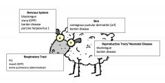
Respiratory tract |
|
|
Simian Immunodeficiency Virus |
(Retroviridae)
Do not appear to cause disease in natural host, but other species of monkey, severe disease results.
Apoptosis-mediated depletion of CD4+ T cells (helper T cells), resulting in immunodeficiency. T cells are highly sensitive to tumor necrosis factor-related apoptosis-inducing ligand (TRAIL), and virus stimulates monocytes to produce TRAIL. |
|
|
Foot and Mouth Disease Virus |
(Picornaviridae)
Seven serotypes, but immunity is serotype specific.
Infects cloven hoofed animals. Cattle, sheep, goats, and swine (and non-domesticated wild mammals).
Vesicular lesions on the muzzle, lips, tongue, and coronary band of the hood. Excessive salivation. Swine get snout and oral lesions, usually subclinical or lameness in sheep and goats.
Hydropic degeneration and necrosis of epithelial cells, sloughing of epithelial surfaces with associated mucosal and epidermal ulceration. Mechanical trauma in cutaneous and oral lesions. - "degloving" of the tongue - separation and loss of hoof wall
Chronically debilitating, poor feed conversion, economic loss. Rare death from viral myocarditis, rarely in swine.
Transmitted by fomites or aerosol, remain viable for up to 48 hours in nasopharynx after contact.
DDx: Encephalomyocarditis |
|
|
Encephalomyocarditis Virus |
(Picorniviridae)
Rats and mice are natural host, excrete virus in feces and urine. Swine, elephants, wide range of vertebrae species (including humans)
Ingestion of contaminated food. Transplacental transmission.
Potentially cause fatal myocarditis. - sudden death, inappetence, vomiting, staggering and dyspnea - subclinical in pigs post weaning - stillbirths and mummies (transplacental)
Soft pale heat with white areas of necrosis. |
|
|
Swine Vesicular Disease |
(Picorniviridae)
Only affects pigs, causes influenza like illness in humans.
Like foot and mouth disease, vesicular lesions of the snout, coronary band, and interdigital space. - lameness, with foot lesions
CNS manifestations - trembling, ataxia, circling, and convulsions
Meningioencephalitis |
|
|
Vesicular Exanthema of Swine Virus |
(Caliciviridae)
Seen in swine.
Vesicle formation on the lips, nostrils, tongue, feet, and udder. Usually mild and runs its course in 10 days.
Ddx: Foot and mouth disease |
|
|
Feline Calicivirus |
(Caliciviridae)
Associated with feline upper respiratory disease complex. - Rhinitis, conjunctivitis, ulcerative lesions on tongue and oral mucosa - Tracheitis and pneumonia can occur - 30% mortality in young kittens
Recovered cats can shed virus for months to years.
Vaccines available.
Virulent systemic-feline calicivirus, highly contagious hemorrhagic fever syndrome. - fever, cutaneous edema, sores or alopecia - diarrhea, icterus, vomiting, pulmonary edema, and coagulation abnormalities - 50% mortality common in adult cats
No vaccine. |
|
|
Rabbit Hemorrhagic Disease |
(Caliciviridae)
Affects domestic and commercial rabbits. Highly contagious.
Causes hemorrhagic fever syndrome. - acute depression, incoordination, seizures, and opisthotonos - foamy nasal discharge (pulmonary edema)
Virus-induced hepatocellular necrosis, resulting in acute liver failure.
Disease of older rabbits. 100% morbidity, 90% mortality. |
|
|
West Nile Virus |
(Flaviviridae)
Affects birds, but can be transmitted to humans, horses, and other mammals.
Causes encephalitis. - inability to fly, ataxia, incoordination - myocarditis
Lymphocytic perivascular cuffing, glial foci, and neuronal satellitosis.
Horses experience anorexia, depression, listlessness, fever, hind limb weakness, flaccid paralysis of lower lip, CNS signs. Neurologic disease in humans and other mammals.
|
|
|
Bovine Viral Diarrhea Virus |
(Flaviviridae)
Affects cattle, 80% worldwide are seropositive.
Wide variety of clinical syndromes. - infertility, abortion, and congenital defects
Mucosal disease, usually less than 2 years of age - High fever, anorexia, diarrhea, and multiple ulcerations and erosions around the mouth and nose - Chronically, cutaneous erosions on the coronary band and interdigital space
More common form is mild. - Mild fever, nasal discharge, a few ulcers on lips, gingiva, or hard palate - Diarrhea may occur - Mild immunosuppression in calves, can result in secondary infection
Transplacental spread, if infected in 2nd trimester, calves are clinically normal but will persistently shed virus.
Atypical, non-cytoplathic form, causes hemorrhagic syndrome. - Thromocytopenia and DIC
|
|
|
Border Disease Virus |
(Flaviviridae)
Affects small ruminants, sheep and goats. Seen in newborn lambs.
CNS involvement and abnormal hairy fleece - Tremors - "Hairy shaker"
Hypomyelination from viral damage to oligodendrogliocytes
Pregnant ewes, subclinical, but can cause abortion. |
|
|
Hog Cholera |
Highly contagious disease. Regional distribution of subgroups.
Fever, leukopenia, and general weakness, CNS signs. - Lethargy, ataxia, muscle incoordination, convulsions - Secondary pneumonia and enteritis
Injure endothelial cells. Hemorrhage. Sharply demarcated splenic infarcts. DIC, petechia Hyaline degeneration of vessels. Perivascular lymphocytic cuffing. Neuronal destruction from ischemia.
Ddx: African swine fever, porcine reproductive and respiratory syndrome, post-weaning multisystemic wasting sydrome, erysipelas, salmonollosis, coumarin poisoning |
|
|
Equine Encephalitis Virus (Eastern, Western, Venezuelan) |
(Togaviridae)
Cause encephalitis in humans and people. Wild birds are natural host, viremia without disease. Mosquitoes are vector (Culiseta primary vector for Eastern, Aedes and Culex for transmitting Eastern to horses and humans)
Eastern is a fatal disease in horses and people. Western more mild in people, but still can be fatal in horses.
Typical signs of encephalitis. - Fever, ataxia, neck rigidity, and variable paralysis - Signs develop rapidly, 12-24 hours
Diffuse meningoencephalitis, neutrophils in EE, and lymphocytes and plasma cells in WE and VE.
Not horse to horse transmission unless in high levels of viremia.
|
|
|
Transmissible Gastroenteritis |
(Coronaviridae)
Affects piglets less than 2 weeks old. Highly contagious, fatal disease. Mild or asymptomatic in adults.
Acute onset of diarrhea, vomiting, and dehydration. Death after a few days of illness.
Intestinal villus atrophy, viral injury to intestinal cells at distal portion of the villi. |
|
|
Porcine Respiratory Coronavirus |
(Coronaviridae)
A deletion mutant of Transmissible Gastroenteritis, only causes a very mild respiratory disease. |
|
|
Porcine Hemagglutinating Encephalomyelitis Virus |
(Coronaviridae)
Affects suckling pigs 2 weeks old or younger.
Causes vomiting and wasting and non-suppurative encephalitis. - anorexia, hyperesthesia, muscle tremor, and vomiting
Vomiting due to infection of vagal ganglion and vagal nuclei in the brain.
Ddx: Transmissible Gastroenteritis |
|
|
Feline Enteric Coronavirus |
(Coronaviridae)
Ubiquitous in feline population.
Can cause persistent infections with the generation of quasispecies variants.
Chronically infected cats develop feline infectious peritonitis. Antibodies against virus, help subsequent spread to macrophages. Disease of young cats, death shortly after weaning.
Infected macrophages spread the virus to multiple sites and cell types, venules in serosal surfaces of abdominal and thoracic viscera, meninges and ependyma of brain and spinal chord. Perivenular inflammation.
Two forms... - Classical wet, peritoneal effusion with few fibrin strands - Granulomatous, disseminated, multifocal pyogranulomas
Fibrinopurulent exudate on serosal surface and meninges. Focal necrosis is occasionally seen. Hypergammaglobinemic. Vascular injury from macrophage-transported virus.
Diagnose with acute phase protein . RT-PCR
|
|
|
Canine Coronavirus |
(Coronaviridae)
Can cause severe diarrhea in highly virulent variants, but more typically causes mild diarrhea and asymptomatic infections.
Loss of absorptive capacity from damage to mature enterocytes. |
|
|
Mouse Hepatitis Virus, Lethal Intestinal Virus of Infant Mice |
(Coronaviridae)
Highly contagious disease of laboratory mice. Affect mouse pups 4-5 weeks old.
Enteropathogenic strain causes intestinal lesions (100% mortality). Vacuolation and desquamation of mucosal cells with syncytia formation and shortened intestinal villi.
Virulent strains cause hepatitis and encephalitis. |
|
|
Sialodacryoadenitis Virus |
(Coronaviridae)
Common in laboratory rats. Highly contagious disease.
Manifests as photophobia, red-brown lacrimation, enlarged salivary glands, and ventral cervical edema.
Necrosis and inflammation of lacrimal glands, submaxillary and parotid salivary glands.
Disease is often self-limiting. |
|
|
Bovine Coronavirus |
(Coronaviridae)
Infects enterocytes of distal portion of intestinal villi. Major cause of diarrhea in calves. - dehydration and diarrhea
Vaccination of dams for prevention. |
|
|
Severe Acute Respiratory Syndrome |
(Coronaviridae)
Emerging infectious disease.
Begins with fever, chills, myalgia, headache, and malaise. Mild respiratory symptoms.
3-7 days. Lower respiratory phase, nonproductive cough.
May progress to dyspnea or hypoxemia.
Binds to common receptor in lungs and lower respiratory tract. Less efficient spread person to person because of low upper respiratory tract. |
|
|
Infectious Bronchitis Virus |
(Coronaviridae)
Domestic chicken is natural host. All ages susceptible, but most severe in chicks. Most important viral disease of chickens.
Respiratory signs. - Gasping, coughing, and nasal discharge in chicks 1-4 weeks - Serous, catarrhal, or caseous exudate present in trachea
Highly contagious, high morbidity. Drop in egg production. Economic loss
Some strains can cause nephritis in chicks, growth stunting. |
|
|
Equine Viral Arteritis |
(Arteriviridae)
Subcutaneous edema of the lower limbs, eyelids, and genitalia. - hydrothorax, hydropericardium, mediastinal edema, and abdominal effusion (ascites).
Virus causes vascular damage to endothelial cells.
Less virulent strains are apathogenic or subclinical. Can still cause edema and occasionally cause abortion storms.
In stallions, scrotal hyperthermia, or can be carriers.
Foals, can get bronchointerstitial edema. |
|
|
Porcine Reproductive and Respiratory Syndrome Virus |
(Arteriviridae)
Characterized by abortion, still births in full term pigs, and weak neonatal piglets.
Adult swine can experience anorexia, fever, edema of the eyelids, periorbital edema, conjunctivitis, cyanosis of the ears, tails, abdomen. Sows, endometritis and myometritis
Neonatal piglets experience dyspnea, diarrhea, periorbital edema, vomiting, lameness, tremors, convulsions.Interstitial pneumonia, intra-alveolar macrophages. Lymphocytic and histiocytic encephalitis, myocarditis. |
|
|
Mouse Pox (Ectromelia) |
(Poxviridae)
Rapidly fatal disease of laboratory mice, highly contagious. Can be latent, then active during stress.
Skin lesions common to pox viruses are not very common.
Internal lesions, tan necrotic foci on liver and spleen. Eosinophilic, intracytoplasmic inclusions within hepatocytes, acinar cells, occasionally epithelium.
Chronic form is characterized by ulcerating lesions of the feet, tail, snout. |
|
|
Swinepox |
(Poxviridae)
Generally mild disease of young pigs.
Lesions on skin of abdomen. Secondary bacterial infections.
Transmission by swine louse, Haematopinus suis |
|
|
Swinepox (picture) |

Skin |
|
|
Myxomatosis |
(Poxviridae)
Highly lethal disease of domestic and laboratory rabbits. Wild North and South American rabbits are natural host, mild skin thickening.
Mechanically transmitted by mosquitoes.
Causes a severe systemic disease that is always fatal. - Lethargy, diffuse edematous swelling of the head, eyelids, and anogenital region
Numerous, large, hyperplastic dermal fibroblasts "myxoma cells", occasional intracytoplasmic inclusions. Necrosis of lymph nodes and splenic germinal centers. |
|
|
Fowlpox |
(Poxviridae)
Infects wild and domestic bird species. Antigenically related to other avian poxviruses.
Biting insects important for transmission.
Lesions occur on non-feather regions of the skin. - oral, esophageal mucous membranes, trachea - "wet pox", usually not lethal
Intracytoplasmic inclusions (Bollinger bodies), stain with lipid stains. |
|
|
Fowlpox (picture) |

Skin |
|
|
Contagious ecthyma |
(Poxviridae)
Common disease of sheep and goats, camels and humans are also susceptible.
Lesions commonly occur around the muzzle, and lightly haired regions (coronary band, ears, udder, perianal region, and vulva) - raised, "proliferative" and scab like
Has been associated with tail dock wounds of young lambs. Young sheep susceptible, spread rapidly. Secondary bacterial infections common.
People can develop skin lesions, vesicular rash. |
|
|
Contagious Ecythma (Contagious pustular dermatitis) (picture) |

Skin |
|
|
Pseudocowpox |
(Poxviridae)
Similar to papular stomatitis and contagious ecthyma.
Blister like red raised lesions, form scabs. - occur on teats, muzzles, mouths of nursing calves
Zoonotic. Lesions on the hand, forearm, or face. |
|
|
Pseudocowpox (picture) |

Skin |
|
|
Papular stomatitis |
(Poxviridae)
Common clinically mild disease associated with oral lesions in young cattle (less than 6 months). Similar to contagious ecthyma in sheep.
Hydropic degeneration of epidermal layers with intracytoplasmic inclusions.
Recover in a few weeks.
DDx: Foot and mouth disease, bovine viral diarrhea, malignant catarrhal fever, rinderpest.
Can cause skin lesions in humans. Small papules on fingers, forearms. |
|
|
Papular stomatitis (picture) |

Stomatitis |
|
|
Bovine herpesvirus-1 |
(Herpresviridae)
Causes infectious bovine rhinotracheitis and infectious pustular vulvovaginitis.
IBR is highly contagious, upper respiratory disease in domestic cattle. Affects animals over 6 months. - Fever, anorexia, coughing, and ocular-nasal discharge, corneal opacity - Nasal discharge, thick and mucopurulent Affects trigeminal ganglion, can remain latent even when cattle is recovered. Predisposition from high protein diet, stress, diverse source of cattle. Stress reactivates virus, transported intra-axonally to site of entry.
Replicates in upper respiratory tract, shed virus in nasal secretions. Usually resolves in 1-2 weeks, tissue necrosis and secondary bacterial infection, leads to severe systemic disease.
DDx: Bovine viral diarrhea, MCF.
IPV characterized by vaginal discharge and frequent urination. Recover within 2 weeks. Secondary bacterial infections can cause metritis. Bulls get lesions on penile, preputial mucosa. - abortion
Intranuclear inclusion bodies in adrenal cortex and liver of aborted fetus. Oral and gastric erosions in newborns, encephalitis.
Transmission vis respiratory or genital route, aborted fetuses and semen are also. Overcrowding.
Vaccination reduces severity. |
|
|
Bovine herpesvirus-1 (picture) |
IBR, respiratory tract. IPV, reproductive tract. |
|
|
Bovine herpesvirus-2 |
(Herpesviridae) Causes mammilitis
Affects skin of the udder and teats. More ulcerative than those caused by poxvirus. - circular ulcers on muzzle and lips of calves from affected cows
Giant cells and intranuclear inclusions
Can develop pseudo-lumpy-skin disease in tropical regions
Transmitted mechanically by suckling calves and insects. Does not spread if intradermal or subcutaneous, only intravenous inoculation causes skin nodules.
DDx: Cowpow, pseudocowpox
Treat with teat dipping or isolation.
|
|
|
Bovine herpesvirus-2 (mammillitis) |

Skin |
|
|
Canine herpesvirus-1 |
(Herpesviridae)
Dogs are natural host. Cause mild tracheobronchitis and occasional genital lesions in adult dogs. - males, balanitis with preputial discharge - females, vaginal petechiae with raised lymphoid nodules
Stress can reactivate latent virus, shedding in oral, nasal,and vaginal secretions.
Causes a fatal, systemic infection of newborn puppies. Infected in utero or during early postnatal period. - multiple hemorrhagic foci in kidneys, lungs, stomach, and adrenal glands - nonsuppurative encephalitis, intranuclear herpetic inclusions within necrotic foci
Warm puppies to reduce viral replication. |
|
|
Herpes B Virus |
(Herpesviridae)
Old World monkeys are natural host.
Oral lesions similar to human cold sores. Latency of virus, potential carries.
Causes fatal encephalitis in humans. Exposure to saliva or monkey bite. |
|
|
Herpes Simplex Virus |
(Herpesviridae)
Humans are natural host. Cold sores are the most common manifestation of infection. - pharyngitis/tonsillitis, keratoconjunctivitis, eczema herpeticum, disseminate neonatal herpes, vulvovaginitis, balanitis - Variable fatal encephalitis in neonates and young adults, compromised mental capacity
Fatal disease in owl monkeys, marmosets, gibbons, similar to herpes T virus.
Type II genital herpes is similar, venereal spread. |
|
|
Equine Herpesvirus 1,4 |
(Herpesviridae)
EHV-1 causes equine viral abortion and EHV-4 causes rhinopneumonitis.
Persistent, latent infection throughout life, reservoir. Transmission of dam to foal, latent virus in foal, reactivation and shedding of the latent virus, horse to horse.
Respiratory disease is usually mild and subclinical. - malaise, inappetence, serous nasal discharge, cough, fever, pharyngitis. - recovery in two weeks if no secondary bronchopneumonia develops
Serious complications of EHV-1 cause late term abortions in pregnant mares, virus activates and spreads to fetus. - herpesvirus septicemia, focal necrosis in the liver and lung - intranuclear viral inclusions - fatal generalized disease
EHV-4 occasionally causes acute myelitis in older horses. - ataxia, posterior paresis - neuronal necrosis, viral damage of blood vessels in spinal meninges
In llamas and alpacas, EHV-1 can cause blindness and encephalitis. |
|
|
Equine Herpesvirus 1,4 (picture) |
EHV-1, reproductive tract. EHV-4, respiratory tract. |
|
|
Equine Herpesvirus-3 |
(Herpesviridae)
Red papules progressing to vesicular and pustular lesions of the vulva and penile mucosa, occasionally on teats, lips, and nares. Lesions heal in a couple weeks.
Venereal transmission, short lived immunity. Do not use for breeding until lesions are completely healed. |
|
|
Equine Herpesvirus-3 (coital exanthema) (picture) |

Reproductive tract |
|
|
Feline viral rhinotracheitis |
(Herpesviridae)
Most cats are positive for FVR virus. Associated with upper respiratory tract disease. - sneezing, coughing, oculo-nasal discharge - ulcerative lesions of the tongue and oral mucosa of kittens and young cats (entire surface can slough) - pneumonia can develop, some strains have affinity for pneumocytes
Epithelial necrosis of nasal turbinate mucosa with occasional intranuclear viral inclusions.
Part of feline upper respiratory disease complex
Rarely abortion with fatal encephalitis in neonatal kittens. |
|
|
Feline viral rhinotracheitis (picture) |
Respiratory tract |
|
|
Avian laryngotracheitis (gallid herpesvirus 1) |
(Herpesviridae)
Affects chickens. Acute, highly contagious upper respiratory disease. - nasal discharge, gasping, dyspnea, and expectoration of blood-tinged mucus - confined to tracheal mucosa - diffuse fibrinohemorrhagic or mucohemorrhagic exudates, plugs can obstruct the tracheal lumen leading to death from asphyxia
Herpetic, intranuclear inclusion bodies in sloughed tracheal epithelial cells. |
|
|
Avian Laryngotracheitis (picture) |

Respiratory tract |
|
|
Marek's Disease (gallid herpesvirus 2) |
(Herpesviridae)
Two types of lesions seen in chickens.
Inflammatory disease, nonsuppurative encephalitis, and peripheral neuritis. - asymmetrical paralysis of wing or lower limb
May also develop visceral lymphoid tumors in liver, spleen, gonad. Polyclonal transformation of T lymphocytes. Contains viral oncogene, MEQ (leucine zippers).
Cutaneous disease causes round, firm lesions at feather follicles.
Virus is shed within feather follicle epithelial cells. Common in young birds < 6 months.
Vaccination in ovo or at 1 day of age reduces neoplasia. |
|
|
Marek's Disease (picture) |
Nervous system and hemolymphatic system |
|
|
Porcine herpesvirus 1 (pseudorabies, Aujeszky's disease) |
(Herpesviridae)
Swine are natural host, adults are usually asymptomatic.
Acute illness and death in neonatal pigs if dams have low antibody titers. Death 24-48 hours after onset of clinical signs in piglets <4 months.
Neurological signs. - paddling, tremors, convulsions, and posterior ataxia
Multisystemic lesions in pigs over 4 weeks of age - neurological and respiratory signs
Transplacental transmission can occur, resorption of fetuses if early, abortion if later.
Virus shed in oral and nasal secretions, semen, and milk.
Pseudorabies is fatal in swine, cattle, sheep, goats, dogs, and cats (rare in horses, humans resistant). Severe encephalitis and extensive neuronal necrosis and neutrophil inflammatory response. - Intense pruritis in cattle, lethal within hours or days - jaw and pharynx paralysis, drooling of saliva - no aggression |
|
|
Porcine herpesvirus 1 (picture) |

Nervous system, respiratory tract, reproductive tract |
|
|
Malignant Catarrhal Fever |
(Herpesviridae)
Severe, sporadic disease of cattle. Can also affect deer and other ruminants.
Long incubation period, then sudden onset. - fever, oculo-nasal discharge, conjunctivitis, erosive lesions in the oral and nasal mucosa, lymphadenopathy - CNS signs, corneal opacity - intestinal disease with diarrhea, blood or mucus in feces - ulcerations of oral mucosa, tongue, and esophagus - lymphoid hyperplasia
Vasculitis, results from deposition of virus-immune complexes within blood vessels
Related to ovine herpesvirus 2 and alcelaphine herpesvirus 1, no clinical disease.
DDx: in sheep, orbivirus (enzootic hemorrhagic disease) |
|
|
Malignant Catarrhal Fever (picture) |

Respiratory, gastrointestinal |
|
|
Canine adenovirus 1 (infectious canine hepatitis) and Canine adenovirus 2 |
(Adenoviridae)
Type 1 causes infectious canine hepatitis. Seen in young dogs less than 1.
Affinity for hepatocytes and endothelial cells. - rapid onset of vomiting, abdominal pain, and diarrhea - cutaneous petechiae, epistaxis, and excessive bleeding from venipuncture sites, DIC - icterus - rarely causes encephalitis in dogs (common in wild foxes)
Grey/white foci in liver, viral induced necrosis of hepatocytes. Edema of gall bladder wall. Intranuclear inclusions in hepatocytes and endothelial cells of glomerulus.
Vaccination (can cause hypersensitivity)
Type 2 causes tracheobronchitis (canine adenovirus pneumonia) . - "kennel cough"
Vaccination available (modified live viral)
|
|
|
Canine adenovirus (picture) |

CAV1, CNS and enteric disease CAV2, respiratory disease |
|
|
Equine adenovirus pneumonia |
(Adenoviridae)
Common in horses, but healthy animals rarely show clinical signs. Occurrence in immunodeficient (signs are an indication). Arabian foals <4 weeks with SCID commonly affected.
Necrotizing bronchiolitis and pneumonia with numerous, large, intranuclear inclusions in epithelial cells. Common in epithelial cells of pancreatic ducts and urinary bladder. |
|
|
Equine adenovirus pneumonia (picture) |

Respiratory tract |
|
|
Egg drop syndrome |
(Adenoviridae)
Affects ducks and geese worldwide, introduced into chickens.
Vertically transmitted virus.
Rapid drop in egg production, loss in egg pigmentation, soft-shelled or shell-less irregular eggs.
Oviduct atrophy and inactive ovaries. |
|
|
Egg drop syndrome (picture) |

Reproductive tract |
|
|
Bovine papillomatosis |
(Papillomaviridae)
Six serologically distinct viruses in domestic cattle.
BVP1 and 2 cause single or multiple warts on the skin of the head and neck. Usually cattle less than 1 year old.
BVP5 and 6 cause papillomas on teats or within teat canal. Can impede milk flow, "rice grain"
BVP 4 causes squamous cell carcinomas of esophagus and rumen, transitional cell carcinomas of urinary bladder, and colon adenocarcinomas. Consumption of bracken fern may lead to immunosuppression favoring neoplasia.
Predilection for epithelial tissue. Usually benign, cauliflower like fibromas and papillomas. Introduced by mechanical abrasion or insect bites. Mature virus in papillomas, not carcinomas. |
|
|
Bovine papillomatosis (picture) |

Skin |
|
|
Equine papillomatosis |
(Papillomaviridae)
Type 1 induces papillomas on muzzle and legs
Type 2 causes papillomas of the genital tract
Affected horses 1-3 years of age.
Lesions typically spontaneously regress after several months. |
|
|
Equine papillomatosis (picture) |

Skin |
|
|
Equine sarcoid (bovine papillomavirus) |
(Papillomaviridae)
Fibrous connective tissue neoplasm in horses, mules, and donkeys caused by bovine papillomavirus 1 and 2.
Common skin tumor on head, legs, or abdomen. Skin abrasions may be involved in pathogenesis. Spontaneously regress. |
|
|
Equine sarcoid (picture) |

Skin |
|
|
Canine oral papillomatosis |
(Papillomaviridae)
Lesions on oral mucosa, sometimes conjuctiva, eyelids, and muzzle.
Initially smooth, but later become rough and cauliflower like.
Regress spontaneously within months |
|
|
Canine oral papillomatosis (picture) |

Skin |
|
|
Feline papillomavirus |
(Papillomaviridae)
Cutaneous fibropapillomas, viral plaques (hyperpigmented, flat warts) in immunosuppressed animals, and early in situ squamous cell carcinomas.
Occurs in young cats on head, neck, and digits. Viral plaques seen in old, immunosuppressed cats. |
|
|
Psittacine polyomavirus |
(Polyomaviridae)
"French molt", severe feather loss, rarely fatal, chronic feather loss in parakeets.
Can cause acute death in other psittacine species such as lovebird, macaws, and cockatoos, finches. "Budgerigar fledgling disease", high mortality. - hepatocellular necrosis, intranuclear inclusions - also seen in the spleen - die with abdominal distension |
|
|
Feline parvovirus (feline panleukopenia) |
(Parvoviridae)
Causes panleukopenia in domestic and wild felids. Also highly pathogenic for raccoons and mustelids.
Most infections are subclinical, but kittens less than one year old may become ill. <3 months of age do not survive, usually death after 12 hours of clinical signs. - vomiting, diarrhea, severe depression, and dehydration - thickened intestinal loops
Attack cells with high mitotic turnover, intestinal crypts and hematopoietic cells, occasionally cerebellar external granular layer (cerebellar hypoplasia) in near term kittens causing tremors and ataxia |
|
|
Feline parvovirus (picture) |

Nervous system, hematopoetic system, neonatal disease |
|
|
Aleutian mink disease virus |
(Parvoviridae)
Seen only in mink with Chediak-Higashi syndome. Neutrophils unable to degrade viral immune complexes. - hypergammaglobulinemia, glomerulitis, and segmental periarteritis |
|
|
Canine parvovirus 2 |
(Parvoviridae)
Two types of disease. Severe, hemorrhagic and necrotizing enteritis in dogs of any age. Necrotizing myocarditis seen in pups less than 3 months.
Evolved from feline parvovirus, capsid mutation. |
|
|
Canine parvovirus 2 |

Enteric disease |
|
|
Porcine parvovirus |
(Parvoviridae)
SMEDI: Stillborn, mummified fetuses, embryonic death, and infertility syndrome. Commonly seen in large commercial swine operations. Parvovirus associated with this entity.
Sows infected during first half of gestation. Fetal mummification most common manifestation. - usually only part of litter is infected - maternal antibody develops and increases fetal resistance
Diagnosis thru serologic methods. |
|
|
Porcine parvovirus (picture) |

Reproductive tract |
|
|
Psittacine Beak and Feather Disease Virus |
(Circoviridae)
Another cause of budgerigar fledging disease, "French molt"
Clinical appearance of "dirty" feathers, abnormal molts, abnormally shaped feathers, retained feather sheaths, hemorrhagic and short clubbed feathers, curled feathers and alopecia.
Necrosis of the beak, palate, and epiglottis in severe cases. May also cause atrophy and necrosis of thymus and bursa of Fabricius, causing immunosuppression.
Globular intracytoplasmic inclusions in feather follicle epithelial cells.
Virus shed in feces, crop secretions, feather dust, and white blood cells. Very stabile in environment, horizontal transmission. |
|
|
Porcine circovirus 2 |
(Circoviridae)
Causes porcine dermatitis and nephropathy syndrome.
Possible Torque teno virus and porcine reproductive and respiratory syndrome virus involvement.
Type III hypersensitivity reaction. Vasculitis and glomeruonephritis. Antibodies against porcine circovirus 2. 12-16 weeks.
Characterized by infarctive lesions of the skin and kidneys.
Also causes post-weaning multisystemic wasting syndrome (co-infection with porcine parvovirus or PRRS).
Immune response increases PCV2 replication. - progressive weight loss, jaundice, dyspnea in young pigs 6-14 weeks of age - primarily generalized lymphadenopathy - hepatomegaly, gastric ulceration, nephritis, and interstitial pneumonia
Lymphohistiocytic inflammation in many organs, lymphoid depletion and granulomatous pneumonia, intracytoplasmic inclusions (grape like) |
|
|
Porcine circovirus 2 (picture) |

Skin and renal.
Reproductive (respiratory) |
|
|
Chicken anemia virus |
(Circoviridae)
Causes subcutaneous and intramuscular hemorrhages, bone marrow aplasia, anemia from decreased erythropoiesis, and leukopenia.
Resistance to disease after 2 weeks of age. Severe disease can develop with dual infections. Transmission horizontally and vertically. |
|
|
Scrapie |
Prion-associated disease
Affects sheep.
Slowly developing syndrome. Clinical signs associated with CNS injury. - tremors of the head and neck, "mad itch" intense pruritus - progressive deterioration with emaciation, weakness, blank staring, ataxia, hindlimb paralysis, and inevitable death
Spongiform encephalopathy, neuronal damage without inflammation. "Holes" in medulla and brain stem from vacuolation.
Route of infection is poorly understood. Infectious tissues include CNS, lymphoid tissue and placenta. |
|
|
Chronic wasting disease |
Prion-associated disease
Affects deer and elk.
Causes spongiform encephalopathy with deposition of scrapie amyloid-immunoreactive plaques.
Only transmissible spongiform encephalopathy. |
|
|
Transmissible Mink Encephalopathy |
Prion-associated disease.
Carcasses of scrapie-infected sheep caused spongiform encephalopathy of mink. Mink to mink transmission.
Hyper-irritability, ataxia, and biting. |
|
|
Bovine spongiform encephalopathy |
Prion-associated disease.
Lesions confined to CNS, spongiform encephalopathy. - frenzy, aggressive behavior, ataxia
Can be transmitted to humans and cause florid plaques in the brain. Ingestion of prion-infected tissues. Transmission between cattle and humans occurs more readily than sheep and humans. |