Reading...
![]()
Play button
![]()
Play button
![]()
Use LEFT and RIGHT arrow keys to navigate between flashcards;
Use UP and DOWN arrow keys to flip the card;
H to show hint;
A reads text to speech;
111 Cards in this Set
- Front
- Back
|
Be able to draw/ explain progression of the airways
(should be able to include characteristic structures and specific epithelial cells in each division |
trachea- bronchi- bronchioles-respiratory bronchioles-alveolar duct- alveolar sac-alveoli
|
|
|
Whcih layers of the tubular airways have a mucosa and a submucosa, cartilage adn an adventitia?
|
trachea and bronchi
|
|
|
Describe the muscle in the trachea
|
not continuous, called trachealis muscle, does not contain muscularis mucosa
|
|
|
Trachea birfurcates into _____to supply each lung
|
primary bronchi
|
|
|
The trachea contains c-shaped pieces of ______ with rings that open dorsally. The ends of the rings are connected by the _____muscle
|
hyaline cartilage, rings connected by the trachealis muscle (smooth)
|
|
|
What are the two main divisions of teh respiratory system?
|
the conducting part and the respiratory (gas exchange part):
|
|
|
What are the components of the conducting part of the respir system?
|
nasal cavity, nasopharynx, larynx, trachea, bronchi, and bronchioles (down to terminal bronchioles)
|
|
|
What are the components of the respiratory (gas exchange) part of the respirt system?
|
respiratory bronchioles, alveolar ducts and alvoli
|
|
|
What is respiratory epithelium? How does it evolve/ change throughout the tract?
|
it is psuedostratified columnar ciliated, with goblet cells. "Respiratory Epithelium" gradually becomes simple epithelial lining in the bronchioles
|
|
|
The largest bronchioles have ____epithelial lining, the terminal and respiratory bronchioles have ____epithelial lining, and ____cells in teh alveolar ducts and alveoli
|
simple columnar to simple cuboidal to simple squamous
|
|
|
What are the functions of the respiratory stystem besides gas exchange?
|
acid-base balance, exretion of toxic compounds and body temp regulation
|
|
|
What are the functions of the respiratory stystem besides gas exchange?
|
acid-base balance, exretion of toxic compounds and body temp regulation
|
|
|
What are the three subdivisions of the nasal cavity
|
vestibular region
respiratory region (largest) olfactory region |
|
|
What are the three subdivisions of the nasal cavity
|
vestibular region
respiratory region (largest) olfactory region |
|
|
What is the classification of epithelium of the vestibule?
|
most rostral part it is skin (stratified squamous, cornified) and then changes to mucous membrane into stratified squamous noncornified epithelium
|
|
|
What is the classification of epithelium of the vestibule?
|
most rostral part it is skin (stratified squamous, cornified) and then changes to mucous membrane into stratified squamous noncornified epithelium
|
|
|
Olfactory epithelium and respiratory epithelium are both classified as psuedostratified columnar but how are they different?
|
the olfactory epithelium is thicker and has more layers. It also lacks goblet cells, it has less cilia that are less organized,
|
|
|
Olfactory epithelium and respiratory epithelium are both classified as psuedostratified columnar but how are they different?
|
the olfactory epithelium is thicker and has more layers. It also lacks goblet cells, it has less cilia that are less organized,
|
|
|
What is the location and appearane of the vomeronasal organ?
|
A tubular organ in the ventral portion of the nasal septum. It has ducts that open into the incisive ducts. Medial wall resembels olfactory epithelium and lateral wall looks like respiratory epithelium,
|
|
|
What is the location and appearane of the vomeronasal organ?
|
A tubular organ in the ventral portion of the nasal septum. It has ducts that open into the incisive ducts. Medial wall resembels olfactory epithelium and lateral wall looks like respiratory epithelium,
|
|
|
What is the function of the vomeronasal organ?
|
sensing compounds that have an effect on sexual behavior (causes the Flehmen resonose in some male animals)
|
|
|
What is the function of the vomeronasal organ?
|
sensing compounds that have an effect on sexual behavior (causes the Flehmen resonose in some male animals)
|
|
|
What is the type of epithelium found in the paranasal epithelium?
|
repiratory epithelium (psuedostratified columnar cilated w/ goblet cells)
|
|
|
What is the type of epithelium found in the paranasal epithelium?
|
repiratory epithelium (psuedostratified columnar cilated w/ goblet cells)
|
|
|
The trachea contains three types of tissues:
|
hylain cartilage rings, smooth muscle, and respiratory epithelium
|
|
|
Name four different kinds of cells that can be found in the trachea
|
Ciliated columnar cells, Goblet cells, Basal cells(serve as germinal cells), Neuroendocrine cells
|
|
|
The trachea contains three types of tissues:
|
hylain cartilage rings, smooth muscle, and respiratory epithelium
|
|
|
What are the other layers of the trachea eep to the epithelium (species dependent)
|
1. lamina propria
2. muscularis mucosa (some species lack this) 3. submucosa |
|
|
Name four different kinds of cells that can be found in the trachea
|
Ciliated columnar cells, Goblet cells, Basal cells(serve as germinal cells), Neuroendocrine cells
|
|
|
What are the other layers of the trachea eep to the epithelium (species dependent)
|
1. lamina propria
2. muscularis mucosa (some species lack this) 3. submucosa |
|
|
From primary bronchi to tertiary bronchi, how can you distinguish bronchi from the trachea in appearance?
|
Wall gets thinner and thinner as you progress down tract and the hyaline cartilage rights become irregular plates to plaques as bronchi become smaller
|
|
|
How do bronchioles differ from bronchi?
|
No cartilage in the walls of the bronchioles
Smaller and have thiner wall not all cells are ciliated |
|
|
What are two important roles of the Clara cells
|
produce surfactant component and help protect the bronchiole linign
|
|
|
How does respiratory bronchiole differ from terminal bronchiole?
|
Resp. Bronchiole is the first palce gas exchange can occur (can see alveoli opening into its lumen)
Lining is simple cubodial |
|
|
Which species hae well developed respiratory bronchioles?
|
dog cat and primate
|
|
|
As airway passages branch, they eventually lose both glands and cilia in the smallest divisions, which will disappear first -glands or cilia?
|
glands disappear first (need cilia to move mucus blanket up toward pharynx
|
|
|
How would you describe the alveolar duct?
|
lining is simple squamous rather than simple cuboidal
many more alveoli "hallway with open doors" b/w openings you see smooth muscle |
|
|
Describe alveolar sacs
|
much like ducts but more round than parallel with alveoli opening into them
|
|
|
Describe alveoli
|
little, round, sac-like chambers that open into respiratory bronchioles, alveolar ducts or alveolar sacs
Can have little pores b/w each other- pores of Kohn walls of tissue b/w them called alveolar septae |
|
|
What makes up the alveolar sepate/ interalveolar septae?
|
rich capillary network
Cells (septal macrophages, fibroblasts) Connective Tissue Matrix (elastic fibers found here) and reticular fibers prevent overdistension |
|
|
The interlobular septae (walls b/w alveolar lobules) are well developed in which species
|
bovine, sheep, and big
|
|
|
Aret there alveioli in avian lungs?
|
No- they have air capillaries instead and gas exchange occurs in lungs vs air sacs b/w blood capillaries and air capillaries
|
|
|
the voice organ in birds is the _______, and is found _____
|
syrinx and is found at the tracheo-bronchial jnxt
|
|
|
Do birds have bronchioles?
|
No, the bronchi divide from primary to secondary into tertiary
|
|
|
Do the lungs in birds inflate during inspiration?
|
very little, air drawn by air sacs instead
|
|
|
What are the two sources of the lung's blood supply?
|
functional supply and nutritional supply
|
|
|
The functional supply to the lung is mainly
|
the pulmonary arteries bringing unoxygenated blood from the right atrium (low pressure
|
|
|
The nutritional blood supply to the lung is via the
|
bronchial arteries carrying oxygenated blood (high pressure)
|
|
|
What are the two cells that make up the simple squamous lining of the the alveolar septae?
|
Type I and Type II pneumocytes
|
|
|
Describe the appearance, location, and function of Type I pneumocytes
|
Very flat cytoplasm spread out over a great distance, covering 97% of the alveolar surface. Main f(x) is to provide a thin surface that O2 can cross from alveolar lumen and CO2 can cross from blood to lumen
|
|
|
Describe the appearance, location, and function of Type II pneumocytes
|
cuboidal cells found mainly at points where alveolar sepate meet each other. Cytoplasm appears foamy. F(x) is to create surfactant that lowers surface tension allowing alveoli to inflate and prevent collapse of cells
|
|
|
What three main components makes up the blood air barrier
|
type I pneumocytes, fusued basal laminae of pneumocyte-endothelial capillary cells in alveolar septae, and the endothelial cells themselves
|
|
|
What are alveolar macrophages?
|
PAMs (pulmonary alveolar macrophages) = activated phagocytic cells = dust cells (become filled with debris from the air
|
|
|
Why/When are PAMs called heart failure cells?
|
These activiated phagocytic cells phagocytosize the backed up RBCs that escape into the lumen of the lung during congestive heart failure
|
|
|
If the alveolar lining is severely damaged, how does it regenerate?
|
The type II pneumocytes are the progenitor (stem) cells and start dividing to replace lining (makes alveolar lining appear cuboidal)
|
|
|
The mucosa of the trachea consists of the following components:
|
(mucosa=the lining layer of the trachea)
respiratory epithelium plus underlying connective tissue (lamina propria) |
|
|
The lamina propria of the trachea contains numerous ____fibers arranged parallel to the long axis of the trachea (which stain____)
|
elastic fibers (acidophilically )
|
|
|
Name four cells that are readily distinguishable in the respiratory epithelium
|
ciliated columnar cell
basal (germinal) cell Goblet cell Diffuse Neuroendocrine System cell (DNES) |
|
|
What cell is important in regulating the tracheal air flow (during asthma)
|
DNES cells (secreting polypeptides and bio active amines-serotonin into blood vessels to act locally)
|
|
|
What type of glands are located in the submucosa of the trachea
|
Submucosal glands are seromucous glands (mixed secretion of mucin, defense peptides, etc)
|
|
|
Where can you find the muscularis mucosae of bronchi
|
b/w the lamina proria and submucosa
|
|
|
In the bronchi the carilage rings are replaced by ____and/or ___
|
plates and or plaques
|
|
|
Progressive changes with successive divisions of bronchi include what kind of changes to:
the diameter of the airway, the epithelium, presence of goblet cells, cartilage plates, the muscle layer |
-airway decreases diameter
-shorter epithelium -(sub)mucosa thinner -less pseudostratification -fewer goblet cells -cartilage plates smaller -muscle layer more prominent |
|
|
Bronchioles contain/lack carilage and have what type of epithelium
|
no cartilage
simple epithelium(columnar to cuboidal in tertiary) no goblet cells smooth muscle in submucosa |
|
|
three types of cells found in the bronchioles are:
|
ciliated epithelial cells, non-ciliated CLARA cells, neuroendocrine cell
|
|
|
Clara cells
|
Tall, non-ciliated cells with apical granules
Produce surfactant Act as reserve stem cells (where lung cancer differentiates from when they undergo neoplastic differentiation) Contains enzymes to detoxify some substances |
|
|
primary and secondary bronchioles have what type of epithelium and the mucosa is ____
|
ciliated simple columnar epithelium
mucosa is highly folded |
|
|
The tertiary (terminal) bronchioles have a thinner/thicker lamina propria and one layer of _____muscle
|
thinner lamina propria
spiral smooth muscle partially ciliated simple columnar or cuboidal epithelium |
|
|
Respiratory bronchiole distinguishing f(x)
|
the level at which gaseous exchange begins
|
|
|
how would you describe a respiratory bronchiole
|
resembling tertiary bronchioles but have alveoli opening to the lumen of bronchioles
|
|
|
Define the alveolar duct and describe the epithelium. What structures are found at tips of the interalveolar septae (entrances to alveoli)
|
Def: a duct composed of numerous adjacent alveoli
Epithelium= simple squamous Tips have smooth muscle bundles/ knobs |
|
|
The alveolar duct leads to the blind-end _____ can be distinguished due to the absence of ____
|
alveolar sac
no smooth muscle knobs |
|
|
Site of gaseous exchange
|
alveoli
|
|
|
Alveolar septum is (thick or thin)
|
very thin and filled wiht capillaries
|
|
|
Which type of alveolar epithelial cell lines the alveolus (and looks like an endothelial cell)
|
Type I pneumocyte
|
|
|
Which alveolar epithelial cell appears vacuolated or granular due to the lamellar bodies in its cytoplasm
|
Type II pneumocyte
|
|
|
The lamellar bodies contain:
|
are vesicles that contain surfactant (phospholipids)
|
|
|
What are the two types of alveolar macrophages
|
septal (tissue) macrophage located in interstitium
Free macrophage- called the pulmonary alveolar macrophage = PAM= dust cell= heart failure cell |
|
|
The nutritional supply in the systemic vessels of the lung is the ___artery from the aorta
|
bronchoesophageal artery
|
|
|
the functional blood supply is the ____artery that are ___pressure
|
pulmonary artery and low pressure
|
|
|
Differences in the anatomical avian respiratory system
|
lungs smaller, don't change in size when breathing, lack diaphragm
|
|
|
The nutritional supply in the systemic vessels of the lung is the ___artery from the aorta
|
bronchoesophageal artery
|
|
|
What are the components of the avian respiratory system
|
bronchi (primary and secondary)
Parabronchi (tertiary bronchi) Atria (anagolus to terminal bronchiole) air capillaries (similar ot alveolar sac) |
|
|
the functional blood supply is the ____artery that are ___pressure
|
pulmonary artery and low pressure
|
|
|
In the avian resp system the ____moves the air in and out of the lungs
|
air sacs in a UNIdirectional air flow
Thin-walled, act like billows |
|
|
Differences in the anatomical avian respiratory system
|
lungs smaller, don't change in size when breathing, lack diaphragm
|
|
|
In avian respiration ther eis a _____volume of air during inspiration and expiration, in one breath the lung gets filled ____
|
constant volume, one breath= two fillings of lung
|
|
|
What are the components of the avian respiratory system
|
bronchi (primary and secondary)
Parabronchi (tertiary bronchi) Atria (anagolus to terminal bronchiole) air capillaries (similar ot alveolar sac) |
|
|
Are the avians more or less efficient at gas exchange due to the thin walls and rich capillary system
|
more efficient
|
|
|
In the avian resp system the ____moves the air in and out of the lungs
|
air sacs in a UNIdirectional air flow
Thin-walled, act like billows |
|
|
Divisions of the airways that have alveoli (respiratory bronchioles, alveolar ducts, alveolar sacs and alveoli) are referred to as ______
|
respiratory portion
|
|
|
In avian respiration ther eis a _____volume of air during inspiration and expiration, in one breath the lung gets filled ____
|
constant volume, one breath= two fillings of lung
|
|
|
Describe how the airways change from trachea to the bronchioles
|
from relatively rigid tubular structure to that of the bronchioles whose diameter is regulated via its higher proportion of smooth muscle.
Glands and goblet cells diminish as airways become smaller. Cilia persist deeper in the airway than mucus-producing elements |
|
|
Are the avians more or less efficient at gas exchange due to the thin walls and rich capillary system
|
more efficient
|
|
|
describe how the epithelium transitions as the airways branch
|
The epithelium transitions from respiratory epithelium, specialized for defense, to the simple squamous lining of the alveoli, specialized for gas exchange across the blood-air barrier between alveoli and the capillary beds
|
|
|
Divisions of the airways that have alveoli (respiratory bronchioles, alveolar ducts, alveolar sacs and alveoli) are referred to as ______
|
respiratory portion
|
|
|
Describe how the airways change from trachea to the bronchioles
|
from relatively rigid tubular structure to that of the bronchioles whose diameter is regulated via its higher proportion of smooth muscle.
Glands and goblet cells diminish as airways become smaller. Cilia persist deeper in the airway than mucus-producing elements |
|
|
describe how the epithelium transitions as the airways branch
|
The epithelium transitions from respiratory epithelium, specialized for defense, to the simple squamous lining of the alveoli, specialized for gas exchange across the blood-air barrier between alveoli and the capillary beds
|
|
|
A mucosal system is one that has an inner___lining (the ______), and that communicates with the external environment.
|
inner wet lining= mucosa
|
|
|
Because the mucosal system is vulnerable to attack by invading microorganisms, defensive cells are usually easily found in its _______layer.
|
connective tissue layer
|
|
|
The mucosa of the respiratory tract has an _______ and a loose ______ layer (lamina propria) that gets progressively______in the smaller airways.
|
epithelium
connective tissue layer = lamina propria thinner |
|
|
Beginning with bronchi a third, ______ layer is also present in the mucosa. The lamina propria of mucosal organs is typically filled with ____cells
|
muscular
defensive cells |
|
|
_________is connective tissue laying under lamina propria of the mucosa. In general it contains____cells and _____ fibers than the lamina propria. It may contain glands
|
Submucosa
less cells and thicker fibers |
|
|
adventitia
|
connective tissue around a tubular organ that is not in a body cavity
|
|
|
Serosa
|
outer layer if it is in a body cavity. The serosa on the lungs and in the pleural cavity is called pleura.
|
|
|
Olfactory epithelium contains _______for cranial nerve I, _____cells that are germinal for the other cell types, and ______cells that are support cells
|
bipolar neurons
basal cells sustentacular |
|
|
Olfactory epithelium is _______but with a thicker layer of nuclei than respiratory epithelium
|
psuedostratfied
|
|
|
What gives the rigidity to the trachea
|
carilage rings and smooth muscle (dorsal side only )
|
|
|
Describe the layers of the trachea
|
Superficial to the cartilage ring - trachealis muscle layer is the submucosa, containing seromucous glands. Superficial to this layer is the mucosa with its typical respiratory epithelium, and a lamina propria containing numerous elastic fibers
|
|

blood air barrier graphic
|

|
|
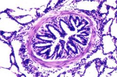
Bronchiole cell
|
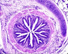
bronchus cell
|