Reading...
![]()
Play button
![]()
Play button
![]()
Use LEFT and RIGHT arrow keys to navigate between flashcards;
Use UP and DOWN arrow keys to flip the card;
H to show hint;
A reads text to speech;
66 Cards in this Set
- Front
- Back
|
Function of the liver
|
Largest gland in the body
Plays a role in digestion Hematopoietic organ during embryonic/fetal developmen |
|
|
Metabolic conversions of the liver
|
After absorption occurs in the gastrointestinal system, all products of digestion are transported in the portal system to hepatic cells
There they may be stored for further processed before entering general circulation |
|
|
Secretory function of the liver
|
Produces bile which is stored in the gall bladder
|
|
|
Excretory function of the liver
|
Converts end products of protein catabolism to urea and uric acid for disposal in the kidneys
|
|
|
Synthetic function of the liver
|
Produces globulin, albumin, prothhrombin, and most other clotting factors
Also produces acute phase proteins, carrier proteins, etc. |
|
|
Storage function of the liver
|
Serves to store starch and glycogen, fat and some protein
|
|
|
Detoxifying function of the liver
|
Extracts harmful substances from the blood
|
|
|
Filtering blood function of the liver
|
Removes foreign particulate matter including the breakdown products of blood cells which come from the spleen
|
|
|
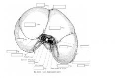
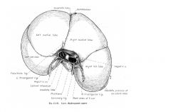
Position/Location of the Liver
|
In adult it is located between the diaphragm cranially and the stomach and intestinal mass causally
Extends across medial plane BULK IN ON THE RIGHT SIDE IN ALL SPECIES |
|
|
the liver is held in position by
|
Pressure of the viscera
Close attachment to the diaphragm |
|
|
External Features of the Liver
|
Reddish brown in color
Firm in consistency by friable Size and weight varies |
|
|
percent body weight of the liver in
carnivores omnivores herbivores |
Carnivores 3-5% body weight
Omnivores 2-3% body weight Herbivores 1-1.5% body weight |
|
|
the liver is covered in peritoneum except at the :
|
Portal/hilus
Fossa for the gall bladder Origins of various ligaments/peritoneal reflections |
|
|
Cranial (parietal) or diaphragmatic
surface of the liver |
Convex
Fits into the concavity of the diaphragm Faces to the left and right sides dorsally |
|
|
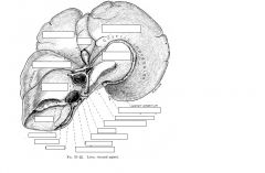
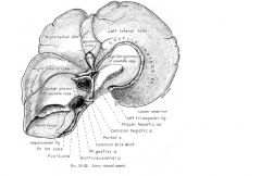
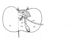
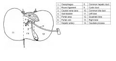
Caudal or visceral surface of the liver
|
Concave
In contact with visceral organs Faces mainly caudoventrally and to the left Contains the portal area (most indented area) - contains hepatic artery, nerves and lymphatic vessels; portal vein and the common bile ducts. |
|
|
Dorsal border of the liver
|
Extended further caudally and dorsally on the right side by the caudate process which carries a deep impression for the cranial pole of the right kidney
Mid line- groove for caudal vena cava To the left of this- notch for esophagus |
|
|
Ventral border of the liver
|
Sharp edged, continues around the edge of the periphery of the organ, except dorsally.
Notch for the round ligament (ligamentum teres) |
|
|
what are the lobes of the liver
|
The liver is divided into lobes by a series of deep fissures that extend inward from the central margin
|
|
|
Left Lateral and Left medial lobes of the liver
|
form 1/2 – 1/3 of liver mass
|
|
|
The quadrate lobe of the liver
|
deep wedge shaped that lies essentially on medial plane
|
|
|
Right ____ and right ___ lobes of the liver
|
medial and lateral
|
|
|
The caudate (caudate/papillary process) lobe of the liver
|
most caudal portion of the liver and is associated with the cranial half of the right kidney.
PAPILLARY PROCESS TENDS TO LIE IN THE LESSER CURVATURE OF THE STOMACH |
|
|
|
|
|
|
|
|
|
|
|
|
|
|
Coronary Ligaments of the liver
|
As the falciform ligament passes up the diaphragmatic surface of the liver, the two layers of peritoneum that compose it separate when the caudal vena cava is reached.
The layers diverge to the left and right to become the left and right laminae of the coronary ligaments which FORM A CONNECTION BETWEEN THE LIVER AND THE IMMEDIATELY ADJOINING PART OF THE DIAHPRAM. |
|
|
Falciform Ligament of the liver
|
Forms a fat filled fold in the dog, but it starts out as a narrow sickle shaped band with a free edge that begins on the VENTRAL WALL OF THE ABDOMEN NEAR THE AMBILICUS.
It passes forward from the abdominal wall TO the diaphragm, gradually increasing in width till it reaches the LIVER where its free border disappears into the UBLIMICAL FISSURE. |
|
|
Right and left triangular ligaments
of the liver |
Laterally the coronary ligaments are continuous with the left and right triangular ligaments and ATTACH THE LIVER FIRMLY TO THE LEFT AND RIGHT TENDINOUS REGIONS OF THE DIAPHRAM.
|
|
|
Hepatorenal ligament of the liver
|
Passes from the CAUDATE PROCESS TO THE VENTRAL SURFACE OF THE RIGHT KIDNEY (and the cecum).
It develops as an offshoot from the right triangular ligament |
|
|
Round (ligamenum teres) of the Liver
|
Is a slight THICKENING OF THE CAUDAL FREE EDGE OF THE FALCIFORM LIGAMENT.
It is the vestige of the UMBILICAL VEIN |
|
|
Lesser omentum of the liver.
|
Passes from the VISCERAL SURFACE OF THE LIVER TO THE STOMACH (HEPATO-GASTRIC) AND DUODENUM (HEPATO-DUODENAL)
|
|
|
Gall Bladder
|
Pear shaped organ
Partly fused to liver ( in the fossa of the gall bladder)- one part of neck and part of body Where it is not fused it has peritoneal covering STORES BILE DURING PERIOD OF DIGESTIVE REST |
|
|
the gallbladder generally lies between
|
THE QUADRATE LOBE MEDIALLY AND THE MEDIAL LOBE LATERALLY
|
|
|
The opening of the bile duct into the duodenum is guarded by .
|
the SPHINCTER OF ODDI, closed except for mealtimes
|
|
|
the gallbladder receives bile via
|
the cystic duct, thus BILE CAN FLOW THOUGH THE CYSTIC DUCT BOTH WAYS.
|
|
|
Histology of gallbladder
|
Simple columnar epithelium
Mucosal crypts Muscularis mucosa Loose connective tissue lamina propria/submucosa Muscularis externa- bundles of muscle fibers random orientation Tunica serosa |
|
|
Bile duct system begins
|
with microscopic canaliculi within the lobules of the liver parenchyma.
|
|
|
canaliculi open into
|
larger ductules that by successive unions within the connective tissue between lobules ultimately form A FEW LARGE HEPATIC DUCTS.
|
|
|
Before or shortly after leaving the liver at the porta these combine to form a
|
single trunk known as the COMMON HEPATIC DUCT.
|
|
|
A tortuous side branch
|
THE CYSTIC DUCT- arises from the common trunk and leads to the pear shaped gall bladde
|
|
|
THE BILE DUCT OR COMMON BILE DUCT-
|
part of the COMMON TRUNK that is DISTRAL TO THE ORIGION OF THE CYSTIC DUCT.
|
|
|
bile duct or common bile duct eventually runs to
|
the duodenum, entering the dorsal or mesenteric border on the MAJOR DOUDENAL PAPILLA.
|
|
|
Blood supply to the liver
|
Receives blood from the HEPATIC ARTERY( BRANCH OF THE CELIAC ARTERY-3/5 O2 supply to organ) and the PORTAL VEIN- 4/5 total blood supply
|
|
|
THE INTRAHEPATIC ARTERIES RUN IN COMPANY WITH THE
|
BRANCHES OF THE PORAL VEIN and the hepatic duct and supply the connective tissue structures before opening into the hepatic sinusoids along with the branches of the portal vein
|
|
|
THE PORTAL VEIN is formed by the union of
|
the tributaries of the digestive tract, pancreas and spleen
|
|
|
ALL BLOOD DELIVERED TO THE LIVER IS COLLECTED BY A SINGLE SET OF VEINS
|
central veins of the hepatic lobules
|
|
|
central veins of the hepatic lobules form a
|
few large hepatic veins that DRAIN INTO THE CAUDAL VENA CAVA as it tunnels through the liver substance.
|
|
|
forms the ventral surface of the fore-gut at the junction of the stomach/duodenum
|
The HEPATIC DIVERTICULUM
|
|
|
GROWS though the mesenteric mesenchymal cells INTO
|
the mesoderm of the SEPTUM TRANSVERSUM.
|
|
|
The ENDODERMAL CELLS OF THE HEPATIC DIVERTICULUM differentiate into
|
Hepatocytes (liver cells) of the bile duct system and gall bladder
MESODERMAL CELLS of the SEPTUM TRANSVERSUM contribute to BLOOD VESSELS |
|
|
Blood stem cells from the
|
yolk sac migrate to the liver to form blood islands, which form blood cells- thus at an early stage the liver is hematopoietic.
|
|
|
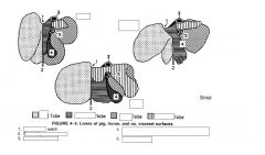
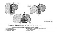
Comparative aspects – DOG
Liver |
Relatively large- 3-4% body weight
Almost entirely within costal area Parietal surface extremely convex Ventral border palpable DEEPLY DIVIDED INTO FIVE CHIEF LOBES BY FISSURES extending from the ventral margin Centrally located papillary process, which protrudes from the dorsal part, most prominent feature on visceral surface |
|
|
Comparative aspects – DOG Gall Bladder
|
Sunk deep between quadrate and right medial lobes
To right of medial plane Appears at visceral surface Common bile duct opens INTO THE DUODENUM about 5-8 CM DISTAL TO PYLORUS, usually in conjunction WITH OR at last NEAR the smaller PEANCREATIC DUCT on the MAJOR DUODENAL PAPILLA. The discharge of bile depends on the duodenum. |
|
|
Comparative aspect- HORSE
Liver |
Smaller than carnivore – 1-1.5% body weight
Long axis oblique- highest point is at level of right kidney and lowest point is on the left side Asymmetrical- 2/3 lies on the right side of median plane Visceral surface lies against the stomach, the duodenum, dorsal diaphragmatic flexure of the colon and the caecal base. Dorsal fixed border of the liver extends between the left and right triangular ligaments and is very irregular-the long free margin (left, ventral and right border) is sharper and is indented by a number of fissures: usually talk of left, quadrate and caudate lobes in the horse- Right lobe is the largest-irregularly quadrilateral in form on the sternal part of the caudate lobe and process. Quadrate lobe is located between the right lobe and the falciform ligament which separates it from the left lobe. Left lobe consists of medial and lateral portion, lateral part id oval in outline and thickest centrally. |
|
|
Comparative aspect- HORSE Bile duct system
|
NO GALLBALDDER
Bile duct opens into the cranial duodenum ON THE PAPILLA SHARED WITH HE MAJOT PEANCREATIC DUCT, ABOUT 13-15 CM DISTAL TO THE PYLORUS. Known as the HEPATOPANCREATIC AMPULLA. |
|
|
Comparative aspect- COW
Liver |
NO DISTINCT PATTERN OF LIVER LOBULATION
CAN BE PALPATED IN CRANIAL RIGHT PARALUMBER FOSSA Lies in contact with right abdominal wall, from the ventral end of seventh rib to the last rib Lies almost entirely in the medial plane Rotated 90 degrees from its position in embryo and most other mammals- THE RIGHT LOBE IS DORSAL AND THE LEFT LOBE IS VENTRAL Displacement due to development of compound stomach on left side of the abdomen Diaphragmatic surface faces dorsally cranially and to the right Falciform ligament is attached along a line from the esophageal impression to the notch for the round ligament Long triangular area on the dorsal surface does not have serous covering due to being in contact with the diaphragm |
|
|
Comparative aspect- COW
Liver visceral surface relates to |
Reticulum
Ruminal atrium Omasum Dudouneum Gall bladder Pancreas |
|
|
Comparative aspect- COW Gall bladder and Bile duct system
|
Gallbladder projects beyond the lateral margin of the right lobe
BILE (COMMON BILE) DUCT TERMINATES IN THE SEOCND BEND OF THE SIGMOIND FLEXTURE OF THE DUENUM ABOUT 60 NM FROM THE PYLORUS |
|
|
Comparative aspect- SHEEP AND GOATS
Liver |
Generally resembles that of cow but smaller
Lobes more distinct Deeper fissure for round ligament Narrower and less blunt shaped caudate process |
|
|
Comparative aspect- SHEEP AND GOATS Gallbladder and bile duct system
|
Elongated gallbladder
Gallbladder fossa more distinct Pancreatic duct joins the bile duct before the reaches the duodenum and no dilatation (ampulla) is present in duodenal wall |
|
|
Comparative aspect- PIG
Liver |
Relatively large and resembles dog in lobation and position
Left lateral lobe usually largest On dorsal part of right lateral lobe is the caudate lobe, clearly marked by a fissure The caudate process does not make contact with right kidney- no renal impression NO PAPILLARY PROCESS Short quadrate lobe is present centrally and lies in the central portal fissure HIGH CONTENT OF INTERLOBULAR FIBROUS TISSUE OUTLINES MINUTE LIVER LOBULES-speckled appearance |
|
|
Comparative aspect- PIG
Liver Three deep fissures that divide it into FOUR PRINCIPLE LOBES- |
LEFT LATERL AND MEDIAL
RIGHT LATERAL AND MEDIAL |
|
|
Comparative aspect- PIG
Gallbladder and bile duct system |
Fundus of gallbladder foes not reach central border of liver
COMMON BILE DUCT OPENS ATH THE DUODENAL PAPILLA ABOUT .5-5 CM FROM PYLORUS |
|
|
Comparative aspect- BIRDS
Liver |
Dark brown
Right and left lobes connected cranially by bridge dorsal to the heart Since no diaphragm lobes of liver embrace the caudal part of heart Larger right lobe carries gallbladder in visceral surface and is perforated by caudal vena cava Left lobe is divided by fissure into caudoventral and caudodorsal parts Most parietal surface is convex and lies against sterna ribs and sternum and is in contact with air sacs Cranioventral part makes contact with spleen, proventriculus, gizzard, duodenum, jejunum and ovary |
|
|
Comparative aspect- BIRDS
Gallbladder and bile duct system |
Two bile ducts one from each lobe enter distal end of duodenum close to pancreatic ducts
Only duct from right liver lobe is connected to gallbladder Some species lack gallbladder Pigeon Budgie Some parrots |