![]()
![]()
![]()
Use LEFT and RIGHT arrow keys to navigate between flashcards;
Use UP and DOWN arrow keys to flip the card;
H to show hint;
A reads text to speech;
22 Cards in this Set
- Front
- Back
|
What is the definition of the mediastinum? |
A thick partition of tissue in and on each side of the median plane -The space between the two pleural sacs wich contains the structures in the thorax except the lungs and pleura |
|
|
How many borders are there in the mediastinum? Name them |
2 -Superior and Inferior -Inferior has 3 parts: Anterior, middle, and posterior |
|

Label the green |
Superior border |
|
|
What are the borders of the superior mediastinum? |
superior to the sternal angle (manubriosternal plane) at the vertebral level of T4-T5 |
|
|
What are the structures inside the superior mediastinum? |
-Thymus -Right and left brachiocephalic veins -superior vena cava -aortic arch -brachiocephalic artery -left common carotid artery -Left subclavian artery -Vagus nerves -Left recurrent laryngeal nerves -Prenic nerves -Trachea -Thoracic duct -esophagus -Sympathetic chain |
|
|
What are the borders of the inferior mediastinum? |
inferior to the sternal angle (T4 to T5) through T2 vertebrae |
|

label the purple |
inferior anterior mediastinum |
|
|
What are the borders of the inferior anterior mediastinum? |
Anteriorly: sternum and transveresus thoracis muscle Posteriorly: pericardium |
|
|
What are the structures in the inferior anterior mediastinum? |
-Remains of the thymus -branches of internal thoracic arteries and veins |
|
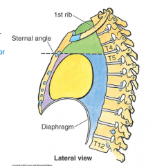
label the yellow |
inferior middle mediastinum |
|
|
What are the borders/relations of the inferior middle mediastinum? |
Anteriorly: body of the sternum, 2nd - 6th costal cartilages Posteriorly: 5th - 8th thoracic vertebrae |
|
|
What are the structures within the inferior middle mediastinum? |
-heart -pericardium -phrenic nerves -primary bronchi -origins of the great vessels (superior and inferior vena cava, ascending aorta, pulmonary arteries and veins) -ligamentum arteriosum -Bronchopulmonary(Hilar) nodes (lymph nodes in the hilum of the heart) |
|

label the blue |
inferior posterior mediastinum |
|
|
What are the borders of the inferior posterior mediastinum? |
Anteriorly: pericardium and diaphragm Posteriorly: T5-T12 vertebra |
|
|
what are the structures in the posterior mediastinum? |
-Thoracic descending aorta -Right bronchial artery -Posterior intercostal arteries -Esophageal arteries -Pericardial arteries -Superior phrenic -Intercostal nerves -posterior interthoracic arteries -Thoacic duct Pretty much if it comes off the thoracic aorta above the diaphragm and below the first rib, it will be considered part of the inferior posterior mediastinum -Azygus vein -Accessory hemiazygus -Hemiazygus -Thoracic veins wrapping with the thoracic arteries -right and left superior intercostal vein -Esophagus -Right and left vagus nerves -Aortic plexus -Anterior vagal trunk -Sympathetic trunk |
|
|
What are the constrictions of the esophagus? |
-Bronchial constriction -aortic constriction -Diaphragmatic constriction |
|
|
Where does the left vagus nerve lie on the stomach? |
the superior portion |
|
|
Where does the right vagus nerve lie on the stomach? |
the posterior side of the stomach |
|
|
What are the main sources of sympathetics in to the abdomen? |
thoracic splanchnic nerves -Greater -Lesser -Least |
|
|
where does the greater thoracic splanchnic nerve come from? |
the sympathetic chain ganglia at T5-T9 |
|
|
where does the lesser thoracic splanchnic nerve come from? |
the sympathetic chain ganglia at T10-T11 |
|
|
where does the least thoracic splanchnic erves come from? |
the sympathetic chain ganglia of T12 |