Reading...
![]()
Play button
![]()
Play button
![]()
Use LEFT and RIGHT arrow keys to navigate between flashcards;
Use UP and DOWN arrow keys to flip the card;
H to show hint;
A reads text to speech;
22 Cards in this Set
- Front
- Back
- 3rd side (hint)
|
Branchial apparatus
consists of = 4ct |
arches
pouches grooves membranes These embryonic structures contribute to the formation of the |
head and neck.
|
|
|
Pharyngeal arches are
x___________ outpouchings |
mesodermal
|
|
|
|
Pharyngeal/Branchial arches are
visible by week __ = |
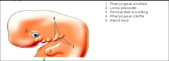
4
|
|
|
|
pharyngeal arches are separated by deep x___________ clefts called
____________ ___________ ________ |
ectodermal
pharyngeal clefts (grooves). |
|
|
|
The x_________ of the pharynx
, which lines the internal surface of pharyngeal arches, passes into evaginations called ______ _______ |
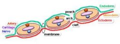
endoderm
pharyngeal pouches |
|
|
|
A typical pharyngeal arch/Branchial arch contains =
4ct |
-artery
-cartilaginous -muscular component -cranial nerve + |
An aortic arch (artery)
A cartilaginous rod that forms the skeleton of the arch A muscular component that differentiates into muscles in the head and neck A definite cranial nerve that supplies the mucosa and muscles derived from the arch |
|
|
pharyngeal arches
( __ -pair) begin to develop early in the __ week as neural crest cells migrate into the future head and neck regions. |
6pr
4th |
|
|
|
By the end of the __ week, four pairs of pharyngeal arches are visible externally.
The fifth and sixth arches are |
4th
rudimentary and are not visible on the surface of the embryo. |
|
|
|
The 1st pair of pharyngeal arches appears as
|
surface elevations lateral to the developing pharynx
|
|
|
|
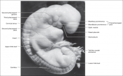
first pharyngeal arch
develops two prominences |
maxillary prominence
mandibular prominence |
|
|
|
maxillary prominence forms =
3ct |
-maxilla
-zygomatic -squamous part of the temporal bone mandibular prominence forms the = |
mandible (lower jaw)
|
|
|
the first pair of pharyngeal arches plays a major role in =
|
facial development
|
|
|
|
second pharyngeal arch
|
hyoid arch
|
|
|
|
___ ? week, the second pharyngeal arch enlarges and overgrows
*** |
During the 5th week
the third and fourth arches forming an ectodermal depression - the _______ _______ |
cervical sinus.
|
|
|
By the end of the __ or __ week, the second to fourth pharyngeal grooves and the cervical sinus have disappeared, giving the =
|
6th or 7th wk
neck a smooth contour. |
|
|
|
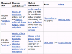
1st Pharyngeal arch gives rise to
2nd arch gives rise to MYO/Skeletal/Nerve/Artery |
.
|
|
|
|
3rd arch
4th arch 6th arch MYO/Skeletal/Nerve/Artery |
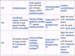
.
|
|
|
|
***
When the branchial arches form during week 4 and 5, they are penetrated by |
-arteries arising from the aortic sac,
which are called the -aortic arches. |
|
|
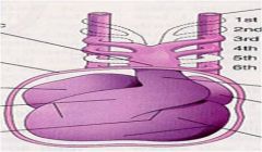
1st aortic arch
2nd aortic arch 3rd aortic arch 4th aortic arch 6th aortic arch |
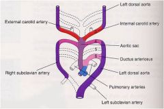
1maxillary
2 stapedial arteries / ext carotids 3 internal carotids 4 (R): proximal part of the right subclavian artery. (L): part of arch of the aorta just proximal to the left subclavian artery 6 (R): proximal part of right pulmonary artery . (L= ductus): proximal left pulmonary artery and ligamentum arteriosum . Ductus arteriosus: ligamentum arteriosum Dorsal aorta (R and L): part of right subclavian, descending aorta below left subclavian |
|
|
|
►►Fate of pharyngeal arches
*** 1st & 2nd arches |
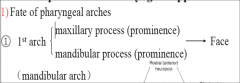
1st
Maxill Mandib = Face 2nd Hyoid = Neck |
|
|
|
Cranial N supplying the PH-Arches are =
|
5
7 9 10 = |
Trigeminal
Facial glossopharengeal Vagus |
|
|
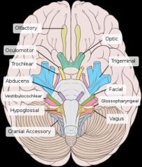
Cranial Nerve Names =
|
OLFACTORY
OPTIC OCCULO TROCHLEAR TRIGEMINAL ABDUCENS FACIAL VESTIBULOCOCHLEAR GLOSSOPHAR VAGUS AXILLARY HYPOGLOSSAL |
|