![]()
![]()
![]()
Use LEFT and RIGHT arrow keys to navigate between flashcards;
Use UP and DOWN arrow keys to flip the card;
H to show hint;
A reads text to speech;
68 Cards in this Set
- Front
- Back
|
What important process is located on the temporal bone? |
-mastoid process |
|
|
What are the important markings on the occipital bone? |
-external occipital protuberance -superior and inferior nucahl lines -occipital condyles -foramen magnum |
|
|
What are the bones of the TMJ? |
mandible maxilla temporal zygomatic sphenoid hyoid |
|
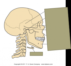
What is this showing? |
Bones of TMJ -temporal bone -mandibular bone -sphenoid bone -zygomatic bone -maxilla -mandible -hyoid bone |
|
|
Which bone is the largest of facial bones? |
-mandible |
|
|
What are the two parts of the mandible? |
-body and rami (2) |
|
|
What two muscles share an attachment site in the area of the angle of the mandible? |
-Masseter -Medial pterygoid |
|
|
What are the important parts on the mandible? |
-coronoid porcess -mandibular condyle -Mandibular notch |
|
|
What is important about the mandibular condyle? |
-it is the convex inferior bony component of TMJ |
|
|
Where is the mandibular notch? |
-extends between the coronoid process and mandibular condyle
|
|
|
Explain the mandibular fossa |
-on the temporal bone -concave (superior) portion of TMJ |
|
|
How does the mandiblular condyles slide when opening the mouth? |
slide anterior and inferior |
|
|
What do the zygomatic bcheeckone form? |
-cheeks and lateral orbits of the eyes |
|
|
What makes up the anterior half of zygomatic arch? |
-temporal process |
|
|
What muscles attach to the sphenoid? |
-pterygoid muscles |
|
|
What suspends the hyoid bone? |
-styloid ligaments |
|
|
What type of joint is TMJ, so what does it contian? |
-synovial joint -articular surfaces made by dense fibrocartilage instead of hyaline cartilage |
|
|
What is important about the TMJ disc ? |
-aneural and avascular -divides the joint into an upper and lower compartment |
|
|
Explain the opening motion of the TMJ |
1: rotation (involving lower compartment) 2: translation ( glide or slide of upper compartment) |
|
|
Explain the posterior region of the TMJ articular disc: |
-convex superiorly/concave inferiorly to accept the condyle |
|
|
Explain the intermediate region of the TMJ articular disc: |
-thinest portion of disc -concave inferiorly/flat superiorly |
|
|
Explain the anterior region of the TMJ articular disc: |
-flat inferiorly/ concave superiorly -accommodate articular eminance |
|
|
What is the role of the articular disc? |
-provides stability to the joint -maximizes congruency within the joint -reduces contact pressure |
|
|
What does the fibrous capsule do and in what directions? |
-provides support to the articulation -firm medially and laterally -lax anteriorly and posteriorly |
|
|
What is the importance of the temporomandibular ligament? |
-lateral ligament -primary stabilizer of TMJ -Horizontal and oblique fibers |
|
|
What is the primary stabilizer of the TMJ? |
lateral ligament (temporomandibular ligament) |
|
|
What are the accessory ligaments of the TMJ? |
-stylomandibular -spenomandibular |
|
|
What are the osteokinematics of protrusion ? |
-anterior translation without significant roation |
|
|
What are the osteokinematics of the lateral excurison? |
-side-to-side translation (normal is 1/2 ") |
|
|
What would be depression and elevation at TMJ? |
-mouth opening (3 adult knuckles) (unable to do 2 knuckles is abnormal)
-mouth closing |
|
|
What are the arthrokinematics of opening the mouth? |
-both heads slide down and forward coupled with bilateral lateral glide (anterior, inferior and lateral) |
|
|
What are the arthrokinematics of closing the mouth? |
-both heads slide up and back coupled w bilateral medial glide (posterior, superior, medial) -exact opposite of opening |
|
|
What are the arthrokinematics of protrusion? |
-mandibular heads glide ant, inf., and laterally (opening) |
|
|
What are the arthrokinematics of retraction? |
both heads posteriorly, superiorly, medial (closing) |
|
|
What are the arthrokinematics or lateral excursion? |
-side-to-side translation of condyle and disc -multi-planar rotation -TMJ opens on contralateral side and closes on the side of excursion |
|
|
Explain lateral excursion in relation to which side opens or closes |
If excursion is to the right then the contralateral or left side is open, while the same side closes |
|
|
What are the two options to describe motion at the TMJ |
-glides anteriorly, inferiorly and laterally (opening, contralateral lateral deviation and protrusion) -gliding posteriorly, superiorly and medially (closing, ipsilateral deviation adn retraction) |
|
|
What are the arthrokinematics of depression/elevation? |
-rotation and translation occur simultaneously -axis of rotation is constantly moving |
|
|
What are the muscles used in opening the mouth bilateral action? |
-lateral pterygoid -digastric (suprahyoid muscle group) -gravity |
|
|
What are the muscles used in closing of the mouth? |
-bilateral action of masseter -medial pterygoid -temporalis |
|
|
What muscles are used in lateral deviation of the mouth? |
-ipsilateral: masseter -contralateral:medial and lateral pterygoid |
|
|
What muscles are used in protrusion of the mouth? |
lateral pterygoid, masseter and medial pterygoid |
|
|
What muscles are used in retraction of the mouth? |
suprahyoid muscles (digastric and genohyoid) posterior fibers of the temporalis |
|
|
TABLE 11-2 LIST OF PRIMARY AND SECONDARY MUSCLES OF MASTICATION |
|
|
|
TABLE 11-3 LISTS OF ACTIONS OF THE MASTICATION MUSCLES |
|
|
|
What are the signs and symptoms of tempromandibular disorders? |
-pain with movement, joint sounds (popping) reduced range of motion (mouth opening), headaches, joint locking, and referred pain to face and scalp |
|
|
What are the causes of tempromandibular disorders? |
-stress or other emotional disturbances, grinding teeth, asymmetric muscle activity, chronic forward head posture, or sensitization of the central nervous system |
|
|
Define ventilation: |
-mechanical process that air is inhaled and exhaled through lungs and airways |
|
|
What is relative intensity defined as? |
-quiet or forced |
|
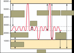
Pg. 439 : Label boxes********* |
Lung volume (mL) Top to bottom left to right Inspiratory reserve volume Expiratory reserve volume residual volume tidal volume inspiratory capacity vital capacity functional residual capacity total lung capacity |
|
|
What occurs during inspiration? |
-intrathoracic volume is increased by the contraction of the muscles that attach to the ribs and sternum -negative air pressure in the lungs increases (further reduced) -alveolar pressure drops below atmospheric pressure, and air is drawn into the lungs |
|
|
What occurs during expiration? |
-by decreasing the lung volume, air pressure increases and the air is forced outwards -quiet expirations is a passive process of elastic recoil of the lungs due to the relaxation of muscles (in healthy people) -forced expiration (cough, blowing out candles)requires activation of expiratory muscles |
|
|
What are the joints of the thorax? |
-manubriosternal joint -sternocostal joint -interchondral joint -costovertebral -costotransverse joints -thoracic intervertebral joints |
|
|
What are the vertical changes during inspiration and expiration? |
-inspiration the vertical diameter of thorax is increased by contraction and lowering of the diaphram -expriation thorax relaxes and diaphram returns to resting position |
|
|
What do ribs 1-6 do in inspiration? |
-pump handle motion |
|
|
Waht do ribs 7-12 do in inspiration? |
-bucket handle motion |
|
|
What are the parts of the diaphram? |
-costal: upper margins of lower 6 ribs -sternal: posterior xiphoid process -crural: thicker, bodies of upper three lumbar vertebrae. Two distinct attachments left and right crus |
|
|
What innervates the diaphram? |
-phrenic nerve (C3-5) |
|
|
What does 60-80 % of the work in inspiration? |
-diaphram |
|
|
What do the scalenes do in ventilation? |
-elevate the ribs and attached sternum |
|
|
What do the intercostals do during ventilation? |
external: inspiration internal: expiration |
|
|
how are intercostales externi fibers directed? |
-external obliques |
|
|
how are intercostales interni fibers directed? |
-internal obliques |
|
|
What is the primary m uscle of inspiration? |
-external intercostals (does its best work at the dorsal and upper regions of thorax) -parasternal fibers of internal intercostals |
|
|
What is concidered the muscles of forced expiration? |
-interosseous fibers of internal intercostals (effective throughout thorax) |
|
|
How do the intercostals assist with rotation? |
external: contralateral roation internal: ipsilateral rotation |
|
|
What are the accessory muscles of inspiration? |
-serratus posterior (inferior and superior) -levator costarum (longus and brevis) -latissimus dorsi -iliocostalis thoracis and cervicis -pectoralis minor -pectoralis major (sternocostal head) -quadratus lumborum |
|
|
What muscles are used in forced expiration? |
-abdominal muscles (flex trunk and depresses ribs, compress abdominal wall, push diaphram upwards, adn decrese intrathoracic volume rectus abdominis external and internal obliques transversus abdominus -transversus thoracis (depress ribs) -intercostales interni (depress ribs) |