![]()
![]()
![]()
Use LEFT and RIGHT arrow keys to navigate between flashcards;
Use UP and DOWN arrow keys to flip the card;
H to show hint;
A reads text to speech;
64 Cards in this Set
- Front
- Back
|
What bones make up the pelvic girdle?
|
The os coxae (hip bones), sacrum, and coccyx.
|
|
|
What bones compose the os coxae?
|
-ilium
-ischium -pubis Fusion of these bones in complete around age 17. |
|
|
Where do the os coxae articulate with one another?
|
At the pubic symphysis anteriorly and the first 3 sacral vertebrae of the sacrum posteriorly.
|
|
|
What is the subpubic angle?
|
The distance between the ischial tuberosities, >80 in females and <70 degrees in males.
|
|
|
What is the sacrum?
|
Composed of 5 vertebrae whose fusion is complete by the 25th year.
|
|
|
What is the coccyx?
|
Composed of 3 to 5 vertebrae, articulates with the sacrum at the sacrococcygeal symphysis, which contains a fibrocartilaginous disc.
|
|
|
What are the two divisions of the pelvic region? What separates these two divisions?
|
The pelvis major and the pelvis minor. Are separated by the pelvic brim.
|
|
|
What is the pelvic brim?
|
A bony structure composed of:
-promontory of the sacrum -anterior border of ala of sacrum -iliopectineal line -pubic crest -superior surface of the pubic symphysis |
|
|
What are the boundaries of the pelvis minor?
|
-sacrum
-coccyx -inner surface of the ischium and pubis -small part of ilium |
|
|
What is the lumbosacral joint?
|
B/t L5 and the sacrum. Large size, allows greater movement and contributes to the lumbar curve.
|
|
|
What is the function of the iliolumbar ligament?
|
It stabilizes L5 on the sacrum by anchoring its transverse processes to the iliac crest, limiting forward motion of L5.
|
|
|
What is the sacrococcygeal joint?
|
B/t S5 an the coccyx. Contains an intervertebral disc which allows for posterior movement of the coccyx during defecation or childbirth.
|
|
|
What stabilizes the sacrococcygeal joint?
|
Supraspinous and interspinous ligaments.
|
|
|
What is the pubic symphysis?
|
B/t the 2 os coxae. United by a fibrocartilaginous disc and numerous liagmentous fibers, including a strong arcuate ligament.
|
|
|
What is the difference b/t the subpubic angles in men and women?
|
<70 degrees in males
>80 degrees in females, to provide enough distance b/t ischial tuberosities to allow space necessary for birthing a child. |
|
|
What is the sacroiliac joint?
|
The strongest joint in the body, has least amount of movement. B/t the lateral surface of segments 1, 2 & 3 of the sacrum and the internal surface of the ilium.
|
|
|
What ligaments stabilize the sacroiliac joint?
|
-interosseous sacroiliac ligament
-posterior sacroiliac ligament -iliolumbar ligament -sacrotuberous ligament -sacrospinous ligament |
|
|
What is the piriformis?
|
O: lateral masses of S2-S4 and the sacrotuberous ligament
I: greater trochanter of the femur Inv: nerve to the piriformis (S1-S2) A: lateral rotator of thigh |
|
|
What is the obturator internus?
|
O: anterolateral wall of pelvis minor and obturator membrane
I: greater trochanter Inv: nerve to the obturator internus (L5-S1) A: lateral rotator of thigh |
|
|
What is the pelvis diaphragm?
|
Forms the floor of the true pelvis and separates the pelvis from the perineum. Comprised of 2 skeletal muscles (levator ani and coccygeus).
|
|
|
What perforates the pelvic diaphragm?
|
Urethra and anal canal, and the vagina in the female.
|
|
|
What is the levator ani?
|
Largest and most important part of the pelvic diaphragm, extends from pubic bone to the coccyx, funnel-shaped. Has 3 parts.
|
|
|
What are the 3 parts of the levator ani?
|
-pubococcygeus
-puborectalis -iliococcygeus |
|
|
What is the origin of the levator ani?
|
Body of the pubis to the ischial spine along the tendinous arch formed by the thickening of pelvic fascia over the obturator externus.
|
|
|
Where is the insertion of the levator ani?
|
Along the midline into the perineal body, wall of the anal canal, anococcygeal ligament and coccyx.
|
|
|
What is the pubococcygeus?
|
The main part originates on the pubis and inserts into the coccyx and anococcygeal ligament.
|
|
|
What is the puborectalis?
|
Originates on the pubis and goes around the anorectal junction and fuses with its counterpart, forming a U-shaped rectal "sling". Medial and inferior to the pubococcygeus.
|
|
|
What innervates the levator ani?
|
-S3-S4 on its pelvic (superior) surface
-inferior rectal n. S2-S4 on its perineal (inferior) surface |
|
|
What are the actions of the levator ani?
|
-supports pelvic viscera
-resists inferior thrust -raises pelvic floor (in forced exhalation, coughing, vomiting, urination and when lifting heavy objects) |
|
|
What is the action of the puborectalis?
|
Increases the angle b/t the rectum and anal canal, preventing defecation and maintaining bowel continence.
|
|
|
What is the coccygeus?
|
Forms the posterior portion of the pelvic diaphragm.
O: ischial spines and sacrospinous ligaments I: inserts on the lateral, anterior sacrum and coccyx Inv: S4-S5 A: supports the coccyx and pulls it forward after childbirth or defecation |
|
|
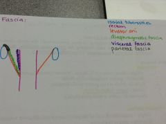
What is the pelvic fascia?
|

Has 3 continuous layers that are in contact with..
1. pelvic diaphragm (diaphragmatic fascia) 2. hollow pelvic organs (visceral fascia) 3. pelvic wall (parietal fascia) |
|
|
What is the retropubic space?
|
Located b/t the pubic bones and anterior surface of the urinary bladder. Contains large amount of subserous fat, which allows for expansion of the urinary bladder.
|
|
|
What is the retrovesical space?
|
Separates the urinary bladder and the rectum in the male.
|
|
|
What is the retrorectal space?
|
A potential space b/t the rectum and the sacrum surrounded by loose connective tissue that allows for expansion of the rectum before defecation.
|
|
|
What are the ureters?
|
Muscular tubes, pass over the pelvic brim just in front of the internal iliac vessels, lie above the levator ani as they approach the bladder.
|
|
|
Where do the ureters enter the bladder?
|
At the posterosuperior angle, in the male the angle is immediately above the seminal vesicless and inferior to the vas deferens.
|
|
|
What prevents retrograde flow of urine?
|
The intramural portion of the ureters in the wall of urinary bladder form a one way flap valve.
|
|
|
What is the urinary bladder?
|
Expandable, 3 layers of smooth (detrusor) muscle in its walls. Size, shape and position and relationships vary with the amount of urine it contains.
|
|
|
What is the trigone?
|
The triangular shaped area that contains the openings of ureters and internal urethral orifice.
|
|
|
What happens to the bladder as it fills?
|
It ascends into the abdomen elevating its peritoneal covering with it. This makes it possible to obtain a sample of urine from a filled bladder by inserting a hypodermic needle just above the pubic symphysis.
|
|
|
What are the rectovesical pouch?
|
A peritoneal recess, separates the bladder from the rectum.
|
|
|
Where is the apex of the bladder? The neck?
|
Apex - anterior end
Neck - junction with the urethra, rests upon the prostate gland |
|
|
What is the internal urethral sphincter?
|
Guards the opening of the urethra. Smooth muscle. In the male, it prevents retrograde flow of semen into the bladder during ejaculation.
|
|
|
What is the male urethra?
|
Has 3 parts
-prostatic urethra (passes through prostate) -membranous urethra (passes through the muscular urogenital UG diaphragm) -spongy (penile) urethra (passes through bulb, body and glans of the corpus spongiosum penis) |
|
|
What are the features of the posterior wall of the prostatic urethra?
|
-urethral crest (long vertical ridge)
-2 prostatic sinuses (where prostatic ducts empty) -seminal colliculus (rounded eminence of crest) -prostatic utricle |
|
|
What is the prostatic utricle?
|
A cul-de-sac structure in the prostatic urethra that is homologous to the uterus and vagina.
|
|
|
What is contained within the urogenital diaphragm?
|
The muscular external urethral sphincter (sphincter urethrae). A voluntary muscle that surrounds the membranous urethra and relaxes only during urination and ejaculation.
|
|
|
What is at the end of the spongy (penile) urethra?
|
External urethral orifice.
|
|
|
What are the vas (ductus) deferens?
|
A thick walled muscular tube that is the continuation of the epididymis. Ascends in the spermatic cord, passes through the inguinal canal, ends by joining the duct of the seminal vesicle to form the ejaculatory duct.
|
|
|
What are the seminal vesicles?
|
"Sperm sac" name is a misnomer, do not store sperm. They produce seminal fluid (pH>7.0) which constitutes the majority of semen. Found on the back of the bladder b/t the vas deferens and the prostate gland.
|
|
|
What are the ejaculatory ducts?
|
Formed by the union of the vas deferens and duct of the seminal vesicle near the neck of the bladder. Pass through the prostate gland to open in the prostatic urethra.
|
|
|
What is the prostate gland?
|
The largest accessory sex gland in the male, size of walnut. Has a prostatic secretion that is milky and alkaline (pH>7.0) which comprises 1/3 of semen. On the lateral surfaces is the prostatic venous plexus that has connections with Batson's veins (vertebral venous plexus).
|
|
|
Why is the prostatic secretion alkaline?
|
The alkaline pH helps to neutralize the acidic fluid produced by the female reproductive tract (vagina).
|
|
|
What carbohydrate is in the prostatic secretion?
|
Fructose, provides an important source of energy to the sperm cells on their journey to hopefully fertilize an ovum.
|
|
|
What type of tissue makes up the prostate gland?
|
Glandular and fibromuscular tissue, surrounded by a dense fascial sheath (capsule) that restricts how much the prostate can enlarge.
|
|
|
What is benign prostatic hypertrophy?
|
In older men, a non malignant enlargement of the prostate gland. Will cause troubles emptying bladder b/c the prostatic urethra is compressed, causing urgency to urinate. Treated with meds or surgery.
|
|
|
What is prostate cancer?
|
Cause of 50% of cancer in male population of the U.S., metastatic cells from a malignant tumor of the prostate gland may enter the prostatic venous plexus and travel to the cranial cavity via vertebral venous plexus (Batson's veins), leading to a secondary, malignant tumor of the brain.
|
|
|
What are the bulbourethral glands?
|
Cowper's glands, pair of pea-sized accessory sex glands, located in the urogenital diaphragm. Ducts pass through the bulb of the penis to open into the spongy urethra.
|
|
|
What do the bulbourethral glands produce?
|
A mucous secretion which is released just prior to ejaculation, for the purpose of lubricating the urethra.
|
|
|
What is the rectum?
|
A continuation of the sigmoid colon begins at S3, follows curvature of the sacrum and coccyx. Has spaces on either side called pararectal fossae.
|
|
|
What parts of rectum are covered in peritoneum?
|
Upper 1/3 is covered by peritoneum on anterior and lateral surfaces. Middle 1/3 is covered only on anterior surface. Lower 1/3 is not covered by peritoneum at all.
|
|
|
What are the pararectal fossae?
|
Spaces on either side of the rectum that permit the rectum to expand when it becomes distended with feces.
|
|
|
What is the anal canal?
|
lkajfldas;lfja;lf
|

