Reading...
![]()
Play button
![]()
Play button
![]()
Use LEFT and RIGHT arrow keys to navigate between flashcards;
Use UP and DOWN arrow keys to flip the card;
H to show hint;
A reads text to speech;
29 Cards in this Set
- Front
- Back
|
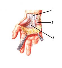
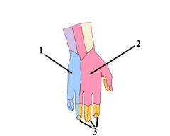
1. Tendon of Palmaris Longus
2. Palmaris Brevis 3. Palmar Aponeurosis |
answer
|
|
|
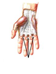
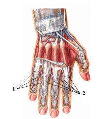
Superficial Transverse Metacarpal Ligaments
|
answer
|
|
|
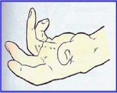
Dupuytren Contracture
Progressive shortening, thickening, and fibrosis of the palmar aponeurosis. Elderly, aging people |
What is this?
Describe the pathogenesis? Occurs in mainly what population group? |
|
|
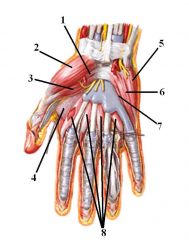
1. Opponens Pollicis
2. Abductor Pollicis Brevis 3. Flexor Pollicis Brevis 4. Adductor Pollicis 5. Abductor Digiti Minimi 6. Flexor Digiti Minimi 7. Opponens Digiti Minimi 8. Lumbricals (1-4) |
Answer
|
|
|
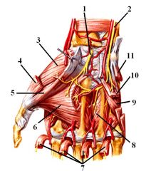
1. Median nerve
2. Ulnar nerve 3. Opponens Pollicis 4. Abductor Pollicis Brevis 5. Flexor Pollicis Brevis 6. Adductor Pollicis 7. Lumbricals 8. Interosseus muscles 9. Opponens Digiti Minimi 10. Flexor Digiti Minimi 11. Abductor Digiti Minimi |
Answer
|
|
|
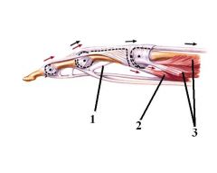
1. Vincula
2. Lumbrical 3. Interosseous muscles |
Answer
|
|
|
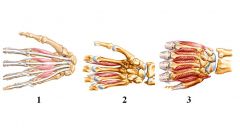
1. Lumbricals
2. Palmar Interosseus 3. Dorsal Interosseus |
Answer
|
|
|
Palmar Aponeurosis
- Attachments |
Flexor Retinaculum
Base of Proximal phalanges |
|
|
Palmar Brevis
- Function - Innervation |
"Wrinkles" skin
(deepens cup of palm for gripping) Ulnar nerve |
|
|
Which intrinsic muscles of the hand are innervated by the Median nerve?
|
Abductor Pollicis Brevis
Flexor Pollicis Brevis Opponens Brevis Lumbricals 1 & 2 |
|
|
Which intrinsic muscles of the hand are innervated by the Ulnar nerve?
|
Abductor Digiti Minimi
Flexor Digiti Minimi Opponens Digiti Minimi Adductor Pollicis Lumbricals 3 & 4 All Interosseus muscles |
|
|
Lumbricals
- Function x2 - Origin - Insertion x2 - Innervation |
Flex @ MCP
Extend @ IP Tendon of FDP 1 & 2 - Median nerve 3 & 4 - Ulnar nerve Lateral side of extensor hood Base of Proximal phalanx |
|
|
Palmar Interosseus
- Function x3 - Origin - Insertion x2 - Innervation - How many are there? |
Adduction
MCP flexion IP extension MCP Lateral side of extensor hood Base of proximal phalanx Ulnar 3 |
|
|
Dorsal Interosseus
- Function x3 - Origin - Insertion x2 - Innervation - How many are there? |
Abduction
MCP flexion IP extension MCP Lateral to extensor hood Base of proximal phalanx Ulnar 4 |
|
|
What structure goes through the space separating Lumbrical from Interosseus muscles?
|
Deep Transverse Metacarpal Ligament
|
|
|
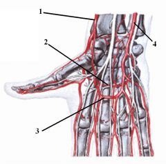
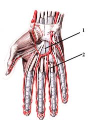
1. Radial aa
2. Deep Palmar Arch 3. Superficial Palmar Arch 4. Ulnar aa. |
Answer
|
|
|
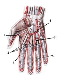
1. Princeps Pollicis
2. Contributions from Deep Palmar Arch 3. Radialis Indicis 4. Superficial Palmar Arch 5. Proper Palmer Digital aa 6. Common Palmar Digital aa |
Answer
|
|
|
1. Dorsal Metacarpal aa
2. Dorsal Digital aa |
Answer
|
|
|
Deep Palmar Arch
- Contribution from where? - Branches x2 - Anastomosis with what? |
Radial aa
Princeps pollicis aa Radialis indicis aa Superficial Palmar arch |
|
|
Superficial Palmar Arch
- Contribution from where? - Branches x2 - Anastomosis with what? |
Ulnar aa
Common Palmar Digital aa. (which will give rise to Proper Palmar Digital aa.) Deep Palmar arch |
|
|
Dorsal Metacarpal Arteries
- Contribution from where? - Branches |
Ulnar and Radial aa.
Dorsal Digital aa. |
|
|
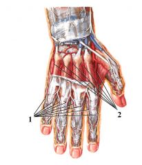
1. Common Palmar Digital nerves
2. Proper Palmar Digital nerves |
Answer
|
|
|
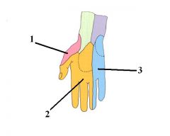
1. Radial
2. Medial 3. Ulnar |
List nerve name (not the root)
|
|
|
1. Ulnar
2. Radial 3. Median |
Nerves (not roots)
|
|
|
1. Dorsal Digital Nerves (Ulnar)
2. Dorsal Digital Nerves (Radial) |
Answer
|
|
|
Does the dermatome pattern match the cutaneous nerve pattern?
|
No
|
|
|
What are the contents of the Carpal Tunnel?
|
Median nerve
FDS tendon FDP tendon Flexor Pollicis Longus tendon |
|
|
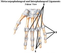
1. Deep Transverse Metacarpal ligament
2. Collateral ligament 3. Palmar Plate |
Answer
|
|
|
What is the uniqueness of the collateral ligament in the hand?
|
While all the ligaments are tight within the hand, the collateral of the MCP is loose until it is extended, at which point it tightens up.
|