![]()
![]()
![]()
Use LEFT and RIGHT arrow keys to navigate between flashcards;
Use UP and DOWN arrow keys to flip the card;
H to show hint;
A reads text to speech;
27 Cards in this Set
- Front
- Back
|
Major location of lymphatic tissue: What are the 3 mucous associated lymphatic tissue?
What 3 organs have lymphatic tissue and have a structure to them? What are the 4 lymphatic organs? |
Mucous associated Lymphatic tissue: 1.GI tract LT 2.Urinary tract LT 3.Respiratory tract LT 3 organs that have lymphatic tissue with structure: 1.Tonsils 2.Peyer’s patches 3.Thymus 4 lymphatic organs: 1.Lymph Node 2.Bone Marrow 3.Spleen 4.Appendix |
|
|
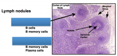
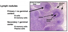
The mucosa (lamina propria) is an easy place for antigen to get into the body -Thus diffuse lymphatic tissue is found here What are lymphatic cells in the diffuse lymphatic tissue? Dense lymphatic tissue or lymphatic nodules have an accumulation of lymphocytes. What are the 2 types of nodules? How do they differ? Secondary nodules are only formed by what cell? So what cells are in the germinal center? |
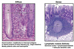
The mucosa (lamina propria) is an easy place for antigen to get into the body -Thus diffuse lymphatic tissue is found here There are mostly plasma cells and eosinophils in diffuse lymphatic tissue -There are also T and B cells at various stages of development Dense lymphatic tissue 2 types of nodules: 1. Primary = does not have a germinal center 2. Secondary = has germinalcenter Only B cells form secondary nodules -so cells in the germinal center are activated B cells = plasma cells or memory b cells |
|
|
Aggregated dense lymphatic tissue can be found in the tonsils -lymphatic tissue is partially encapsulated What 3 things can be found in aggregated dense lymphatic tissue in the tonsils? What are 2 functions of lymphatic tissue in the tonsils? |

Aggregated dense lymphatic tissue in tonsils 1. Group of nodules surrounded by simple squamous epithelium 2. Indentations of connective tissue called crypts 3. Efferent lymphatic vessels Filtration and Lymphocyte production (mostly B cells) |
|
|
Aggregated dense lymphatic tissue can be found in the Peyer's patch (lines the gut, found in ileum) -lymphatic tissue is partially encapsulated When there are antigens in the lumen, what does the Peyer's patch do? |
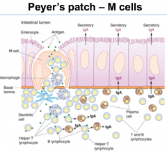
Aggregated dense lymphatic tissue can be found in the Peyer's patch (lines the gut, found in ileum) -lymphatic tissue is partially encapsulated Peyer's patch has M cells transcytose antigens from the lumen to the submucosal side to the lymphatic cells |
|
|
How many lymph nodes in the body? What type of tissue is lymph? What is lymph comprised of? What is edema? What does the lymph circulation connect with? |
About 350 lymph nodes Lymphis fluid connective tissue Lymph is the liquid in interstitial space in between cells (lots of water and other material flowing in with water) edema = accumulation of lymph in a particular tissue Connects with the blood circulation |
|
|
What are 3 functions of lymph nodes? |
Lymph nodes: 1. Filters lymph from one node to the next (eliminates microorganisms and particulate matter) 2.Phagocytosis of particles 3. Allows recognition ofantigen presented by APC to lymphoctyes and generate T and B cells Before lymphocytes enter the blood, they stop at the lymph node -B cell goes from red bone marrow to lymph node to blood -T cells go from red bone marrow to thymus to lymph node to blood |
|
|
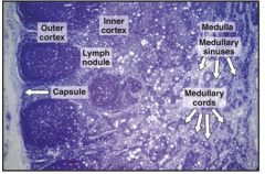
Lymph nodes are fully encapsulated by CT What are the 3 zones in lymph nodes? They are bean shaped with an indentation called? Where are the afferent and efferent vessels? |
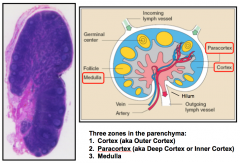
Lymph nodes are fully encapsulated by CT 3 zones: Cortex, paracortex, and medulla Indentation = hillum Has afferent vessels at the top and efferent vessel from the hillum |
|
What extends process through the lymph sinuses and slow down the flow of lymph and filter it? The lymph node also has? |
Macrophages and reticular cells Has blood vessels and capillaries |
|
|
Some (about 10%) of lymphocytes come into the lymph node through? Most (90%) of lymphocytes come into the lymph node through? What are they? What are 2 functions of the above? |
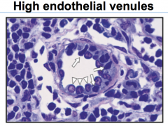
Somelymphocytes (B cells and T cells ) come into the lymph node through lymph vessels Most get to lymph nodes through high endothelial veins (HEV) -Blood vesselsthat terminate in the lymph node 1. Allow lymphocytes to leave blood and enterinto lymph nodes 2. Allow fluid transport from the blood to the lymph node and back |
|
|
When lymphocytes arrive at the lymph nodes, where do the B and T cells go? |
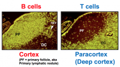
B cells go to outer cortex of nodules T cells go to the inner/deep (paracortex) T cells are not nodular |
|
|
B cells in the cortex |
|
|
T cells in the paracortex do not have? What do they have? |
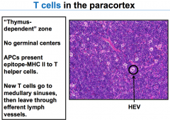
T cells in paracortex: Have no germinal centers, no nodules, no B cells Has HEV, T cells, and APCs (not B cells), macrophages |
|
|
Dense reticular meshwork in the lymph node sinus includes what 4 cells? |
1.Follicular dendritic cells 2.Dendritic cells = 3rd type of APC 3.Macrophages 4.Reticular cells |
|
|
Follicular dendritic cells are not derived in the bone marrow They are in lymph nodes to trap __ and present it to __ In the outer cortex of lymph nodes, FDCs have long processes with Fc receptors which to bind? The above binds antigen and passes it to? |
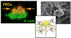
Follicular dendritic cells are not derived in the bone marrow. They are in lymph nodes to trap antigens and present it to B cells (they are not APCs!) In the outer cortex of lymph nodes, FDCs have long processes that are covered with Fc receptors which bind to antibodies Antibodies on the FDCs bind antigen and pass it to antibodies of B cells (also have Fc receptors) |
|
|
Where do most of the lymphocytes enter? Why? Where do T cells and B cells go? How do they leave the lymph nodes? |
Innercortex is where most lymphocytes enter because that is where HEVs are present - T cells stay in inner cortex - B cells form nodules in outer cortex When they leave they go out through the efferent lymphatic vessels which is an afferent vessel for the next one - Ultimately they are transferred to the blood |
|
|
|
|
|
Thymus is a lymph epithelial organ and site of? Thymus has 2 large lobes each consisting of? |
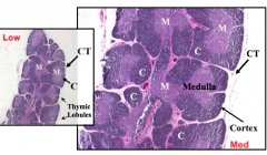
Thymus:lymph epithelial organ and site of T cell differentiation and proliferation Thymus has 2 large lobes, each consists of lobules containing cortex and medulla and are partitioned by CT trabeculae -also have lymphatic vessels and blood vessels (young thymus) |
|
|
After puberty, the thymus shrinks and what other features appear in the thymus? |
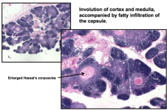
Afterpuberty, thymus shrinks and there is 1. fat in the tissue 2. enlarged hassal'scorpuscles 3. has extensive connective tissue |
|
|
In the thymus are epithelioreticular cells which do what 2 things? Thymic cortex stains darkly because? |
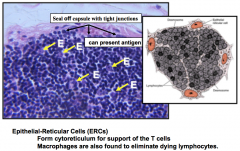
Inthe thymus are epithelioreticularcells which support the thymus andinteract with T cells and form tight junctions to form a barrier (darkly stained) = inactivated T cells -lighter cells are epithelioreticular cells for support |
|
|
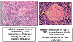
The thymic medulla is a heterogenous mixture of what 5 things? What are Hassall's corpuscles and what do they do? |
|
|
|
T lymphoblasts leave bone marrow and enter what part of they thymus? What occurs in this part of the thymus? When this occurs where is it sent to next? What occurs in this part of the thymus? What % leaves the thymus? |
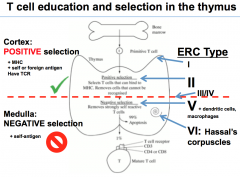
Tlymphoblastic leaves bone marrow and goes to the cortex of the thymus(inactive=dark) Positive selection occurs in cortex: Selects T cells that have a functional T cell receptor (not activated) and develops within the cortex If it survives it goes to the medulla Negative selection in the medulla: Immunotoleranceoccurs - removes self reactive T cells 1% leaves thymus -ERC are involved to form barrieresof thymus and form hassall'scorpuscles |
|
|
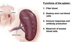
What are the 4 functions of the spleen? |
|
|
|
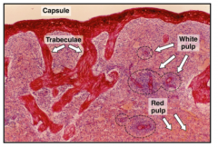
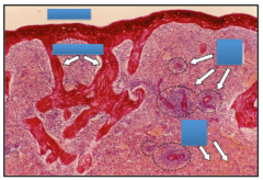
Spleen has -thickconnective tissue capsule -that breaks into trabecula witharteries -that run throughout the splenic pulp: red pulp and white pulp |
|
|
White blood cells = lymphocytes = White pulp |
|
|
Red pulp consists of what 2 things? Gaps between __ and__ allow blood cells to transitbetween splenic cords and sinuses |
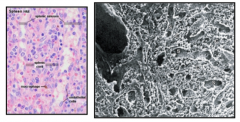
Splenicsinuses: blood filled vessels Splenic cords: loose meshwork of reticularcells, fibers, other cell types Gapsbetween endothelium (rod shaped =stave cells) and discontinuous basal lamina allow blood cells to transitbetween splenic cords and sinuses |
|
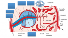
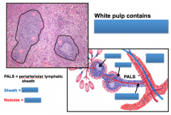
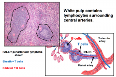
Spleen: Trabecular artery branches into the? The sheath around it are? The nodules around it are? All of this together is? What are a key site for the interaction between lymphocytes and antigen in the blood? Blood is emptied into the splenic cord which has? These cords are filled with macrophages that filter blood cells before they reenter the blood via the? |
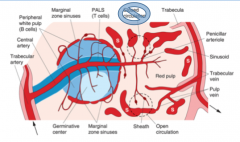
Spleen: Trabecular artery branches into the central artery The sheath around it are T cells(PALS) and the nodules around it are B cells All together is white pulp Marginal zone sinuses are a key sitefor the main function of the spleen: interaction between lymphocytes andantigen in the blood Blood is emptied into the spleniccord which has open circulation (just end spilling blood into the red pulp) The cordsare filled with macrophages that filter the blood cells before they reenter theblood via the venous sinuses and to thetrabecular veins back into the blood stream |
|
|
The spleen has what 2 main functions? |
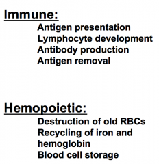
Spleen has immune functions and hemopoieticfunction |