Reading...
![]()
Play button
![]()
Play button
![]()
Use LEFT and RIGHT arrow keys to navigate between flashcards;
Use UP and DOWN arrow keys to flip the card;
H to show hint;
A reads text to speech;
31 Cards in this Set
- Front
- Back
|
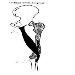
Biceps Femoris: Long Head
•Long Head Origin: Ischial Tuberosity (Lower/posterior aspect) •Common Insertion: Head of the Fibula (posterior/lateral) •Function: Ties into Sacrotuberous ligament, contraction tenses the ligament to stabilize SI-Joint. Biceps Femoris: Short Head •Short Head Origin: Posterior Femoral Shaft (lower 1/3) •Common Insertion: Head of the Fibula (posterior/lateral) •Function: Flexion of the knee, emphasized with external rotation of the tibia/lower leg. Biceps Femoris: Function •Latin = Two headed muscle of the thigh. •The primary function of the hamstrings is flexion of the knee and extension of the hip. The muscle has a slightly different function relative to rotation. •Aids in external rotation of the tibia. •Lateral Stabilizers of knee •Combined with Glute max: Creates posterior pelvic pull, which increases tension on TL fascia. •Prior to heel strike: hams contract creating posterior rotary motion of ilium on sacrum, the tension through ligament creates stability. •Provides posterior knee stability at heel strike. •If the muscle is weak, it can stress the other knee flexors (and external rotators of the tibia/lower leg). Weakness can also contribute to tension in the opposing muscles – the knee extenders (and internal rotators of tibia/lower leg). |
Hamstrings: Biceps Femoris
|
|
|
Semimembranosus: Attachments
•Origin: Ischial Tuberosity •Insertion: Medial Condyle of Tibia (Posterior Aspect) Semitendinosus: Attachments •Origin: Ischial tuberosity •Insertion: Pes Anserine Tendon (Proximal, Medial Shaft of Tibia). Medial Hamstrings: Function •Semimembranosus:(Latin = half membranosus) •Semitendinosus:(Latin = half tendinous) •The primary function of the semimembranosus, along with the semitendinosus, is flexion of the knee and extension of the hip and is emphasized with internal rotation of the tibia/lower leg. •Provide medial knee frontal plane stability. •Acts as medial knee stabilizer as the Popliteus provides lateral stability. •Medially rotates the femur at the hip. •Eccentrically decelerates knee flexion and internal rotation while regulating pelvis tilt. •If the muscle is weak, it can stress the other knee flexors and hip extenders (and internal rotators of the tibia/lower leg). Weakness can also contribute to tension in the opposing muscles – the knee extenders and hip flexors (and external rotators of the tibia/lower leg). |
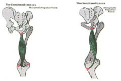
Medial Hamstrings: Semimembranosus & Semitendinosus
|
|
|
Gemellus Superior:
•Origin: Ischial spine (Outer surface) •Insertion: Greater trochanter fossa Gemellus Inferior: •Origin: Ischial tuberosity (superior aspect) •Insertion: Greater trochantor (medial surface) Gemellus: Function: •(Latin: gemellus = twin) •Work together with Piriformis. •One of the ‘deep six lateral rotators’ that is located under the Gluteus Maximus muscle. •Together they stabilize the head of the femur in the Acetabulum during weight bearing. •Deep to sciatic nerve. •They are both external rotators of the hip. If they are weak, it can put stress on the other external rotators of the hip and create tension in the opposing internal rotator muscles. |
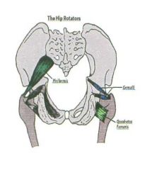
Hip Rotators: Gemellus
|
|
|
Piriformis: Attachments
•Origin: Sacrum (anterior surface) •Insertion: Greater Trochantor (Superior/posterior border) Piriformis: Function •(Latin – ‘pear shaped muscle’) •One of the ‘deep six lateral rotators’ that is located under the Gluteus Maximus muscle. •Extension or neutral: External rotator of the hip/femur. •Flexed past 90º: Internal rotator of the hip. •At 90º: Abductor of the hip. •Keeps femoral head in contact with the Acetabulum: pulls it in. •Direct attachment to the anterior surface of the sacrotuberus ligament = SI-Joint stability. •Relationship with sciatic nerve. •If the muscle is weak in extension it can put stress on the other external rotators of the hip. •If it is weak in flexion, it can put stress on the internal rotators of the hip. |
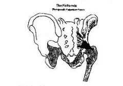
Hip Rotators: Piriformis
|
|
|
Quadratus Femoris: Attachments
•Origin: Ischial Tuberosity (Superior/lateral) •Insertion: Greater Trochantor (Intertrochanteric crest) Quadratus Femoris: Function •(Latin – ‘four sided muscle of the thigh’) •One of the ‘deep six lateral rotators’ that is located under the Gluteus Maximus muscle. •Work together with Piriformis. •Primary function: External rotation of hip. •Together they stabilize the head of the femur in the Acetabulum during weight bearing. •Deep to sciatic nerve. •If this muscle is weak, it can put stress on the other external rotators of the hip and create tension in the opposing internal rotator muscles. |
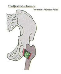
Hip Rotators: Quadratus Femoris
|
|
|
Psoas Minor: Attachments
•Origin: Anterior bodies of L1 & L2 •Insertion: Superior ramus of the pubis (most posterior/lateral aspect) Psoas Minor: Function •Roughly 40% of population has a psoas minor. •Upward rotation of the pelvis (the opposite action of the psoas major). •Flexion of the lumbar spine. |

Hip Flexors: Psoas Minor
|
|
|
Psoas Major Attachments:
•Origin: Bodies and TP’s of vertebrae T12-L5. •Insertion: Lesser Trochantor of Femur. •Thoracic Fibers Origin: Bodies and TP’s of T12-L1. •Lumbar Fibers Origin: Bodies and anterior surfaces of TP’s L2-L5. • Diaphragmatic Fibers: Origin – Right Crus: upper 3 lumbar bodies & left Crus: upper 2 lumbar bodies. Psoas Major Function: •Greek: For Muscle of the loin •Often categorized as a muscle of the trunk as it lies under the abdominal muscles and digestive organs (anterior to spine) and attaches to the spinal column. •Along with the Iliacus muscle, the psoas is considered part of the ‘core’ trunk muscles. These muscles are usually referred to as one muscle – the ‘iliopsoas’ as they share the same attachment at the femur. •Flexes the spine, and abducts & externally rotates the hip (each fiber is emphasized in a different degree of hip flexion, much less for the diaphragmatic fibers and more for the lumbar fibers). •Hip flexion with origin fixed. •When the lumbar spine is flexed, the fibers cross anterior to the axis creating a role in flexion of the lumbar spine. •When the spine is extended, fibers are posterior to the axis creating an extension moment. •Hip external rotation. •Insertion fixed: Lateral bending of lumbar spine to same side and opposite rotation. •Major role in lumbar stabilization & hip flexion. •Accentuates anterior pelvic tilt. •The bridge between the spine and the leg while the lat is the link between the spine and humerus. •Cura of the diaphragm: L2 & L3 vertebrae: aid intra abdominal pressure. •If the psoas major is weak, it can stress the other hip flexors and external rotators. A weak psoas major can also contribute to tension in the lower back muscles. Activating these muscles can increase stability in the trunk and lumbar spine and help to take tension out of the lower back. |
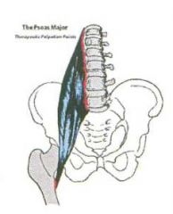
Hip Flexors: Psoas Major
|
|
|
Iliacus: Attachments
•Origin: Anterior Surface of Iliac Crest (attaches inside bowl of ilium) •Insertion: Lesser Trochantor Iliacus: Function •Latin: Pertaining to the loin/groin •Hip flexion and external rotation of the femur. •Flexes pelvis on femur and extends lumbosacral joint when foot is on the ground. •Anterior rotation of pelvis independent of the psoas. •With femur fixed: medially rotates pelvis. •Flexes the pelvis (anteriorly) and externally rotates the hip. •Is usually combined with the psoas and referred to as the ‘iliopsoas’ as they share the same attachment at the femur. These are the strong ‘core’ muscles. •The ‘iliopsoas’ is a major hip flexor and stabilizer of the lumbar spine. •If the psoas major is weak, it can stress the other hip flexors and external rotators. A weak psoas major can also contribute to tension in the lower back muscles. Activating these muscles can increase stability in the trunk and lumbar spine and help to take tension out of the lower back. |
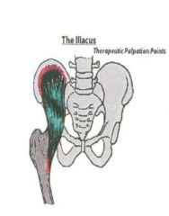
Hip Flexors: Iliacus
|
|
|
Sartorius: Attachments
•Origin: ASIS (inferior portion of ASIS) •Insertion: Pes-Anserine (most anterior/lateral on tibia, at the supero-medial tibial shaft). Sartorius: Function •Latin: Sartor = tailor; ‘tailor’s muscle’. The muscle is called the “tailor’s muscle” because of the position a tailor would sit in while sewing (bringing one foot to rest on the opposite knee. This position emphasizes/shortens the Sartorius muscle. •Longest muscle in the body. •Flexes, abducts and externally rotates the hip. Because it attaches below the knee, it also aids in knee flexion. •Strengthened in soccer style kicking motion. •Medial knee stabilizer. •If tight: will increase pelvic tilt, creating increased extension of lumbo-sacral joint. •When the foot is fixed: anterior pelvic tilt/hip flexion. •If this muscle is weak, it can put stress on the other external rotators of the hip, hip flexors and knee flexors. Activating the muscle can help to strengthen movements into these positions. |
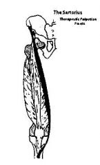
Hip Flexors: Sartorius
|
|
|
Rectus Femoris: Attachments
•Origin: AIIS & Groove of Upper brim of Acetabulum just above femoral head (reflected head fibers) •Insertion: Base of patella and tendon attachment to tibial tuberosity. Rectus Femoris: Function •Latin: rectus = straight, femur = thigh (Crosses 2 joints: hip & knee). •It is the only hip flexor of the quadriceps muscles. It also extends the knee. •Reflected head attaches just to the side of the hip attachment. This fiber attachment is emphasized in hip internal rotation; otherwise the rectus femoris functions in neutral (no rotation) as a hip flexor. •Eccentrically controls knee flexion. •With foot fixed: anteriorly rotates ilium. •Key function in shock absorption. •Most active in knee extension when the hip is neutral or extended. •When the Hip is flexed, the Rectus losses its efficiency as a knee extender. •Girth: Increases fascia latae tension within the envelope to stabilize hip and knee. •Works as a cable: The moment arm lengthens during hip flexion, giving it the MAd, as it gets further away from the hip socket. During knee flexion the rectus femoris lengthens at the hip and shortens at the knee. •If the muscle is weak, it can put stress on the other hip flexors. Weakness in the rectus femoris can also contribute to tension in the hip extenders and lower back. Activating the muscle can help to increase strength through hip flexion and knee extension, and can take tension out of the lower back, extenders of the hip and flexors of the knee. |
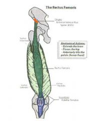
Hip Flexors: Rectus Femoris
|
|
|
TFL: Attachments
•Origin: Iliac Crest: Anterior portion of outer lip (under lip of ASIS or posterior-lateral) •Insertion: Into ilio-tibial tract just below joint capsule. (Indirectly attaches to lateral femoral condyle & tibial tubercle due to IT-Band). TFL: Function •Latin: tensor = stretcher, fascia = band, lata = broad •TFL: Kind of like the “lateral quad.” •Hip flexion, abduction, and internal rotation of the hip. •Inserts into the ilio-tibial tract making it a bi-articular muscle (crossing hip & knee). •Large role in stabilizing the pelvis in the close chain (active lateral collateral ligament). •Regulates tension as pelvis moves into posterior rotation. •Sits anterior/lateral in oblique plane. •If weak, the opposite side pelvis can drop laterally, medial knee compression, and stress the ankle. •If the TFL is weak, it can put stress on the other hip flexors, abductors and internal rotators. Weakness can also contribute to tension in the opposite muscles (hip extenders, external rotators and adductors). |
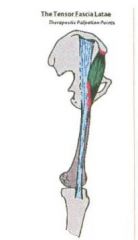
Hip Flexors: Tensor Fascia Lata (TFL)
|
|
|
Pectineus: Attachments
•Origin: Superior border of ramus of pubis. •Insertion: Pectineal line of femur. Pectineus: Function •Latin - comb like •Primary function of the adductor group as a whole is adduction of the hip. •Pectineus aids in flexion & external rotation. Adductor muscle closest to the hip and has the greatest component of hip flexion of all the adductors. •If the muscle is weak, it can put stress on the other adductors, hip flexors and external rotators of the hip. Weakness in the muscle can also contribute to tension in the opposing muscles (abduction, extension and internal rotation). •Activating the muscle can strengthen movement into adduction, flexion and external rotation and help to take tension out of the opposing muscles. |
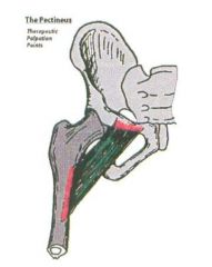
Hip Adductors:
Pectineus |
|
|
Adductor Magnus: Vertical Fibers
•Origin: Ischial Tuberosity & Ramus of Ischium •Insertion: Adductor Tubercle and lower medial femur •Function: Hip extension with internal rotation of the femur. Weight bearing: regulate against the anterior pull from the adductors and hip flexors that flex the hip. Works with all adductors in frontal plane stability during weight bearing. Adductor Magnus: Oblique Fibers •Origin: Ischial tuberosity and ramus of ischium. •Insertion: Linea Aspera (Middle 1/3) •Function: Adduction, hip extension with external rotation. When the foot is on the ground, it controls in both frontal and sagittal plane stability. Anterior and inferior rotatory movement of the ilium in weight bearing: increasing lumbo-sacral extension. Adductor Magnus: Function •Latin: adduct = to bring together; magnus = largest of the adductor group. Lies underneath the Pectineus, Adductor Brevis and Adductor Longus, and is therefore referred to as ‘the floor of the adductors’. •The primary function of the adductor group as a whole is adduction of the hip. The Magnus has two separate fibers: the oblique fibers externally rotate and adduct the hip; the vertical fibers internally rotate and extend the hip. •If weak, it can stress the other adductors and extenders of the hip. Weakness can also contribute to tension in the opposing muscles (hip flexors & hip abductors). Activating can help to increase strength in adduction and extension of the hip. |
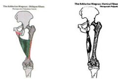
Hip Adductors: Adductor Magnus
|
|
|
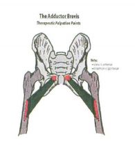
Adductor Brevis: Attachments
•Origin: Inferior Ramus of Pubis •Insertion: Pectineal line & middle portion of the linea aspera. Adductor Brevis: Function •Latin: adduct = to bring together; brevis = shortest of the adductor group. •Primary function is adduction of the hip. It also aids in flexion of the hip. The fibers of the brevis are emphasized in external rotation as well as internal rotation. •Works with all adductors in frontal and sagittal plane stability. •If weak, it can put stress on the other adductors and hip flexors. Weakness can also contribute to tension in the opposing muscles (abductors and extensors). •Activation can strengthen these movements and help to take tension out of the opposing muscles. |
Hip Adductors: Adductor Brevis
|
|
|
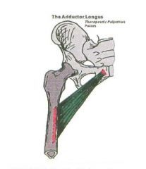
Adductor Longus: Attachments
•Origin: Pubic Tubercle •Insertion: Linea Aspera (lower portion) Adductor Longus: Function •Latin: adduct = to bring together; longus = longest of the adductor group. •Primary function is adduction of the hip. It is emphasized in external rotation of the hip. •Adduction and hip flexion. •Most activity as a frontal plane stabilizer in single limb support. •Works with all adductors in frontal and sagittal plane stability. •If weak, it can stress the other adductors of the hip. Weakness can also contribute to tension in the opposing muscles (hip abduction with internal rotation). Activation can increase strength in adduction—particularly when the hip is externally rotated—and help to take tension out of the opposing muscles. |
Hip Adductors: Adductor Longus
|
|
|
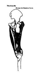
Gracilis: Attachments
•Origin: Inferior Ramus of Pubis & Ramus of Ischium •Insertion: Pes Anserine (Upper/Medial Tibia) Gracilis: Function •Latin = slender. Long and tendinous muscle in between the medial hamstrings and the other adductors. •Primary function is hip adduction and it is emphasized in internal rotation of the hip. Because is crosses the knee joint it also aids in flexion of the knee. •Adduction with hip extension, knee flexion and medial rotation. •Medial knee stabilizer. •Bi-articular motion creating simultaneous stability at the hip and the knee during weight bearing. •If weak, it can put stress on the other adductors and flexors of the knee. Weakness can also contribute to tension in the opposing muscles – the abductors and the knee extensors. |
Hip Adductors: Gracilis
|
|
|
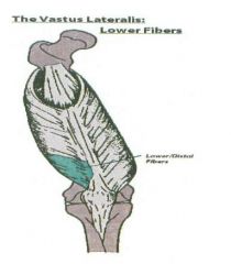
Vastus Lateralis: Upper Fibers
•Origin: Capsule of hip joint, Greater trochantor (anterior-inferior border) & Linea Aspera (Upper 1/3, lateral/posterior lip). •Insertion: Patella (Superior/Lateral) & Patellar Tendon into tibial tuberosity. Vastus Lateralis: Middle Fibers •Origin: Linea Aspera (Middle 1/3) •Insertion: Patella (Superior-Lateral) & Patellar Tendon into tibial tuberosity. Vastus Lateralis: Lower Fibers •Origin: Linea Aspera (Lower 1/3) •Insertion: Patella (Superior-Lateral) & Patellar Tendon into Tibial tuberosity. Vastus Lateralis: Function •Latin: vastus = vast, lateral = to the side. •Most active in knee extension up to 150º. •Provide tension in fascia latae envelope to create knee and hip stability. •Proprioceptive response to hip internal rotation. •Extends the knee, emphasized in internal rotation of the hip. •Each fiber has a slightly different emphasis relative to degree of flexion of knee and hip. The upper fibers involve more hip and knee flexion and the lower fibers involve slight hip and knee flexion. •Aids in internal rotation of the hip when the foot is fixed. •If weak, it can put stress on the other knee extenders and hip internal rotators. Weakness can also contribute to tension in the knee flexors, particularly the ones that externally rotate the hip. |
Knee Extensors: Vastus Lateralis
|
|
|
Vastus Medialis: Upper Fibers
•Origin: Linea Aspera (Upper 1/3) •Insertion: Patella (Superior/Medial) & Patellar Tendon into Tibial Tuberosity. Vastus Medialis: Middle Fibers •Origin: Linea Aspera (Middle-Posterior 1/3) •Insertion: Patella (Superior-Medial) & Patellar Tendon into Tibial Tuberosity. Vastus Medialis: Lower Fibers •Origin: Linea Aspera (Down supracondylar line, just above medial condyle). •Insertion: Patella (Superior-Medial) & Patellar Tendon into Tibial Tuberosity. Vastus Medialis: Function •Latin: vastus = vast, medial = middle. •Extends the knee. Emphasized in external rotation of the hip when foot is fixed. •Each fiber has a slightly different emphasis relative to degree of flexion of knee and hip. Upper fibers involve more hip & knee flexion. Lower fibers involve slight hip & knee flexion. •Terminal knee extension: screw home mech. •With femur fixed is highly involved in knee extension. •Weight bearing: provides frontal plane stability at the knee. •3 divisions having a proprioceptive response to hip external rotation. •If weak, can put stress on the other knee extenders and external rotators of the hip. Weakness can also contribute to tension in the knee flexors, particularly the ones that are emphasized in hip internal rotation. |
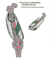
Knee Extensors: Vastus Medialis
|
|
|
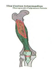
Vastus Intermedius: Medial Fibers
•Origin: Femur (Upper 2/3, Anterior-Medial surface) •Insertion: Patella & Tibial Tuberosity Vastus Intermedius: Lateral Fibers •Origin: Femur (Upper 2/3, Anterior-Lateral surface) •Insertion: Patella & Tibial Tuberosity Vastus Intermedius: Function •Latin: vastus = vast, intermedius = under the middle. Muscle is located under the rectus femoris in the middle of the thigh. •Has fibers on the medial and lateral aspect of the anterior shaft of the femur; therefore has slightly different emphasis in knee extension on the different fibers relative to the amount of hip flexion. •Knee Extension. •Highly involved in eccentric control of knee flexion and shock absorption. •If weak, can put stress on the other knee extenders. Can also contribute to tension in the knee flexors. |
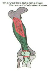
Knee Extensors: Vastus Intermedius
|
|
|
Articularis Genu: Attachments
•Origin: Femur (Anterior-Inferior/Supra-Patellar Tendon) •Insertion: Supra-patellar bursa (Superior border of patella/synovial membrane) Articularis Genu: Function •Latin: articularis = joint or turning point, genu = knee. •Primary function is assisting the quads in knee extension & helping the eccentric control of hip flexion. •Attaches to supra-patellar bursa and moves it as the knee extends. •Provides an interlinking mechanism for hip flexor strength. •Helps get rid of slack in extension & flexion. •If weak, it can put stress on the other knee extensors and hip flexors. •Activation can help increase strength through knee extension and hip flexion. |
Knee Extensors: Articularis Genu
|
|
|
Gluteus Maximus: Coccygeal Fibers
•Origin: Coccyx (Posterior-Lateral portion) •Insertion: Gluteal tuberosity of femur: approx. 2 inches Gluteus Maximus: Iliac Fibers •Origin: Iliac Crest (Lateral) & Gluteal Line (Posterior) •Insertion: Gluteal tuberosity of femur: approx. 2 inches Gluteus Maximus: Sacral Fibers •Origin: Sacrum (Posterior-lateral portion) •Insertion: Gluteal tuberosity of femur: approx. 2 inches Gluteus Maximus: Function •All fibers extend & abduct the hip. Each fiber has a slightly different emphasis relative to rotation of the hip. •Links: Opposite lat for SI compression to ‘cinch’ lumbo-pelvic unit for stability. •Compresses SI-joint with same side Multifidus & Erectors. •Knee flexed: Gain MAd since hamstrings effort is in knee flexion. •Increased requirement in knee flexed gait pattern. •If weak: Role in SI-joint stability due to loss of compressive force with multifidus, Latissimus and pull on sac-tub. •Overwork hams or lower lumbars. |
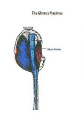
Hip Extensors: Gluteus Maximus
|
|
|
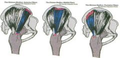
Gluteus Medius: Anterior Fibers
•Origin: Ilium (Outer surface below iliac crest, anterior 1/3) •Insertion: Greater Trochantor (Lateral/Superior & Posterior Border) •Function: Aid in internal rotation and abduction of the hip. Gluteus Medius: Middle Fibers •Origin: Ilium (Outer surface below iliac crest, middle 1/3) •Insertion: Greater Trochantor (Lateral/Superior & Posterior Border) •Function: Abduct the hip without any rotation. Gluteus Medius: Posterior Fibers •Origin: A Ilium (Outer surface below iliac crest, posterior 1/3) •Insertion: Greater Trochantor (Lateral/Superior & Posterior Border) •Function: Aid in external rotation & abduction of the hip. Gluteus Medius: Function •The Greek word for ‘end’ is gloutos,& the Latin word for ‘middle’ is medius. The Gluteus Medius is the ‘middle of the ‘end’ muscles’ •Primary ‘abductor’ muscles of the hip – moving the leg away from the body. Each fiber has a slightly different function relative to rotation. •You may find that you have strength in one direction, but weakness in the opposite. This is why it is important to work the different fibers of the muscle •Lateral hip stabilizer: Pulling femur up and in to the Acetabulum. •If weak: Lose hip stability and increase stress on spine in weight bearing. •If weak: Results in compressive forces on head of femur in Acetabulum: DJD. •Posterior weakness: pelvis will rotate backwards on opposite side. •Posterior: main complaint for SI-Joint •Note: Cannot abduct only one hip at a time. •Gluteus Medius middle fibers bring leg back to neutral from internal or external position (like rectus abdominis). |
Hip Abductors:
Gluteus Medius |
|
|
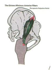
Gluteus Minimus: Anterior Fibers
•Origin: Ilium (Anterior ¼ of Outer Middle Portion) •Insertion: Greater Trochantor (Anterior Border) •Function: Aid in internal rotation. Gluteus Minimus: Lateral Fibers •Origin: Ilium (Anterior 2nd ¼ of Outer Middle Portion) •Insertion: Greater Trochantor (Anterior Border) •Function: Aid in external rotation. Gluteus Minimus: Function •The Greek word for ‘end’ is gloutos, and the Latin word for ‘smallest’ is minimus. Gluteus Minimus is the ‘smallest of the ‘end’ muscles’ •Abducts & flexes the hip. Each fiber has a slightly different emphasis relative to rotation. •Located under the gluteus medius muscle. |
Hip Abductors: Gluteus Minimus
|
|
|
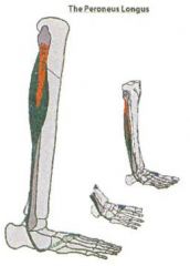
Peroneus Longus: Attachments
•Origin: Fibula (Upper 1/3 Posterior/Lateral) •Insertion: 1st Metatarsal (Lateral Base) & 1st Cuneiform (Lateral Base) Peroneus Longus: Function •Greek: peroneus = pin, buckle; longus = Latin for long. Located on lateral side of fibula. Function: Plantar flexion. All Peroneus muscles are also everters of the foot (turn foot out). •Stabilizes 1st met. To the lesser tarsus as plantar stability of the 1st ray is created. •Transfers weight from medial to lateral into propulsion through the pulley mechanism provided by the cuboid. •Decelerates ankle Dorsiflexion and aiding heel lift. •Assists in plantarflexion and heel lift. •If increased pronation, the base of the 1st met. Will drop lower than the cuboid losing its mechanical advantage to plantarflex 1st ray. •Resultant torque on 2-4 metatarsals. •If the peroneus longus is weak, it can put stress on the other everters and plantar flexors. |
Lower Leg:
Peroneus Longus |
|
|
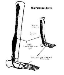
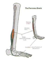
Peroneus Brevis: Attachments
•Origin: Fibula (Middle 1/3 Posterior-Lateral) •Insertion: 5th Metatarsal Peroneus Brevis: Function •Greek: peroneus = pin, buckle; brevis = Latin for short. •Function: Slight plantar flexion (for exercise the foot is in neutral). The brevis everts the foot while the ankle is in a neutral position. •Stabilizes the 5th met. Against the cuboid and the cuboid to the calcaneus creating lateral foot stability. •Decelerate mid-tarsal joint oblique axis foot stability. •If the peroneus brevis is weak, it can put stress on the other everters of the foot. •Activating the muscle can help to strengthen eversion, while helping to take tension out of the opposing muscles. |
Lower Leg:
Peroneus Brevis |
|
|
Peroneus Tertius: Attachments
•Origin: Fibula (Lower Anterior 1/3) •Insertion: 4th & 5th Metatarsal (Dorsal, medial surface at base of 5th met.) Peroneus Tertius: Function •Greek: peroneus = pin, buckle; tertius = Latin for third. •The peroneus tertius everts and dorsal flexes the foot. •If the peroneus tertius is weak, it can put stress on the other everters, and dorsal flexors of the foot. |
Lower Leg:
Peroneus Tertius |
|
|
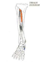
Tibialis Anterior: Attachments
•Origin: Lateral condyle and upper ½ of lateral surface of tibia (also along interosseous membrane) •Insertion: Plantar-medial surface of base of 1st met; Plantar-medial surface of 1st cuneiform Tibialis Anterior: Function •Latin: tibia = shin bone, anterior = front: muscle on the front of the shin bone. •Dorsiflexes and inverts foot with its pull on the base of the 1st metatarsal. •Decelerates the foot following heel strike. •Prevents foot from landing in a pronated position. •When foot is pronated: MAd of Anterior Tibialis is decreased as the Peroneus Tertius MAd increases causing further pronation: Thus ineffective in a pronated foot. •If weak, it can stress the other dorsal flexors and inverters of the foot. Can also contribute to tension in the opposing muscles (everters and plantar flexors of the foot). |
Lower Leg:
Tibialis Anterior |
|
|
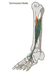
Tibialis Posterior: Attachments
•Origin: Proximal posterior shaft of the tibia, proximal fibula and interosseous membrane •Insertion: Navicular, all cuneiforms, cuboid, and bases of 2-4 metatarsals Tibialis Posterior: Function •Latin: tibia = shin bone, posterior = back: muscle on the back of the shin bone. •Located under the Gastrocnemius and soleus. •Primary Function: Plantar Flexion & inversion of the foot (turn foot in). •Tendon provides a medial and posterior stabilizing force on the lesser tarsus and metatarsal bases. •Decelerates subtalar joint pronation and tibial internal rotation in contact phase. •Re-supinates the subtalar joint while externally rotating the tibia in mid-stance. •Helps lock the foot by stabilizing the lesser tarsals in a posterior medial direction as the Peroneus longus and brevis stabilize in a posterior lateral direction. •Note: if either is weak, the stability is lost. •Decelerates forward momentum of the tibia to assist in knee extension and heel lift. |
Lower Leg:
Tibialis Posterior |
|
|
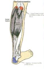
Gastrocnemius: Medial Fibers
•Origin: Medial Femoral Condyle (Posterior aspect) •Insertion: Calcaneus (Medial aspect) Gastrocnemius: Lateral Fibers •Origin: Lateral Femoral Condyle (Posterior aspect) •Insertion: Calcaneus (Lateral aspect) Gastrocnemius: Function •Greek: gaster = stomach, kneme = leg…‘belly of the leg’. •Primary Function: Plantarflexion & Knee Flexion. •Medial fibers emphasized in internal rotation of the lower leg. Lateral fibers emphasized in external rotation of the lower leg. •Eccentrically decelerates internal rotation of both the femur and the lower leg. •Eccentrically decelerates pronation while initiating Supination at the subtalar joint into propulsion. •Maintains a knee flexion tension as the body moves over the decelerating tibia, thus preventing hyperextension. •Initiates upward/forward trunk acceleration by flexing the knee and creating heel lift. •Supinates foot while externally rotating the leg during propulsion. •Plantarflexes ankle to move body upward and forward. •If weak, can stress the other knee flexors/plantar flexors of the foot. Can also contribute to tension in the opposing muscles (knee extensors/dorsal flexors of the foot. |
Lower Leg:
Gastrocnemius |
|
|
Soleus: Attachments
•Origin: Posterior-medial Epicondyle of fibula, upper 1/3 of medial fibula, Middle 1/3 of medial border of tibia. •Insertion: Achilles tendon into calcaneus. Soleus: Function •Latin: solea = sole fish shaped (located underneath the Gastroc). •Primary Function: Plantarflexion of Ankle. •Medial Fibers: Aid in inversion of the foot/ankle and the lateral fibers aid in eversion of the foot/ankle. •Eccentrically decelerates subtalar joint pronation and internal leg rotation into mid-stance. •Stabilizes lateral forefoot against the ground through subtalar Supination and ankle plantarflexion while peroneal stabilize 1st ray. •Extends knee by decelerating forward momentum of the tibia. •Assists in heel lift as it stops ankle Dorsiflexion. •If weak, can put stress on the other plantar flexors of the ankle. Can also contribute to tension in the opposing muscles, the dorsal flexors of the ankle. |
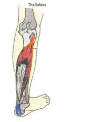
Lower Leg:
Soleus |
|
|
Popliteus: Attachments
•Origin: Lateral Femoral Condyle (Lateral aspect; outer margins of lateral meniscus) •Insertion: Tibia (Posterior/Superior/Medial 1/3, triangular area). Popliteus: Function •Latin: poples = ham of the knee (deepest muscle in the back of the knee). •The region behind the knee is referred to as the ‘”popliteal region,” hence its name. •Primary Function: When the foot is not fixed is internal rotation of the lower leg (tibia) on the thigh (femur). When the foot is fixed the primary function is external rotation of the femur on the tibia. This muscle also helps to stabilize the knee. •Externally rotates femur on fixed foot: aiding in Supination. •Active lateral knee stabilizer with the biceps femoris and the IT-tract through the attachments with the arcuate ligament and the posterior horn of the lateral meniscus. •Pulls lateral meniscus back out of the way of the lateral femoral condyle during flexion and internal rotation. •With PCL: prevents excessive forward motion of the femur on the tibia. • If weak, can put stress on the other tibial internal rotators and hip external rotators. Can also contribute to tension in the tibial external rotators and hip internal rotators. |
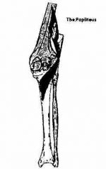
Lower Leg:
Popliteus |