![]()
![]()
![]()
Use LEFT and RIGHT arrow keys to navigate between flashcards;
Use UP and DOWN arrow keys to flip the card;
H to show hint;
A reads text to speech;
63 Cards in this Set
- Front
- Back
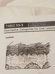
Thinning of tissue layers with loss of normal skin furrow, with shiny and translucent appearance |
Atrophy |
|
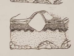
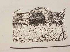
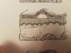
>0.5 Circumscribed blister containing clear, watery fluid or blood |
Bulla |
|
<1 Flat, nonpalpable |
Macule |
|
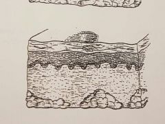
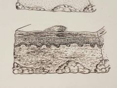
Elevated solid mass, deeper and firmer than papule |
Nodule ex. Tori |
|
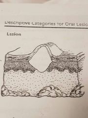
Palpable, circumscribed, solid elevation |
Papule |
|
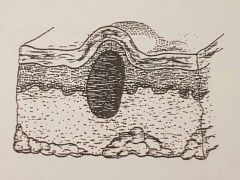
Similar to vesicle, lesion filled with pus |
Pustule ex. Acne |
|
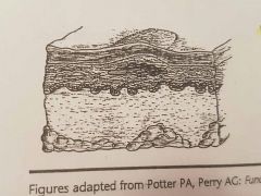
>.5 Discrete, slightly elevated are of altered texture or coloration |
Plaque ex. Canadidiasis |
|
Deep loss of epithelial layer; may extend to connective tissue layers |
Ulcer ex. Recurrent aphthous ulcer |
|
Circumscribed blister, filled with clear, watery fluid |
Vesicle ex. Herpes labial |
|
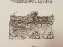
Elevated area of superficial localized edema; irregularly shaped |
Wheal ex. Type 1 hypersensitivity |
|
|
Corrugated |
Wrinkled |
|
|
Fissure |
Cleft or groove with prominent depth |
|
|
Papillary |
Resembling small, nipple-shaped projections or elevations found in clusters |
|
|
Coalescence |
2 parts of a whole fuse together to make 1 |
|
|
Diffuse |
Lesion with borders that are not well defined. Making it impossible to detect exact parameters |
|
|
Multilocular |
Lesion that extends beyond confines of one distinct area |
|
|
Unilocular |
Having 1 compartment or unit that is well defined or outlined as in a simple radicular cyst |
|
|
Well circumscribed |
A lesion with borders that are specifically defined |
|
|
Perdunculated |
Attached by stem like base similar to a mushroom |
|
|
Sessile |
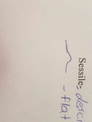
Described base of lesion that is flat or broad instead of stemlike |
|
|
Palpation |
Eval of lesion with fingers ex. Soft, firm, semifirm, fluid firm |
|
|
Erythema |
Abnormal redness of mucosa or gingiva |
|
|
Pallor |
Paleness of the skin or mucosa |
|
|
Erythroplakia |
Lesion that appears smooth red patch or granular red and velvety patch |
|
|
Leukoplakia |
White, plaque like lesion on the oral mucosa that can not be rubbed off or diagnosed as a specific disease |
|
|
Color of lesions |
Red pink salmon white blueblack gray brown black |
|
|
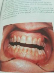
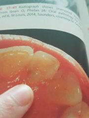
Dentinogenesis imperfecta |
|
|
Melanin Pigmentation |
|
|
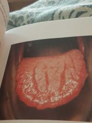
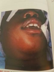
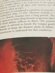
Fissured Tongue |
|
|
Tori |
|
|
Lobulated torus palatinus (palatal tori) |
|
|
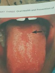
Medial Rhomboid glossitis |
|
|
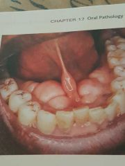
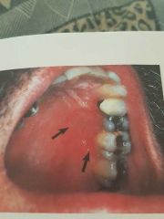
Periapical pathology |
|
|
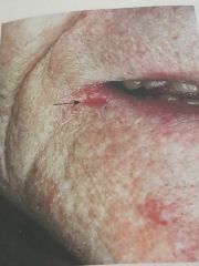
Angular Cheilitis |
|
|
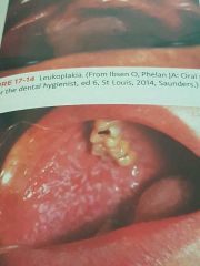
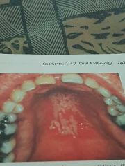
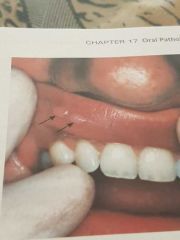
Leukoplakia |
|
|
Lichen planus |
|
|
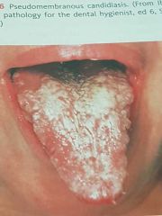
Pseudomembranous candidiasis |
|
|
Chronic Hyperplastic Candidiasis |
|
|
Aphthous Ulcer AKA Canker Sore |
|
|
Cellulitis |
|
(Tongue) |
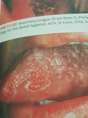
Black hairy tongue |
|
|
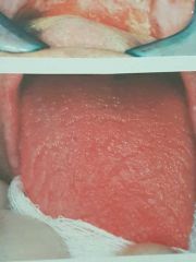
Geographic Tongue |
|
(2 answers) |
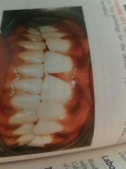
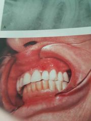
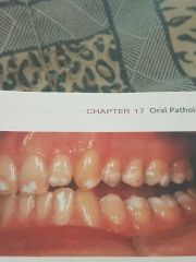
Fissured Tongue, and Attrition of the teeth |
|
|
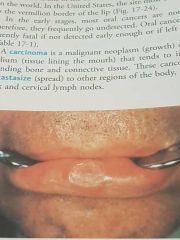
Squamous cell carcinoma |
|
|
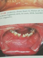
Leukemia |
|
|
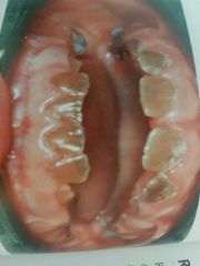
Tobacco chewer's white lesion |
|
Both photos = same thing |
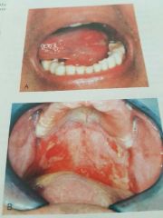
Radiation mucositis |
|
|
PostRadiation xerostomia |
|
|
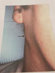
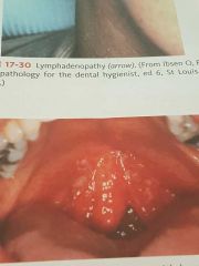
Lymphadenopathy |
|
|
Candidiasis in a patient with HIV |
|
|
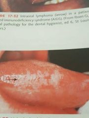
Intraoral lymphoma |
|
|
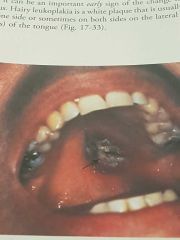
Hairy leukoplakia |
|
Both pictures = same thing |
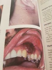
Kaposi's sarcoma |
|
|
Herpes simplex ulceration on hard palate in HIV pt. |
|
|
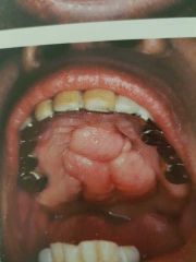
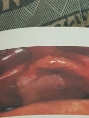
Papillary lesion |
|
Discoloration of teeth caused by ___ |
Tetracycline ingestion |
|
|
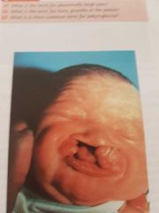
Cleft lip and palate |
|
|
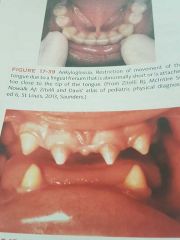
Ankyglossia (tongue tied) |
|
|
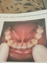
Partial anodontia |
|
|
Dens in dente |
|
|
Hypocalcified amelogenesis imperfecta |
|
|
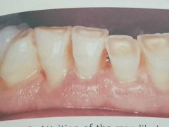
Attrition |
|
|
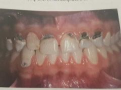
Meth mouth |