Reading...
![]()
Play button
![]()
Play button
![]()
Use LEFT and RIGHT arrow keys to navigate between flashcards;
Use UP and DOWN arrow keys to flip the card;
H to show hint;
A reads text to speech;
4 Cards in this Set
- Front
- Back
|
What are some degenerative disorders of the spleen? What can cause them? (6 degenetative diseases)
|
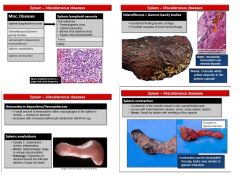
1. Splenic Lymphoid necrosis - nerosis of lymphocytes of the germinal centers following viral infections (parvovirus, equine herpes virus, BVDV), or stress, toxins, aging
-it is associated with necrosis of other lymphoid organs (lymph nodes, bone marrow, thymus...) 2. Siderotic plaques (siderofibrosis) - brown/yellow plaques, mostly along splenic margins -represent iron and calcium deposits in connective tissue of the capsule -associated with old age and have no clinical significance 3. Haemosiderosis - small amounts of hemosiderin are normal (RBC turnover). Can be increased in cases of hemolytic anemias due to increased breakdown of erythrocytes 4. Amyloidosis - part of generalized amyloidosis; grossly normal or mildly enlarged, orange colored HISTO: accumulation of amorphous pink material in germinal centers (sometimes requires special stains (Congo Red) 5. Senile atrophy - old dogs and old horses may have smaller, paler spleens with thickened capsule HISTO: might be paucity of white pulp & sinuses are collapsed 6. Splenic contraction - causes: circulatory shock (hypovolemic, cardiogenic, septic) or parasympathetic response (flight) -incompletely contracted areas of the spleen are easily confused with haematomas and infarcts -cause by failure of smooth muscle to contract resulting in incomplete evacuation of blood |
|
|
What can cause uniform splenomegaly with a bloody consistency?
|
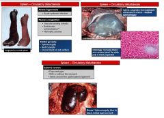
Active Acute Hyperemia - in septicemias and bacteremias, microbes are transported to the splenic red pulp, where they are rapidly phagocytosed by the splenic macrophages
GROSS: splenomegaly, moderately turgid, cyanotic with blue/black capsule. On cut surface exudes blood 2. Passive congestion -barbituate euthanasia, anesthesia or sedation -acute passive congestion is seen most dramatically at necroscopy in horses and dogs that have been euthanized or anesthetized by intravenous injection of barbituate -through to be the result of splenic relaxation of smooth muscle trabeculae and capsule GROSS: spleen is extremely enlarged, cut surface is notably congested & bulges and oozes blood HISTO: as a result of distension of the red pulp by blood, the normal archtecture of the splenic parenchyma is almost obliterated by masses of blood cells, chiefly erythrocytes 3. Torsion - occurs in mainly pigs & dogs -torsion of the spleen & stomach together occurs in dogs, usually deep-chested ones -in dogs & pigs spleen is loosely attached to the stomach by the gastrosplenic ligament vs. ruminants where it is firmly attached to the rumen GROSS: spleen is uniormly and markedly enlarged and blue-black from cyanosis -often folds back on itself in the shape of the letter "C" |
|
|
What is an example of a disease with acute active hyperemia in the spleen?
|
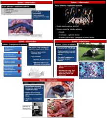
EXAMPLE: Anthrax
-caused by Bacillus anthracis -grows in an aerobic to facultative anaerobic environment -ingested spores replicate locally in the intestinal tract, spread to regional lymph nodes, and then disseminate systemically, through the blood stream, resulting in septicemia -the bacterium produced endotoxins that damage the endothelia -the spleen in a case of anthrax is uniformly enlarged, is dark red to bluish-black, and contains abundant unclotted blood because of extensive liquefactive necrosis HISTO: infiltration of inflammatory cells (mostly neutrophils), vascular sinuses are massively distended by blood DIAGNOSE: by pripheral blood smear without opening the carcass b/c of risk of contamination of spores -would see lots of gram positive rod shaped bacteria with square ends and a pink capsule stained by methylene blue or Giemsa |
|
|
In what species do hematomas occur? Why do they occur?
|
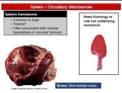
HEMATOMAS (INDUCED BY LYMPHOID HYPERPLASTIC NODULES)
-hematomas are usually associated with hyperplastic lymphoid nodules (arising from the PALS of the white pulp) and occur regionally or focally in the splenic parenchyma -occur idiosynchratically in the canine and less common in cat spleens & are NOT associated with general antigenic stimulation -in these species the circulation in the marginal zone is OPEN, the blood exits via open-ended arterial capillaries to percolate among lymphocytes of the marginal zone and reenters through similar open venous capillaries -distortion of the marginal zone area by hyperplastic lymphocytes disrupts the normal blood flow, resulting in accumulation of pooled blood that is unable to find its way into sinusoids or red pulp vascular spaces (reticular meshwork), thus forming a hematoma that durther distorts the splenic architecture HEMATOMAS (INDUCED BY SPLENIC VASCULAR NEOPLASMS) -can occur secondarily to splenic neoplasms of vascular origin -bleeding into the red pulp, which is confined by the splenic capsule, produces a dark red soft bulging, usually solitary mass of varying size (2-15 cm) -if the capsule over the hematoma ruptures, hemoperitoneum, hypovolemic shock, circulatory failure and death can ensue |