![]()
![]()
![]()
Use LEFT and RIGHT arrow keys to navigate between flashcards;
Use UP and DOWN arrow keys to flip the card;
H to show hint;
A reads text to speech;
62 Cards in this Set
- Front
- Back
|
What are the two fluid filled chambers of the eye, and what type of humor do they have?? |
-anterior chamber (in front of lens) -aqueous humor -posterior chamber (behind the lins)= vitreous body -vitreous humor |
|
|
What are images focused through? |
cornea, pupil of the iris, lens, and then on the retina. |
|
|
What is the retina? |
-neural part of the eye that converts a visual image into nerve action potentials |
|
|
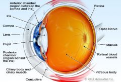
Anatomy of the eye |
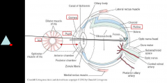
-conea -pupil of the iris -iris -lens -retina -anterior chamber -posterior chamber -optic nerve |
|
|
How many layers of neorons in the retina? |
7-10 |
|
|
How is the actual transduction accomplished? |
-photoreceptors: that contain pigments that can be altered very rapidly by light to generate a depolarization or hyperpolarization of a nerve cell membrane |
|
|
What are the two types of photoreceptors? |
-rods
-cones |
|
|
How many rods per eye? |
130 million |
|
|
What are rods sensitive to, cannot differentiate between, and what do rods contain? |
-dim light (night vision, scotopic vision) -cannot differentiate color -Rhodopsin (Vit A) |
|
|
What does the total loss of rods result in? |
-night blindness = legally blind |
|
|
What can cause night blindness? |
decrease in dietary Vitamin-A |
|
|
How many cones per eye? |
7 million |
|
|
What are cones sensitive to? |
-different frequencies |
|
|
What frequencies are cones sensitive to, what are they called? |
REE, GREEN, BLUE Iodopsins |
|
|
What does color blindness result from? |
-absence of one of the photopigments of cones (usually red or green) -more common in males than females |
|
|
Why is blue not affected in color blindness? |
-blue pigment genes are on different chromosomes |
|
|
Where are cones most numerous? |
fovea |
|
|
What is the fovea? |
-small depression in the retina in which the primary visual image is focused |
|
|
Where is the primary visual image focused? |
fovea |
|
|
What area of the eye has the highest visual acuity? |
-fovea |
|
|
What is the Macula lutea |
area surroundiing the fovea that has a yellowish appearance due to the presense of yellow pigment |
|
|
what occurs when localized degeneration takes place in the macula in unknown orgin? |
macular degeneration: may resut in severe visual impairment |
|
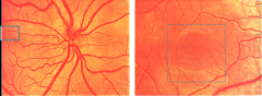
What is this picture? |
Macula |
|
|
How do the action potentials get conducted out of the eye after reaching the retina? |
-retinal ganglion cells |
|
|
What do rential ganglion cells form? |
-optic nerve, chias, and optic tracts |
|
|
What is the optic disc? |
(papilla) -point at which retinal ganglion cell axons leave the eye and retinal blood vessels enter |
|
|
What forms the blind spot in the visual fields? |
optic disc |
|
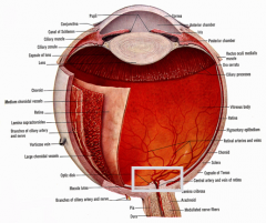
What is the square showing? |
optic disc |
|
|
Why is the optic disc examined after head trauma? |
-it can indicate the presence of increased intacranial pressure from hydrocephalus or other sources |
|
|
What can cause swelling of the optic nerve, and what is this? |
-increased cranial pressure -papilledema |
|
|
What is glaucoma? |
-increased pressure from the fluid chambers of the eyes on the optic disc |
|
|
What is perimetry? |
-quantitiation of the visual field -usually done by a machine that shows brief flashes of light at different points within the visual field as the patient looks at a central point |
|
|
What is the visual pathway starting with the retina to the optic chiasm? |
-retina -axons from retinal ganglion cells -optic nerve -optic chiasm |
|
|
What happens at the optic chiasm? |
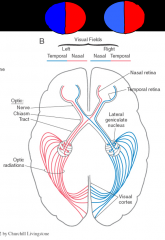
-half of the axons carrying info from the retina of each eye cross thereby dividing the visual field (what you see) into halves, vertically |
|
|
Explain the visual pathways:*
|
The right half of the visual field in each eye gets routed to the left hemisphere, and the left half of the visual field from each eye gets routed to the right hemisphere |
|
|
What is important so we can have depth perception/ binocular vision? |
-that the divisions of the visual field into left and right halves and that each eye overlaps |
|
|
How does the brain create depth perception? |
-by comparient the overlapping images in the occipital lobe, it creates three dimensional perception of space |
|
|
Were do the optic chiasm axons travel, and what do they contact? |
-around the brainstem near the crus cerebri -contact the lateral geniculate nucleus of the thalamus |
|
|
What does the lateral geniculate nucleus do? |
-puts together the entire left or right visual field from both eyes |
|
|
What is the geniculocalcarine tract? |
-from lateral geniculate neurons to primary visual cortex in the occipital lobe |
|
|
Where is the primary visual cortex located? |
-either side of the calcarine sulcus of the occipital lobe (posterior cerebral artery territory) |
|
|
How is the visual field (retinal surface) represented, where is the macula represented, and the surrounding retina? |
topographic (retinotopic) fashion in the area around the calcarine sulcus -posteriorly -anteriorly |
|
|
Where can lesions in the visual pathway take place? |
retina optic nerve optic tract visual cortex |
|
|
What will happen if there is a lesion in the optic nerve? |
loss of vision in one eye |
|
|
What will happen if there is a lesion at or beyond the chiasm? |
both eyes may be affected |
|
|
What will happen if there is a lesion that includes the primary visual cortex ? |
contralateral hemianopsia with macular sparing *the macula is said to be spared because of some (collateral circulation) from the middle cerebral arteries (or anastomoses) |
|

What occurs at 1? |
1. Monocular blindness |
|
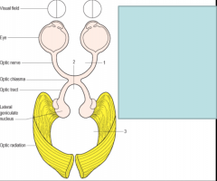
What occurs at 2? |
2. bitemporal hemianopia |
|

What occurs at 3? |
3. Homonymous hemianopia |
|
|
What is the pupillary light reflex? |
a light is shine in one eye and observed for pupillary constriction |
|
|
What is involved in the pathway that you test for pupillary light reflex? |
-ciliary ganglion, oculommotor nerve (CNIII), optic nerve, and some nuclei in the brainstem near the superior colliculus of the tectum |
|
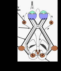
What are the arrows pointing at? |
Cilliary ganglion Oculomotor nerve optic nerve lateral geniculate brainstem- Pretectum |
|
|
What is the most common impairment of the pupillary reaction to light? |
-in patients with deteriorating conscious level after head injury |
|
|
What will happen to the pupil of the eye that has a disrupted optic nerve when doing the pupillary light reflex? |
-it will not produce constriction when the light is shined in the blind eye -but will when the light is shined into the normal eye -swinging the light back to the blind eye will then result in dialation of both pupils |
|
|
what is pupillary constriction under the control of? |
-oculomotor nerve III |
|
|
What is pupillary dialation under the control of? |
-sympathentic nervous system -ex. in response to fear |
|
|
In the picture know what the cornea, pupil of the iris, lens, and retina are: |
|
|
|
What does the anterior chamber vs. posterior chamber of the eye have in them? |
anterior: aqueous humor posterior: vitreous humor |
|
|
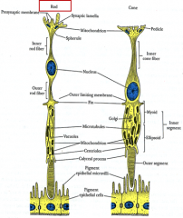
Retina: |
the neural part of the eye; consists of highly organized layers of neurons & neuroglia cells |
|
|
What are photoreceptors? |
the cells that detect light |
|
|
What is the chemical in rods vs cones? |
rods: rhodopsin (vitamin A) = light cones: iodopsin = color |
|
|
What is the visual pathway? |
Retina -> optic nerve -> optic chiasm -> optic tract -> brain stem (cranial nerves III, IV & VI controling movement of the eyes) -> thalamus (lateral geniculate nucleus) -> priamry visual cortes in the occipital lobe |