![]()
![]()
![]()
Use LEFT and RIGHT arrow keys to navigate between flashcards;
Use UP and DOWN arrow keys to flip the card;
H to show hint;
A reads text to speech;
20 Cards in this Set
- Front
- Back
|
What is the definition of apoptosis? |
- a process seen in multicellular organisms, by which specific cells are killed and removed for the benefit of the organism |
|
|
Programmed cell death is essential for embryonic development. How? |
- sculpting of limbs - neuronal development - negative selection of T cell lymphocytes that recognise self; |
|
|
Why is PCD important for neuronal development? |
- excessive production of neurons at first; - signals from target tissue promote neuronal survival |
|
|
Why is PCD important for negative selection of T cell lymphocytes? |
- immune cells are made in embryonic thymus gland; - receptors on T cells recognise abnormal proteins/pathogens; - during embryogenesis variety of T cells generated recognising normal and abnormal proteins |
|
|
PCD is used after development for removal of redundant structures? |
- there is an increased number of milk secreting cells formed in pregnancy - PCD after - metamorphosis |
|
|
PCD is important for regulation of cell numbers. Problems with it lead to pathogenic conditions? |
- degenerative disorders - Alzheimers or autoimmune diseases - diseases of over-proliferation - cancer |
|
|
PCD also limits collateral tissue damage by eliminating specific cells that are damaged beyond repair as result of: |
- DNA damage (when repair mechanisms cannot cope) - accumulation of misfolded proteins (causes ER stress and cell death; linked with neurodegenerative disorders) - cells infected by certain viral agents (limits spread of infection) |
|
|
What is the difference between necrosis and apoptosis? |
- Necrosis is always pathological - Apoptosis can be pathological but is usually physiological (but has the same morphological features in either case) |
|
|
What are the reversible morphological features of necrosis? |
- cell swelling - plasma membrane alterations (blebbing, loss of microvili) - mitochondrial changes (swelling, formation of small amorphous densities) - ER dilation (polysome formation) - Nuclear changes (disaggregation of fibrils) |
|
|
What are the irreversible morphological features of necrosis (due to severe injury)? |
- Extensive cell swelling and loss of plasma membrane integrity - mitochondrial changes (marked swelling, formation of large amorphous densities) - dilation of organelles - myelin formation (phospholipid masses) - nuclear changes (pyknosis, karyorrhexis) |
|
|
What are the initial morphological features of apoptosis? |
- cell shrinkage (dense cytoplasm, organelles are tightly packed) - minor membrane blebs - chromatin condensation (peripheral aggregation of dense chromatin mass under nuclear membrane; various shapes (crescent or circular dense mass) |
|
|
What are the morphological features of apoptosis? |
- extensive membrane blebbing - cell fragments via blebbing into apoptotic bodies (membrane-bound portions of cytoplasm and organelles; with or w/o nuclear fragments) - phagocytic ingestion and degradation |
|
|
What are the biochemical features of apoptosis (3)? |
- activation of caspases - DNA and protein breakdown - membrane alteration and recognition by phagocytes |
|
|
What are caspases? |
- found in animals - suicide proteases - cysteinyl-aspartate-specifc proteases |
|
|
What are the features of caspases? |
- cysteine at the active site - cleaves target proteins at specific aspartic acids - synthesised as inactive procaspase - activated by proteolytic cleavage at own aspartic residues to generate 2 subunits - 2 released subunits associate with another 2 subunits to form the active tetrameric caspase |
|
|
What domains do initiator caspases contain? |
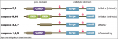
- CARD - caspase recruitment domain - DED - death effector domain |
|
|
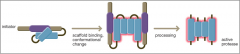
How are initiator caspases activated? |
- exist as monomers - interact with adaptor/scaffold protein causing procaspases to aggregate - procaspase form posses some proteolytic activity - activation through proximity or conformational change - released subunits from active tetrameric protease and activate effector caspases |
|
|
How are initiator caspases activated (image)? |
|
|
|
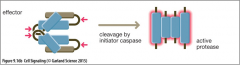
How are effector caspases activated? |
- exist as inactive dimers - cleaved by active initiator caspases - released subunits form active tetrameric proteases - execute the proteolysis seen during the demolition phase of apoptosis by targeting a range of cellular proteins |
|
|
How are effector caspases activated (image)? |
|