![]()
![]()
![]()
Use LEFT and RIGHT arrow keys to navigate between flashcards;
Use UP and DOWN arrow keys to flip the card;
H to show hint;
A reads text to speech;
147 Cards in this Set
- Front
- Back
|
Osteology |
Study of bones |
|
|
Individual bones |
Anatomical structures |
|
|
Entire skeleton |
Physiological organ (Functions like a stomach/heart) |
|
|
Axial skeleton |
Upright part of body (trunk) Skull, spine, ribs, sacrum |
|
|
Appendicular Skelton |
The extremities Hands, fingers, scapula, pelvis |
|
|
Functions of skeletal system: |
Rigid framework Lever for locomotor function of muscles Protection of viscera and organs Contains hemopoietic tissue for production of RBC, WBC, and platelets Stores magnesium, calcium, & phosphorus |
|
|
Description of a bone: |
Hard but resilient Highly vascular Living Mineralized Consists of cells embedded in a fibrous matrix filled with minerals |
|
|
Fibrous matrix |
Filled with minerals. Fibrous matrix = elasticity |
|
|
Mineral salts |
= hardness and rigidity |
|
|
Wolf's law: If stress on bone increases, |
Bone density increases |
|
|
Wolf's law: If stress on bone decreases... |
Bone density decreases |
|
|
Wolf's law: If stress on a bone is excessive... |
Bone density decreases |
|
|
Bone layers: (3) |
Compact bone Spongy bone/cancellous bone Trabeculae |
|
|
Compact bone |
Outer surface, dense and hard |
|
|
Spongy bone/cancellous bone |
Softer, interior bone |
|
|
Trabeculae |
Calcified tissue that is arranged in lines along areas of stress within the bone (in spongy/cancellous bone) surrounds open spaces full of red bone marrow |
|
|
Description of a bone: |
Hard but resilient Highly vascular Living Mineralized Consists of cells embedded in a fibrous matrix filled with minerals |
|
|
Types of bones: (5) |
Long bones Short bones Flat bones Irregular bones Sesamoid bones |
|
|
Fibrous matrix |
Filled with minerals. Fibrous matrix = elasticity |
|
|
Mineral salts |
= hardness and rigidity |
|
|
Wolf's law: If stress on bone increases, |
Bone density increases |
|
|
Wolf's law: If stress on bone decreases... |
Bone density decreases |
|
|
Wolf's law: If stress on a bone is excessive... |
Bone density decreases |
|
|
Bone layers: (3) |
Compact bone Spongy bone/cancellous bone Trabeculae |
|
|
Compact bone |
Outer surface, dense and hard |
|
|
Trabeculae |
Calcified tissue that is arranged in lines along areas of stress within the bone (in spongy/cancellous bone) surrounds open spaces full of red bone marrow (Individual to the person) |
|
|
Trabeculae |
Calcified tissue that is arranged in lines along areas of stress within the bone (in spongy/cancellous bone) surrounds open spaces full of red bone marrow |
|
|
Long bones: |
Support weight and facilitate movement. -femus, humerus, radius, fibula, tibia, metatarsal, phalanges, metacarpal, ulna |
|
|
Short bones: |
Cube shaped. Are as long as they are wide.. Tarsals and carpals |
|
|
Short bones: |
Cube shaped. Are as long as they are wide.. Tarsals and carpals |
|
|
Flat bones: |
Protect internal organs: Sternum, scapula, ribs, cranial bones |
|
|
Short bones: |
Cube shaped. Are as long as they are wide.. Tarsals and carpals |
|
|
Flat bones: |
Protect internal organs: Sternum, scapula, ribs, cranial bones |
|
|
Irregular bones |
Complex shapes. Sacrum (ischium, ilium, pubis) and vertebrae |
|
|
Short bones: |
Cube shaped. Are as long as they are wide.. Tarsals and carpals |
|
|
Flat bones: |
Protect internal organs: Sternum, scapula, ribs, cranial bones |
|
|
Irregular bones |
Complex shapes. Sacrum (ischium, ilium, pubis) and vertebrae |
|
|
Sesamoid bones |
Reinforce tendons and protect them from stress and wear: patella |
|
|
Name the 6 regions of a long bone: |
Diaphysis Epiphysis Metaphysis Meduallary canal Periosteum Epiphyseal growth plate |
|
|
Diaphysis: |
Main shaft of the bone |
|
|
Epiphysis |
The area at the end of the bone |
|
|
Epiphysis |
The area at the end of the bone |
|
|
Metaphysis |
Flared end of the diaphysis |
|
|
Epiphysis |
The area at the end of the bone |
|
|
Metaphysis |
Flared end of the diaphysis |
|
|
Meduallary canal |
Hollow center of diaphysis, contains marrow and arteries |
|
|
Periosteum |
Thin, fibrous membrane covering all of the bone except the articulate surfaces |
|
|
Periosteum |
Thin, fibrous membrane covering all of the bone except the articulate surfaces |
|
|
Epiphyseal growth plate |
In a growing bone the epiphysis is cartilagenous |
|
|
Types of connective tissues:(4) |
Cartilage Tendons Bursae Ligaments |
|
|
Types of connective tissues:(4) |
Cartilage Tendons Bursae Ligaments |
|
|
Connective tissue : cartilage |
Rigid tissue that is not as hard as bone. Relatively non-vascular: gets nutrition from surrounding tissue fluid -during dvpt many bones are first formed from cartilage |
|
|
Types of connective tissues:(4) |
Cartilage Tendons Bursae Ligaments |
|
|
Connective tissue : cartilage |
Rigid tissue that is not as hard as bone. Relatively non-vascular: gets nutrition from surrounding tissue fluid -during dvpt many bones are first formed from cartilage |
|
|
Types of cartilage: |
Hyaline cartilage, white fibrocartilage, elastic cartilage |
|
|
Types of connective tissues:(4) |
Cartilage Tendons Bursae Ligaments |
|
|
Connective tissue : cartilage |
Rigid tissue that is not as hard as bone. Relatively non-vascular: gets nutrition from surrounding tissue fluid -during dvpt many bones are first formed from cartilage |
|
|
Types of cartilage: |
Hyaline/articular cartilage, white fibrocartilage, elastic cartilage |
|
|
Hyaline/articular cartilage |
-composed the "temporary" skeleton -found in the epiphyseal growth plate -makes up articular cartilage -provides: elasticity to absorb shock & smooth surface for movement. with age becomes calcified/ ossified (less resilient the older we get). non-vascular(doesn't have own blood supply) and a-neutral (no nervous tissue) |
|
|
White fibrocartilage |
-has great tensile strength and some elasticity -able to resist considerable pressure Locations: within the IV disc Menisci of knee Glenoid and acetabular labrum Articular discs (TMJ, A-C, S-C) Articulating surface of the clavicle *found more in a dense later (pad) not thin it's more thick than articular cartilage |
|
|
Elastic cartilage |
Maintains the shape of a structure -found in the external ear, tip of the nose, the Eustachian tube and the larynx |
|
|
Tendons |
-Connective tissue attaching muscle to bone -may be encased in tendon sheaths: fibrous sleeves containing lubricating fluid.(lays over the tendon, helps move it) -may be in the form of an aponeurosis: a broad, flat tendinous sheet of tissue |
|
|
Bursae |
Fluid filled sacs found in areas of high friction. Lined with a synovial membrane and filled with fluid. -may be natural bursa or acquired bursa. |
|
|
Synovial membrane |
A thick, vascular connective tissue that secretes synovial fluid. |
|
|
Synovial fluid |
A thick, clear fluid that lubricates the articular cartilage and reduces friction and helps the joint move freely |
|
|
Ligaments |
Connective tissue that connects one bone to another. -flexible enough to allow joint movement. -rigid enough to prevent the bones from separating to create stability for the bone. -more dense, less movement than tendon |
|
|
Properties of connective tissue: |
Viscoelasticity Elasticity Viscosity |
|
|
Properties of connective tissue: |
Viscoelasticity Elasticity Viscosity |
|
|
Viscoelasticity |
Property that exhibits both viscosity and elasticity characteristics when undergoing deformation. |
|
|
Elasticity |
The ability to return to original shape after being deformed |
|
|
Elasticity |
The ability to return to original shape after being deformed |
|
|
Viscosity |
Ability of a material to resist a deforming force. Can't change no matter the force/stress Ex: honey |
|
|
Visco-elastic materials are sensitive to: |
1. How long the deforming load is applied 2. The speed at which the deforming load is applied 3. The magnitude of the deforming force (how much is applied) 4. The temperature of the tissue at the time of deformation (internal variable) |
|
|
Stress |
Force applied to the tissue |
|
|
Subluxation |
A partial dislocation of a joint |
|
|
Sprain |
A partial or complete tear of a ligament. Mild: few fibers torn, function intact. Moderate: partial tear with some loss of function Severe: rupture of ligament with loss of function |
|
|
Strain |
Over stretching of muscle fibers |
|
|
Tendonitis |
Inflammation of a tendon |
|
|
Tenosynovitis |
Inflammation of a tendon sheath |
|
|
Draw the stress-strain curve |
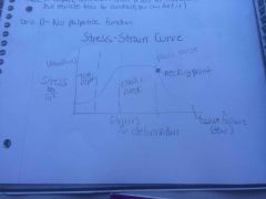
Back (Definition) |
|
|
Bursitis |
Inflammation of the bursa |
|
|
Capsulitis |
Inflammation of a joint capsule |
|
|
Athrology |
The study of joints |
|
|
Joint |
Articulation between 2 bones. -movement occurs at the articulation |
|
|
Strain |
The deformation that occurs to the tissue as a result of stress |
|
|
Joints have to: |
Support the body weight and provide stability |
|
|
Important things about joints: |
-a joint that provides a great deal of stability will provide little motion -the more functions a joint performs, the more complex it will be. |
|
|
Elastic range |
Tissue returns to original size and shape when stress is removed |
|
|
Plastic range |
Tissue will not return to original shake after stress removal, but is still intact |
|
|
Failure |
Rupture of a tissue |
|
|
Toe region |
Area where crimps in the collagen are being taken out |
|
|
Draw the stress-strain curve |
Back (Definition) |
|
|
10 common connective tissue pathologies: |
Fracture Dislocation Subluxation Sprain Strain Tendonitis Tenosynovitis Synovitis Bursitis Capsulitis |
|
|
Fracture |
A break in the continuity of a bone |
|
|
Dislocation |
A complete separation of two articular surfaces of a joint |
|
|
Stress |
Force applied to the tissue |
|
|
Subluxation |
A partial dislocation of a joint |
|
|
Sprain |
A partial or complete tear of a ligament. Mild: few fibers torn, function intact. Moderate: partial tear with some loss of function Severe: rupture of ligament with loss of function |
|
|
Strain |
Over stretching of muscle fibers |
|
|
Tendonitis |
Inflammation of a tendon |
|
|
Tenosynovitis |
Inflammation of a tendon sheath |
|
|
Draw the stress-strain curve |

Back (Definition) |
|
|
10 common connective tissue pathologies: |
Fracture Dislocation Subluxation Sprain Strain Tendonitis Tenosynovitis Synovitis Bursitis Capsulitis |
|
|
Capsulitis |
Inflammation of a joint capsule |
|
|
Athrology |
The study of joints |
|
|
Joint |
Articulation between 2 bones. -movement occurs at the articulation |
|
|
Sprain |
A partial or complete tear of a ligament. Mild: few fibers torn, function intact. Moderate: partial tear with some loss of function Severe: rupture of ligament with loss of function-complete tear |
|
|
Joints have to: |
Support the body weight and provide stability |
|
|
Important things about joints: |
-a joint that provides a great deal of stability will provide little motion -the more functions a joint performs, the more complex it will be. |
|
|
Joint classifications: (3) |
1. Fibrous (synarthrodial)
2. Cartilagenous (amphiarthrodial)
3. Synovial (diarthrodial) |
|
|
Synovitis |
Inflammation of the synovial membrane -causes swelling, fluid production in joint |
|
|
Plastic range |
Tissue will not return to original shake after stress removal, but is still intact |
|
|
Failure |
Rupture of a tissue |
|
|
Toe region |
Area where crimps in the collagen are being taken out |
|
|
Draw the stress-strain curve |
Back (Definition) |
|
|
Joints have to: |
Support the body weight (movement) and provide stability |
|
|
Important things about joints: |
-a joint that provides a great deal of stability will provide little motion -the more functions a joint performs, the more complex it will be. ***more stability=less motion ***less stability = more motion |
|
|
Dislocation |
A complete separation of two articular surfaces of a joint |
|
|
Fibrous joints |
-no movement -thin layer of periosteum between 2 bones -purpose to provide shape and strength to the bone |
|
|
3 types of fibrous joints: |
1.) sutures 2.) gomphosis 3.) syndesmosis |
|
|
Articulation |
2 different structures coming together |
|
|
Sutures |
Type of fibrous joint Thin layer of connective tissue that holds bones together. -found between bones in skull -no movement |
|
|
Gomphosis: |
-resembles a hole and peg -roots of the teeth in the mandible and maxilla |
|
|
Syndesmosis |
Large amount of fibrous tissue holds 2 bones together -distal tibia-fibula and radius-ulna |
|
|
Articulation |
2 different structures coming together |
|
|
Sutures |
Type of fibrous joint Thin layer of connective tissue that holds bones together. -found between bones in skull -no movement |
|
|
Gomphosis: |
-fibrous joint -resembles a hole and peg -roots of the teeth in the mandible and maxilla |
|
|
Syndesmosis |
Fibrous joint Large amount of fibrous tissue holds 2 bones together -distal tibia-fibula and radius-ulna |
|
|
Cartilaginous joints |
2 bones united by a pad of cartilage -allows a small amount of movement |
|
|
Types of cartilaginous joints: (2) |
1.) synchondrosis 2.) symphysis |
|
|
Types of cartilaginous joints: (2) |
1.) synchondrosis 2.) symphysis |
|
|
Synchondrosis |
-cartilaginous joint -continuous layer of hyaline cartilage (epiphyseal growth plate) -the first sternocostal joint -purpose: to allow for bone growth while providing stability |
|
|
Types of cartilaginous joints: (2) |
1.) synchondrosis 2.) symphysis |
|
|
Synchondrosis |
-cartilaginous joint -continuous layer of hyaline cartilage (epiphyseal growth plate) -the first sternocostal joint -purpose: to allow for bone growth while providing stability |
|
|
Symphysis |
-cartilaginous joint -occur in the midline of body -hyaline cartilage covers the bones involved but btw is a fibrocartilage pad - ex: the joints btw the vertebra and the pubic symphysis |
|
|
Synovial joints |
-Freely movable No direct connection btw the bone ends |
|
|
5 characteristics of a synovial joint: |
1.) articular surfaces are lined with hyaline cartilage 2.) bones are indirectly connected by a fibrous joint capsule 3.) synovial membrane covers the inner surface of the capsule but not the articular cartilage 4.) synovial fluid is secreted by the synovial membrane to lubricate and nourish the articular cartilage 5.) a joint space is present btw the two bone ends |
|
|
Classifications of synovial joints: (4) |
1.) nonaxial/ plane joints 2.) uniaxial joints 3.) biaxial joints 4.) triaxial joints |
|
|
Classifications of synovial joints: (4) |
1.) nonaxial/ plane joints 2.) uniaxial joints (moves in 1 plane) 3.) biaxial joints (Mvmt in 2 planes) 4.) triaxial joints (3 planes) |
|
|
Nonaxial joints |
No axis. No plane of motion. -plane joints that permit gliding btw 2 or more bones -inter carpal joints |
|
|
Uniaxial joints |
Moves in one plane around 1 axis 1.) hinge -IP joints , fingers, elbow, knee(mod. Hinge)
2.) pivot (trochoid) ring and a peg - atlanto-axial joint of C1 & C2 - radial head moves around ulna |
|
|
Biaxial joints |
-allow movement in 2 planes around 2 axes 1.) condyloid: concave surface moves over convex surface - MCP's of fingers 2.) saddle: each joint surface is concave and convex - CMC joint of the thumb - only one joint! |
|
|
Triaxial joints |
Allows movement in 3 planes around 3 axes
1.) ball and socket: ball-like convex surface fits into a concave socket -hip and shoulder joints |
|
|
Joint positions: |
1.) locked or closed pack position 2.) unlocked or loose pack position |
|
|
Locked/ closed pack position: |
-position of most contact btw bones - position of Max tautness (tense) in capsule and ligaments -position of greatest stability -usually at end-range ***greatest amount of stability that joints can achieve. Usually end of ROM. *** |
|
|
Unlocked/ loose pack position |
-position in which articular surfaces are free to move -ligaments and capsule are slack - any position other than closed pack |
|
|
Why bone ends don't actually touch in synovial joints? |
So they don't wear each other out and don't limit their movements |