Reading...
![]()
Play button
![]()
Play button
![]()
Use LEFT and RIGHT arrow keys to navigate between flashcards;
Use UP and DOWN arrow keys to flip the card;
H to show hint;
A reads text to speech;
44 Cards in this Set
- Front
- Back
|
How much of a RBC is water and hemoglobin?
|
Water 60% + Hemoglobin 40%
|
|
|
What is the lifespan of a RBC?
|
90-120 days
|
|
|
What granules do neutrophils contain?
|
Primary (or Azurophilic) Granule: Larger, denser
-contain lysosomal enzymes and are likely to be primary lysozymes -contain peroxidase, lysozyme, hydrolytic enzymes Secondary (or Specific) Granules: contain enzymes with strong bactericidal actions ie. collagenase, lactoferrin, liposome |
|
|
What is the function of neutrophils?
|
-first line of defense against microbial infection
-phagocytic, produce antimicrobial substances (lytic enzymes, bactericidal enzymes) -also modulate the inflammatory response by producing cytokines, chemokines |
|
|
What is the function of eosinophils?
|
-control parasitic infestation (MAJOR ROLE)
-role in allergic responses -regulate allergic & acute inflammatory processes: ✸inactivate histamine, inhibit histamine release ✸chemokines, cytokines -phagocytic: antigen-antibody complexes |
|
|
What granules do eosinophils contain?
|
-contain coarse cytoplasmic granules
-spherical, oval, or ellipsoid granules with electron dense core -major granule proteins: Major Basic Protein, eosinophil peroxidase, eosinophil derived neurotoxin, eosinophil cationic protein -lipid mediators (e.g. platelet activating factor), leukotrienes, hydrolytic enzymes, reactive O2 intermediates |
|
|
What is the function of basophils?
|
-major role in allergic & inflammatory reactions
-lipid metabolism, blood coagulation -possess IgE surface receptors -limited phagocytic & bactericidal activity -related to connective tissue mast cells – possibly the same myeloid precursor and may be the circulating form of the common mast cell |
|
|
What granules do basophils contain?
|
-contain coarse cytoplasmic granules – basophilic
-contain histamine, heparin, serotonin, hyaluronic acid, hydrolytic enzymes, chemotactic factors (same as tissue mast cells) |
|
|
Describe the structure & function of monocytes
|
-reach functional maturity after leaving bloodstream
-kidney or horseshoe shaped nucleus, deeply indented -paler nucleus than lymphocytes, loose chromatin strands -cytoplasm stains blue-grey w/ ground glass appearance -differentiate into macrophages and dendritic cells |
|
|
What is the function of dendritic cells? Where are they found?
|
(not present in blood, except for a small # of immature cells)
-antigen presenting cells, activate T-cells -present in small quantities in tissues that are in contact with the external environment (skin, intestines etc.) |
|
|
What is the function of macrophages?
|
-APC – initiate immune response
- highly phagocytic -produce cytokines |
|
|
What are platelets? What is their structure?
|
-they have no nucleus
-they are fragments of large megakaryocytes STRUCTURE: -small ovoid anucleated bodies -approx.2µm diameter & disk shaped |
|
|
What is the lifespan of a platelet?
|
Lifespan: 8-10 days
|
|
|
What is the granule composition in a platelet?
|
Dense granules – ADP, serotonin (a vasoconstrictor), calcium
Alpha granules – thrombospondin, fibrinogen |
|
|
What are the functions of platelets?
|
-Aggregation– associated with endothelial cell injury, formation of a platelet plug
-Coagulation– initiated at larger sites of injury; formation of a clot (thrombus) |
|
|
Where does haemopoiesis occur in embryonic life?
|
Begins in 1st weeks of life in the yolk sac
Stem cells migrate into the liver & spleen Liver & spleen play a major role in haemopoiesis during mid stages of gestation As bone develops → bone marrow becomes major haemopoietic organ Primary & secondary lymphoid organs then established |
|
|
Describe the development of RBCs
|
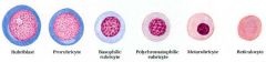
-derived from progenitor cell (Pro-erythroblast)
-erythropoietin (EPO) is produced by the kidney & regulates the # of RBCs produced Rubriblast → Prorubricyte → Basophilic rubricyte →Polychromatophilic rubricyte → Metarubricyte → Reticulocyte |
|
|
What are reticulocytes?
|
Reticulocytes – are immature RBCs, composing about 1% of the red cells
-Developed & matured in the red bone marrow & then circulate for a day in the bloodstream b4 developing into mature RBCs -No nucleus, have a reticular (mesh-like) network of ribosomal RNA, visible with methylene blue stain |
|
|
Describe Granulopoiesis
|
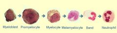
Myeloblast
- Large rounded cell -Spherical nucleus; nucleoi ↓ Promyelocyte - Large cell - Azurophilic granules appear ↓ Myelocyte - Specific granules appear - Mitotic ability lost ↓ Metamyelocyte - Nucleus becomes kidney shaped ↓ Band -Segmented form -Further maturation -Change in nuclear appearance |
|
|
Where is red marrow found? Where is yellow marrow found?
|
Red Marrow –found in flat bones
→Skull, ribs, sternum, pelvis, vertebrae, head of long bones Yellow Marrow –found in shaft of long bones -As the animal ages, adipose tissue replaces the marrow in the long bones |
|
|
Describe the structure of bone
|
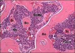
STRUCTURE
Stroma →Collagen fibers →Reticular fibers – reticular cells, fibroblasts, fat cells, osteogenic cells -Stromal elements impact on cell differentiation in the bone morrow Blood Vessels – arterioles, venules, sinusoids Sinusoids: -Wider than blood capillaries -Lined by endothelial cells -Gaps btwn endothelial cells allow cell traffic into sinusoids -Newly formed cells enter the circulation -Macrophages line the external sinusoidal surface -Megakaryocytes & RBC precursors adjacent to sinusoids Free Cells (parenchyma) – heterogenous population of cells Stem cells → active haemopoiesis |
|
|
What are the functions of the thymus?
|
-differentiation of T lymphocytes (thymocytes)
-development of immunological tolerance |
|
|
Describe the structure of the thymus
|
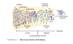
The stroma of the thymus consists of a prominent connective tissue capsule, which invaginates into the parenchyma as septa & divides the thymus into lobules. That parenchyma is organized into a cortex & medulla
CORTEX -darker peripheral zone -composed of tightly packed differentiating thymocytes surrounded by a meshwork of epithelial reticular cells and macrophages MEDULLA -lighter central zone -consists of epithelial reticular cells and the presence of larger lymphocytes, which are not as densely packed -medulla also contains the characteristic Hassal’s corpuscles, which are composed of concentrically arranged dead and dying reticular cells, macrophages neutrophils, and nuclear material whose origin is unknown (seen in older animals post puberty) |
|
|
What makes the blood thymus barrier?
|
-endothelial lining of blood capillaries
-basement membrane -layer of perivascular connective tissue -second basement membrane and layer of reticular epithelial cells |
|
|
What is the function of thymic reticular epithelial cells?
|
-direct the differentiation of T cells, producing thymic hormones including thymopoietin, thymosin α and thymulin
|
|
|
Describe the structure of the Bursa of Fabricius ("cloacal tonsil")
|
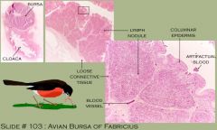
-organ peculiar to birds
-is a blind, sac-like structure on the caudodorsal side of the cloaca -like the tonsil, it has a number of blind, crypt-like invaginations -looks similar to the thymus -it is involved in conferring immunocompetance to the avian equivalent of B-cells in birds -free surface is covered with stratified squamous epithelium -most of the cells in it are lymphocytes -it lacks the lobulation and extensive connective tissue of the thymus |
|
|
What are Harderian glands?
|
-found in birds
-a pair locates between the eye and the nasal cavity -involved in the local immune response of the conjunctiva and upper respiratory tract -a central duct drains the gland secretion to the eye (acts as an accessory to the lacrimal gland) -play a role in terminal B cell differentiation and Ig class switching |
|
|
What do Harderian glands look like?
|
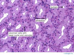
Capsule: thin connective tissue
-lobes divided by septa -main ducts -compound tubuloacinar gland, unequal size tubular secretory units -massive accumulations of lymphoid cells, mostly plasma cells in the interstitium, particularly in the center area of a lobule |
|
|
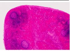
Describe the structure of the lymph nodes.
|
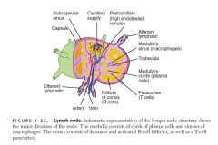
-they are encapsulated kidney shaped structures that have a concave side with a hilum
-the blood vessels and nerves enter and exit at the hilum, and the efferent lymphatic exits from this region -beneath the stromal capsule, the parenchyma is organized into cortex & medulla STROMA -the stoma consists of a dense connective tissue capsule that surrounds each lymph node and sends collagenous trabeculae into the node to divide its parenchyma into incomplete compartments ❀reticular cells produce reticular fibers that anastamose with the trabeculae and form an extensive network ❀the delicate reticulum filters the lymph and suspends the lymphocytes and macrophages CORTEX Trabeculae span the outer capsule into the medulla, separating the cortical follicles of B lymphocytes ♦primary or inactive follicles are dense with stored lymphocytes awaiting antigen presentation ♦secondary, or active follicles have pale germinal centers with the clusters of lymphocytes. Here, B cells proliferate and produce antibodies in response to antigen T cells are found in the paracortex between the follicles and medulla MEDULLA -medulla has medullary cords (lymphocytes, plasma cells, macrophages) and medullary sinuses |
|
|
How to B & T lymphoctes enter & exit the lymph nodes?
|
Lymph nodes are supplied by their own capillary system. B & T lymphocytes enter & exit the nodes via high endothelial venules located in the paracortex
|
|
|
How does lymph flow through the lymph node?
|
-lymph enters the node via afferent lymphatic vessels, which are located on the convex side of the organ
-from the afferent lymphatics, lymph passes through the subcapsular sinus to the peritrabecular sinuses. -it then enters the medullary sinuses and exits the lymph node via the efferent lymphatic vessels at the hilum |
|
|
What is the function of the lymph nodes?
|
-lymph nodes serve as filters clearing lymph of foreign particles before circulating to other areas of the body
-antigen uptake is carried out by macrophages, dendritic cells -antigen uptake leads to lymphocyte formation →clonal expansion of B cells →plasma cells → antibody production →stimulated T cells proliferate in thymus dependent zone →leave node as recirculating lymphocytes |
|
|
How does the lymph node of a pig differ?
|
-structure is reversed
-inner cortex & outer medulla -few cells in efferent lymph |
|
|
Describe the histology of hemal nodes
|
-unique to ruminants
-found along major muscular arteries; derived from these vessels during embryonic development, as cul-de-sacs, blind pouches off the artery -can be thought of as “accessory spleens” -architecture resemble that of a lymph node with diffuse lymphatic tissue, lymph follicles and sinuses, except the sinuses are filled with blood -there is a distict capsule, and hemal nodes are served at the hilus by an afferent arteriole -no lymph vessels, no typical medulla -in young animals hemal nodes display a distinct cortical accumulation of lymphocytes but few nodules are present |
|
|
Describe the location & function of mucosa-associated lymphoid tissue
|
LOCATION
-located in subepithelial mucosal connective tissue *bronchial, gut, nasal, conjunctiva– associated *lymphatic nodules – solitary or aggregates *organ-like aggregated lymphatic nodules e.g. tonsils, Peyers patches FUNCTION: -provide means to allow early encounter of foreign antigens, leading to antibody production |
|
|
Describe the location of tonsilar lymphoid tissue
|
-lingual→ base of tongue
-palatine→ caudolateral oropharynx -nasopharangeal → roof of the nasopharynx |
|
|
Describe the structure of tonsilar lymphoid tissue
|
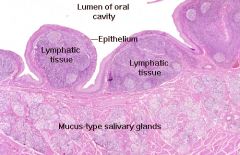
STRUCTURE:
-covered by epithelium: smooth or invaginated (fossulae) – with lymphoid tissue filling in the spaces between crypts -covered by stratified squamous epithelium -lymphatic nodules: surrounded by diffuse dense tissue (T cells); plasma cells (activated B cells that have moved out of follicles) -efferent lymphatic vessels but no afferent vessels -blood vessels -there is connective tissue investment on the attachment side -the mucus type salivary glands below the lymphatic tissue are small salivary glands whose ducts open onto the surface of the tonsil -see lymphocytes, macrophages & plasma cells in the tonsil |
|
|
Where are Peyer’s patches found?
|
-found predominantly in the ileum and at the ileocecal junction
|
|
|
What are caecal tonsils?
|
-aggregates of lymphatic tissues form nodular units at the caecum in chickens
-each nodular unit is demarcated by thin connective tissue |
|
|
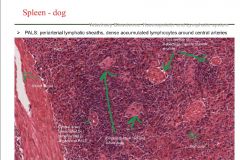
Describe the structure of the spleen
|
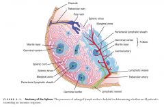
-has a hilus, efferent lymphatic (NO AFFERENT LYMPHATIC), blood vessels, & trabeculae
CAPSULE -connective tissue & smooth muscle -smooth muscle allows the spleen to contract and eject stored erythrocytes when needed STROMA -consists of the following: ❀ a dense connective tissue capsule that contains smooth muscle cells ❀ trabeculae that branch off of the capsule and partially partition the parenchyma of the splenic pulp ❀ a delicate meshwork of reticular connective tissue that filters the blood Splenic parenchyma consists of white and red pulp WHITE PULP (Dense lymphoid tissue) Composed of compact lymphatic tissue (nodules) in a special cylindrical arrangement called PALS (periarterial lymphatic sheaths) surrounding a central artery. * Nodular lymphatic tissue in PALS: B cells * Diffuse lymphatic tissue in PALS: T cells RED PULP (loose lymphoid tissue) -consists primarily of erythrocyte filled sinusoids and macrophages in a reticular fiber network; most filtration occurs here -can also contain smooth muscle fibers (depending on species) Free cells: reticular cells, RBCs, macrophages, plasma cells & lymphocytes ✸The sinusoids vary in size and are separated by pulp (ie Billroth cords) ✸RBCs and platelets are exposed to macrophages in the pulp cords; macrophages phagocytize worn-out or damaged cells ✸Splenic sinusoids are lined by loosely arranged endothelial cells which have numerous fenestrations. These sinusoids are surrounded by a poorly developed basal lamina ✸Phagocytosis is carried out primarily by macrophages located outside the sinusoids -the macrophages extend finger-like projections into the sinusoids, which push through the discontinuous basement membrane between the endothelial cells |
|
|
What are ellipsoids?
|
Ellipsiods (sheathed capillaries):
-leaky capillaries – prominent endothelium -terminal ends surrounded by reticular sheath of macrophages -found around white pulp (well developed in dog, horse, pig) |
|
|
Describe the circulation of blood through the spleen
|
Blood enters from the splenic artery via the splenic capsule at the hilus → gives rise to trabecular arteries in the fibromuscular trabeculae→ leave the trabeculae, become invested by a periarterial lymphatic sheath (PALS), and are known as central arteries → branch but maintain their lymphatic sheath until they leave the white pulp to form several straight penicillar arteries → enter red pulp as a capillary network surrounded by macrophages, called the macrophage sheathed capillaries → either drain directly into splenic sinusoids (closed circulation) or terminate in open-ended vessels within the splenic cords of the red pulp (open circulation) → splenic sinusoids are drained by pulp veins, which are tributaries of trabecular veins → drain into splenic vein, which exits
DOGS: open & closed circulation CATS, OX, PIGS, and HORSE: open, no sinusoids |
|
|
What is the function of the spleen?
|
-Antibody production → humoral immunity against blood-borne antigens
-Filter blood ✸dispose of defective blood cells ✸preserve iron for re-use; bilirubin recirculated to liver -Storage of cells – red blood cells, platelets Haemopoiesis – in fetal life |
|
|
Are there any consequences to a splenectomy?
|
-can be removes without any long term problems
-can make an animal susceptible to blood borne infections |