Reading...
![]()
Play button
![]()
Play button
![]()
Use LEFT and RIGHT arrow keys to navigate between flashcards;
Use UP and DOWN arrow keys to flip the card;
H to show hint;
A reads text to speech;
31 Cards in this Set
- Front
- Back

What is image a slide 1? What are the distinguishing features?
|
Oocyte. Large nucleus
|
|

What is image b slide 1?
|
Red Blood Cell
|
|

What is image c slide 1?
|
Neurons
|
|
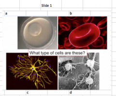
What is image d slide 1?
|
Macrophages
|
|
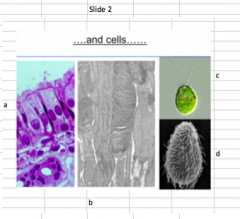
What is image a slide 2 What are the names of the surfaces? Where are they found?
|
They have apical and basolateral surfaces. They have cilia, so they are found in our airways
|
|

What is image b slide 2?
|
Photoreceptors in our retina
|
|

What is image c slide 2? What is special about this?
|
Chylemedamonis Hymadidi, a free swimming algae. It is a model organism and has flagella. It is multicellular
|
|

What is image d slide 2 What is on the outside? Where is this found?
|
Tetrahymena and have cilia on the outside. Cilia. Found in the centrosome (grows out of there)
|
|

What is a slide 3?
|
Sperm
|
|

What is B slide 3?
|
Spiderkea (bacterium)
|
|
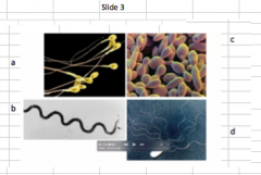
What is C slide 3?
|
A model organism, yeast
|
|

What is D slide 3?
|
Bacteria with a lot of flagella
|
|
|
Are cells different in size or the same?
|
Different
|
|
|
Rank from smallest to largest: Epithelia cell, oocyte, yeast, bacterium, sperm
|
Bacterium, yeast, sperm, epithelia cell, oocyte
|
|
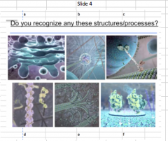
What is a in slide 4?
|
Golgi apparatus (vesicles budding from it)
|
|

What is b in slide 4?
|
Centrosome
|
|

What is c in slide 4?
|
Kinesin (microtubule bound with motor)
|
|

What is d in slide 4?
|
Actin filament building
|
|

What is e in slide 4? What is happening?
|
Microtubule. Depolarization of it, which is how it disappears, called dynamic instability
|
|

What is f in slide 4? What is the thing bound to it and what does it make?
|
Plasma membrane that is enriched for a protein. A protein bound to the lipid of membrane. Lipid raft
|
|
|
What is dynamic instability and where does it occur?
|
The disassocation of microtubule
|
|
|
What does MTOC stand for?
|
Microtubule organizing center, responsible for arranging the cell in a specific array
|
|
|
What is next to the centrosome?
|
Golgi apparatus
|
|
|
What are the two that secretes vesicles?
|
Golgi apparatus and the ER
|
|
|
What is the organizational heirarchy / orders of magnitude leading up to a cell?
|
Molecule in nanometers, complex, structure organelle and then cell
|
|
|
How are function and geometry related in a cell?
|
The geometry of a cell affects the function, ex. Cancer cell where geometry is off
|
|
|
What is Wiskott-Aldrich Syndrome? And what are the symptoms? (4 of them)
|
A rare X-linked recessive disease. Symptoms include: Eczema, thrombocytopenia (low platelet count that causes lots of bleeding that doesn't stop), immunodeficiency (repeated infection cause), and a risk of autoimmune diseases like cancer
|
|
|
What causes Wiskott-Aldrich Syndrome? Who gets it?
|
A regulator of actin (WASp), is mutated. Only males get it. Actin is important because a macrophage needs WASp to move forward. Think: Actin involved in movement. Immune functions depend on actin organization
|
|
|
What can we actually see by light microscopy?
|
Light is involved in this, so the spectrum of light is involved. The spectrum is 400-700. We can see them as two structures, and blurry is one. The rule of thumb: D ~ 1/2 wavelength.
|
|
|
What is the rule of thumb for light microscopy and what does it mean?
|
If we see something that is 250nm apart, we'll see it as two since 1/2 wavelength. BUT, if it is 200 nm apart, we can only see it as one, meaning there is a minimal distance required between 2 distinguishable objects
|
|

What is this disease? What are the symptoms and the genetic category of it? (dominant/recessive/autosomal/x-linked) |
This is Wiskott-Aldrich Syndrome. It's a x-linked recessive disease that's characterized by eczema, low platelet count, immunodeficiency, and increased risk for autoimmune diseases like cancer. |