Reading...
![]()
Play button
![]()
Play button
![]()
Use LEFT and RIGHT arrow keys to navigate between flashcards;
Use UP and DOWN arrow keys to flip the card;
H to show hint;
A reads text to speech;
64 Cards in this Set
- Front
- Back
|
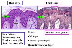
What are the characteristics of thin skin?
|
SHEA butter goes on the thin skin
Sebaceous glands Hair Eccrine Apocrine sweat glands |
|
|
What are the characteristics of THick skin?
|
Just Eccrine sweat glands
Sweaty palms and feet |
|
|
What is epithelium Cellular type?
|
SSK
Simple Stratified Karatinized |
|
|
Epidermis is to Epithelium as Dermis is to
|
Connective Tissue
|
|
|
CHaracteristics of Epidermis
|
Avascular
No Lymphatic capillaries Where does the vasculature and lymphatics begin? |
|
|
Dermis characteristics
|
Vascular
Lymphatics Cells with abundant ECM |
|
|
What is ECM
|
ECM= fibers + "ground substance"
|
|
|
What are ECM pimary fiber types, What is ground substance?
|
Collagen and Elastic fibers
Proteoglycans, adhesive glycoproteins |
|
|
What are proteoglycans?
|
Core protein with surrounding GAGs
|
|
|
What are GAGs
|
Repeating disaccharide units (most are sulfated)
Hyaluronic acid is nOt sulfated |
|
|
What in Ground substance has the negative charge?
What is the result of the charge? |
Proteoglycans over-all, but the hi negative charge is b/c of the sulfated Gags.
Attract Na+ and H2O follows to create a hydration shell |
|
|
Proteoglycans form aggreagates by securing to what structure? How do they bond?
|
Proteoglycans non-covalently boind to Hyaluronic Acid- become large "biological sponges'
|
|
|
Where is Versican? Where is Aggrecan?
|
Versican is in General Connective Tissues
Aggrecan is in Cartilage (going to be on exam) |
|
|
What is the role of Myofibroblasts in wound healing?
|
responsible for wound contraction
|
|
|
What is the collagen deposition pattern in wound healing?
|
Type III collagen, then type I
Type I is scar tissue |
|
|
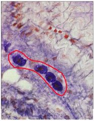
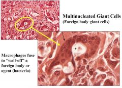
What is a multinucleated Giant Cell?
|
Macrophages fuse to wall of a foreign body or agent (bacteria)
|
|
|
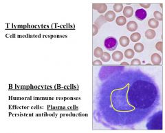
What is the difference in T and B cells
|
T lymphocytes- Cell mediated Response
B Lymphoscytes- Humoral Immune responses. Require Effector Plasma Cell secretion of antibodies |
|
|
Two types of Fat
|
Unilocular for E storage
Multilocular (BAT) for thermogenesis |
|
|
What is the suggested characteristic order to consider when classifying general connective tissues?
|
# of cells (lots/little)
Fibers (Arrangement/ Type) Spaces between (loose/dense) |
|
|
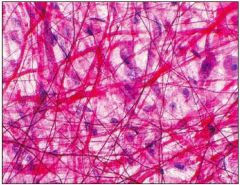
What is indicative of Dense Irregular CT?
|
Collagen bundles in all different directions, Large course bundles
|
|
|
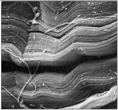
What feature is characteristic of Dense Regular CT?
|
Box car nuclei (looks like antelope canyon in SEM)
|
|
|
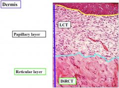
What are the two layers of Dermis?
|
Papillary layer and Reticular layer
|
|
|
What are the charachteristics of the papillary layer?
|
Has fingerlike projections up into the epidermis. Lots of different cell types and shapes, This is LOOSE CT, where all the transport is occuring to feed the epithelium
|
|
|
What are the characteristics of the Reticular layer?
|
DENSE IRREGULAR Lattice like network, packed with course thick dense collagen bundles
Has larger blood vessels that can't be squished |
|
|
Remember:
Epidermis: SSK Dermis: LCT (papillary) & DiRCT (reticular) Where else can DiRCT be found? What about DRCT? |
DiRCT- Joint capsules, Perichondrium, Periosteum
DRCT- Tendons (handle all force in 1 direction, hence dense regular) Type I collagen w/ versican in both. |
|
|
What are the three Cs and E that don't have lymphatics?
|
Cartilage, Cornea, CNS, Epidermis
(fluid drains out of these areas to those that have lymphatic capillaries |
|
|
What are the three types of cartilage?
|
Hyaline
Elastic (confined to the head, neck, respiratory tract, and ear) Fibrocartilage |
|
|
What is the importance of Hyaline cartilage in the embryo and immature skeleton?
|
They make templates for long bones and epiphyseal plates.
|
|
|
What is the importance of Hyaline cartilage in the Adult?
|
Articular cartilage of the synovial Joints
|
|
|
What are the two functions of the perichondrium?
|
Nutrient supply to cartilage
Chondrogenic stem cells |
|
|
Describe the evolution of Stem Cells to Hyaline Cartilage
|
Mesenchyme -
Chondrogenic cells + SOX9= Chondroblasts + surrounding matrix (lacunae)= Chondrocytes+ appositional growht= hyaline cartilage |
|
|
What is the most common type of growth in mature cartilages?
|
Appositional
|
|
|
Isogenous groups represent what?
|
Inidcative of early cartilage formation & epiphyseal plates
Interstitial growth (from within) |
|
|
What is the collagen/PG in Cartilage? What is the collagen/PG in bone and perichondrium?
|
Cartilage: TypeII/ Agrican
Perichondrium and bone: Type I/Veriscan |
|
|
What is the ECM of cartilage?
|
The same as Dermis
Fibers and ground substance |
|
|
What types of skeletal problems might an embryo or child have if the hyaline cartilage ECM is defective?
|
Problems with formation of long bones and elongation of the long bones during growth.
|
|
|
What is achondroplasia? Why does it happen?
|
Short long bones with thickened diaphyseal walls,
can develop nice bones bu they don't elongate properly. |
|
|
Why is the skull of an individual with achondroplasia shaped the way it is?
|
Jaw and mandible form from endochondral ossification, skull doesn't need a cartilaginous model.
|
|
|
How can you differentiate Articular cartilage with epiphyseal plate cartilage?
|
Look for perichondrium, also, epiphyseal plate cartilage will have a strip of clustered isogenous groups thickened in the middle. Articular cartilage will have trails of isogenous groups headed toward the surface of the cartilage.
|
|
|
Where does articular cartilage receive nutrition?
|
from synovial fluid on the joint side, from BV in bone and perichondrium on bone side
|
|
|
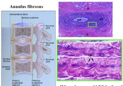
Where is fibrocartilage?
What type of joint? |
IVD
Symphysis type joints Labrum of Acetabulum Menisci, Articular disks. Can be deposited in fracture repair. |
|
|
What is the major difference between hyaline cartilage and fibrocartilage?
|
Fibrocartilage is type I Cartilage,
It is a hybrid of DCT and hyaline cartilage There won't be any perichondrium |
|
|
What can be seen in both fribro/hyaline cartilage?
|
Chondrocytes
Lacunae Isogenous groups |
|
|
What collagen and PG is in hyaline vs fibrocartilage? What structures have the same collagen as fibrocartilage?
|
Hyaline: Type II and agrican PG
Fibro; Type I and Versican, bone, perichondrium |
|
|
In a stained image of fibrocartilage, where will agrican be found
|
directly surrounding the lacunae of the chondrocytes (stains darker, but disappears as changes to Versican and Type I
|
|
|
What places would fibrocartilage be found in the Lower extremity?
|
Labrum of the acetabulum
Meniscis, Articular disks |
|
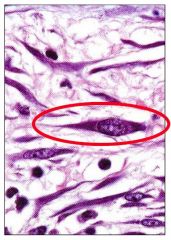
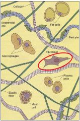
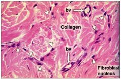
Cell type?
|
|
|
Tissue Type?
|
DiRCT
|
|
Tissue Type
|
DiRCT
|
|
Tissue Type?
|
DRCT
|
|

Tissue Type
|
DRCT
|
|
Identify the Layers
|
Don't forget epidermis
|
|
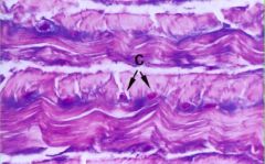
Tissue Type?
|
Loose Connective Tissue
|
|
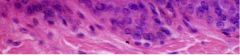
Cell type?
|

|
|
|
Cell type?
|
|
|

Cell type?
|
|
|
Cell type Top? Bottom?
|
|
|
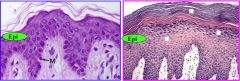
Skin types? Contents?
|
|
|
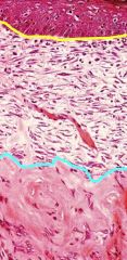
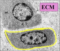
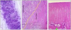
Where are dermis and epidermis divided? What is the layer of dermis?
|
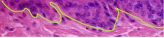
Yellow line,
Papillary layer |
|

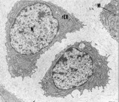
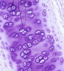
Cell type?
|
Chondrocytes in Lacunae
|
|
Cell type?
|
Chondrocytes in lacunae
|
|
What on earth is this?
|
|
|
What tissue type is this?
|
Hyaline cartilage of new bone growth or epiphyseal plate
|
|
What are these four different types of cartilage? Can you name their Collagen type and PG?
|
All are type II and agrecan except Fibrocartilage which has type I and versican.
|