![]()
![]()
![]()
Use LEFT and RIGHT arrow keys to navigate between flashcards;
Use UP and DOWN arrow keys to flip the card;
H to show hint;
A reads text to speech;
46 Cards in this Set
- Front
- Back
|
How is the body (shaft) of a bone called? |
Diaphysis |
|
|
How is the extremety of a bone called? |
Epiphysis |
|
|
What is the region between the diaphysis and epiphyses called? |
Metaphysis |
|
|
What do metaphyses contain in growing individuals? |
Epiphyseal plate - a layer of hyaline catrilage that allows growth. |
|
|
What is the name of an ossified epiphyseal plate? |
Epiphyseal line |
|
|
What is the superficial layer of a bone's extremety? |
A layer of articular cartilage |
|
|
What is the significance of articular cartilage on bones? |
It reduces friction and absorbs shock at the articulation point between two bones. |
|
|
What is periosteum? |
A superficial layer of tough connective tissue together with its blood supply that covers bones where articular cartilage is absent |
|
|
What are the fucntions of periosteum? |
Protection Osteogenic cell residence Repair assisstance Tendon attachments. |
|
|
What are the two bone tissue types? |
1) Cortical/compact bone 2) Spongy/trabecular/cancellous bone |
|
|
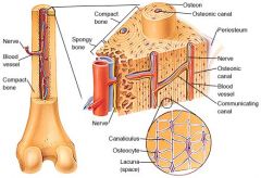
What are the structural features of compact bone? |
1) Consists of osteons between outer and inner circumferential lamellae. 2) Osteons are ring-like structures with central canal in the middle and osteocytes residing along lamellular lines. 3) The stronger of the two. |
|
|
Describe the structure of bones. |
|
|
|
Whar are the 4 types of bone cells? |
Osteoblasts Osteocytes Osteoclasts Osteoprogenitor cells |
|
|
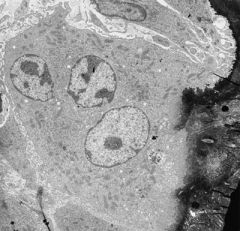
What are the microscopic features of osteoblasts? |
1) Pale activated nucleus 2) Lots of rER and Golgi 3) Evidence of collagen production |
|
|
What are the microscopic features of osteocytes? |
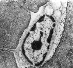
1) Deactivated nucleus 2) Presence of canaliculi |
|
|
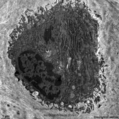
What are the microscopic features of osteclasts? |
1) Ruffled border 2) Several nuclei |
|
|
What is the bone matrix made of? |
1) Collagen type 1 - provides resilience and wholeness 2) Bone salts, e.g. Calcium Hydroxyapatite (Ca10(PO4)6(OH)2) |
|
|
How is blood supplied to bones and nutrients distributed? |
Blood is delivered to bones by nutrient arteries through nutrient foramina. Nutrients then diffuse through canaliculi of ostecytes. |
|
|
What is the approximate total amount of calcium in the body? |
+- 1kg |
|
|
Describe the distribution of calcium in the body. |
1) 99% in bones 2) Rest - soft tissue, teeth and ECF |
|
|
What are the 3 types of calcium in circulation? |
1) Free ionised, unbound calcium - 47%. Physiologically important, acts like a ligand. 2) Protein bound (mostly to albumin) - 47%. 3) Complexed calcium, bound to phosphate and citrate - 6%. |
|
|
What is the normal range of total calcium concentration? |
2.20 - 2.60 mmol/l in the population Narrower in individuals |
|
|
What is the formula for adjusted calcium? |
Ca(adj) = Ca(tot) + 0.2(normal alb - alb) Normal albumin = 40 or 45 alb = recorded value of albumin |
|
|
What are the major organs involved in calcium metabolism and homeostasis? |
Liver Kidneys Bone paarthyroid glands |
|
|
What are the major hormones involved in calcium metabolism and homeostasis? |
Parathyroid hormone (PTH) 1.25 dihydroxyvitamin D, also called 1.25 dihydroxycholecalciferol (1.25 DHCC or calcitriol) |
|
|
What is PTH? |
An 84-amino acid, single chain peptide hormone, secreted by chief cells of parathyroid glands. Its production is stimulated by low ionised calcium or high phosphate concentrations. |
|
|
How is vitamin D made in the body? |
1) Cholecalciferol is made in the skin (UV + diet) 2) 25-hydroxycholecalciferol is then made in the liver 3) Finally, 1.25 dihydroxycholecalciferol is made in the kidneys |
|
|
Where and how is calcium absorbed? |
Absorbed in the gut (duodenum and jejunum) by either passive diffusion or cell-mediated active transport, which can be influenced by 1.25 DHCC. |
|
|
How does PTH control calcium metabolism? |
1) G-protein coupled receptors in the parathyroid glands and renal tubules sense changes in calcium concentration. 2) A drop in calcium concentration stimulates PTH production. 3) PTH and 1.25 DHCC increase calcium resorption from bone and PTH increases calcium reabsorption in the kidney. |
|
|
Where in the nephron is calcium reabsorbed? |
1) 65% is reasbsorbed in the PCT together with other solutes like Na. 2) 15% is reabsorbed in the DCT. 3) 20% is reabsorbed in the thick ascending limb of Henle's loop. * 2 and 3 are controled by PTH. |
|
|
What are the causes of hypocalcaemia? |
1) PTH problem: deficiency due to hypoparathyroidism caused by neck surgery, Mg deficite or idiopathic. 2) 1.25 DHCC problem: deficiency due to malabsorption or insufficient UV or renal failure and inability to synthesise active 1.25 DHCC. |
|
|
What are the causes of hypercalcaemia? |
1) PTH problem: hyperthyroidism due to adenoma (high Ca and PTH on blood test) malignancy - production of PTH related peptide (high Ca but suppressed PTH) 2) 1.25 DHCC problem: inadequate dose |
|
|
What is the distribution of phosphate in the body? |
1) 85% in bone matrix. 2) Rest in predominantly intracellular: membranes, nucleic acids, enzyme cofactors, ATP. 3) 1% in ECF, 30% of which in inorganic. |
|
|
What are the organs involved in phosphate metabolism? |
1) Kidney 2) Gut 3) Bone |
|
|
What are the main hormones involved in phosphate metabolism? |
1) PTH 2) Fibroblast growth factor 23 (FGF 23) 3) 1,25(OH)2 Vitamin D |
|
|
What are the significats points about phosphate absorption in the gut? (4 points) |
1) Less regulated than that of Ca. 2) Entire small intestine is involved. 3) Rate of absorption is increased by 1.25(OH)2 Vitamin D 4) Phosphate is more plentiful in diet than calcium. |
|
|
Where in the kidney is PO4 reabsorbed? |
1) 75% in the PCT 2) 25% in the DCT |
|
|
What happends when phosphate levels rise too high? |
1) When PO4 rises Ca falls. 2) Then PTH and FGF 23 levels increase. 3) Both hormones induce PO4 excretion in the urine. |
|
|
What are the 4 phases of bone remodelling? |
1) Quiescence 2) Resorption 3) Reversal 4) Formation |
|
|
How much of bone is being remodeled at any one time? |
10% |
|
|
How long does a single remodelling cycle last? |
About 6 months: 10 - 14 days of resorption and approx. 150 days of formation. |
|
|
What do osteocytes secrete in quiescence phase to inhibit wnt signalling and osteoclast activation? |
Sclerostin |
|
|
Which molecules are responsible for osteoclast formation? |
1) Early differentiation: PU-1 and M-CSF - maturation of early osteoclast precursor from stem cells. 2) Later differentiation: C-fos, nuclear factor kappa B (NfkB), receptor activator of NFkB (RANK), and its ligand (RANKL). |
|
|
Outline the process of osteoclast maturation. (6 points) |
1) Nearby osteocytes undergo apoptosis. 2) Lining cells form canopy with blood vessels. 3) Sclerostin is no longer released. 4) IL-3 stimulates pre-osteoblasts and M-CSF stimulates pre-osteocytes. Both are released by stromal cells. 5) Pre-osteoblasts proliferate, secrete factors, e.g. Wnt, and express RANK-L on their surfaces and extracellulary. 6) RANK receptors on osteclasts are stimulated, causing them to fuse into mature osteoclasts. |
|
|
Which molecule inhibits RANK - RANKL interaction and what is it released by? |
Osteoprotegerin (OPG) is the RANKL inhibitor released by osteoblasts, and also other cells in the body, to prevent osteoclast formation and thus decrease bone resorption. |
|
|
Outline the process of bone resorption. (3 points) |
1) A tight seal is formed by mature osteoclasts over the bone surface. 2) Osteoclast release HCl to digest hydroxyapatite and proteolytic enzymes to break down the matrix. 3) Osteoclasts undergo apoptosis heralding the beginning of bone formation. |