Reading...
![]()
Play button
![]()
Play button
![]()
Use LEFT and RIGHT arrow keys to navigate between flashcards;
Use UP and DOWN arrow keys to flip the card;
H to show hint;
A reads text to speech;
149 Cards in this Set
- Front
- Back
|
AbdominoPelvic Regions
|
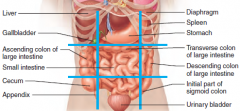
- Rt hypochondriac - (below rib cartilage) Right lobe of liver
- Epigastric – left liver lobe, most of stomach - Lt hypochondriac – small portion of stomach and transverse colon - Rt lumbar – gallbladder, ascending colon, - Umbilical (belly button) –small intestines, most of transverse colon - Lt lumbar – portion of descending colon, some small intestines - Rt iliac (Inguinal) - cecum, appendix - Hypogastric (Pubic) - small intestines, urinary bladder, rectum - Lt iliac - portion of descending colon, some small intestines |
|
|
Ribosomes
|
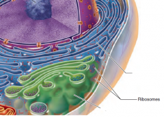
Actual site of protein synthesis
Non-membranous(rRNA, mRNA, tRNA) |
|
|
Endoplasmic Reticulum
|
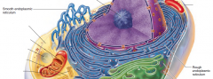
Rough ER provides an area for storge and transport of proteins
Membranous Smooth ER is a site of steroid and lipid synthesis |
|
|
Golgi
|
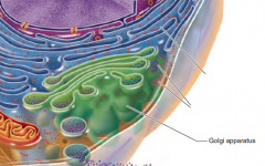
Packages proteins and other substances for export.
Membranous "UPS" of cell |
|
|
Lysosomes
|
Digest worn out cell organelles and foreign substances.
Membranous |
|
|
Peroxisomes
|
Contain oxidase enzymes to detoxify
Membranous clean up ... |
|
|
Mitochondria
|
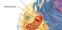
Oxidize foodstuffs to Produce ATP
Membranous |
|
|
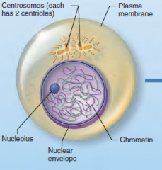
Centrioles
|
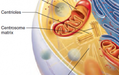
Direct formation of mitotic spindle during cell division
Non-Membranous |
|
|
Nucleus
|
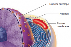
Contains nuclear envelope, nucleoli, chromatin, and distinct compartments rich in specific protein sets
Gene-containing control center of the cell Contains the genetic library with blueprints for nearly all cellular proteins Dictates the kinds and amounts of proteins to be synthesized |
|
|
Plasma Membrane
|

Double layer of lipids (phospholipids, cholesterol...) with embedded proteins. Proteins may extend entirely through the lipid bilayer or protrude on only one face. Most externally facing proteins and some lipids have attached sugar groups.
External cell barrier, acts in transport of substances into or out of the cell. Maintains a resting potential. Externally facing proteins act as receptors (for hormones, neurotransmitters...), transport proteins, and in cell-to-cell recognition. |
|
|
Nuclear Envelop
|

Selectively permeable double membrane barrier containing pores
Encloses jellylike nucleoplasm, which contains essential solutes Outer membrane is continuous with the rough ER and is studded with ribosomes Pore complex regulates transport of large molecules into and out of the nucleus |
|
|
Cyoskeletal
|
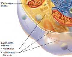
The “skeleton” of the cell
Dynamic, elaborate series of rods running through the cytosol Consists of microtubules, microfilaments, and intermediate filaments |
|
|
Cell cycle phases
|
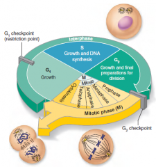
1. Interphase 22hrs
2. Mitosis/cell division- starts when cell volume increases 1.5hrs 3. Cytokinesis- pinching in 2 of the cell .5hrs |
|
Name the phase and describe it.
|
Interphase
G1Phase - Growth phase S Phase -DNA is replicated (92 chromasomes) G2 Phase - Prep for mitosis. Starts bulging Nucleus still present |
|
|
Mitosis
|

Asexual
1. Prophase 2. Metaphase 3. Anaphase 4. Telaphase |
|
|
Chromasomes vs Chromatid
|
Each chromasome has 2 chromatids
|
|
Name the phase and describe it.
|
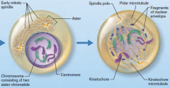
-Nuclear membrane begins to break apart(egg yolk breaks)
-Chromosomes become visible -Centrioles separate -Spindle fibers form 'chromatin (loose spaghetti) spirals to form chromosomes(spagetti spun on fork) |
|
Name the phase and describe it.
|
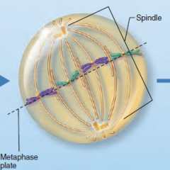
-Chromosomes attach to spindle
-Chromasomes line up across the center. -Centrioles separate (* astors) |
|
Name the phase and describe it.
|
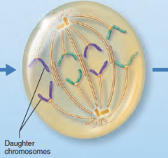
-Spindle fibers shorton pulling centromeres apart
-Sister Chromatids separate to opposite poles becoming chromosomes -Shortest phase |
|
Name the phase and describe it.
|
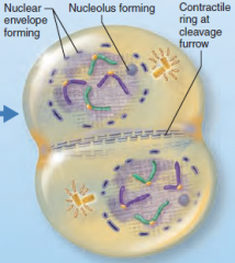
-Chromosomes gather at opposite ends
-Nuclear membrane forms enclosing chromosomes -Cytoplasm pinches inward to prepare for cytokinesis |
|
|
Effects of Solutions of Varying Tonicity
|
- Isotonic – solutions with the same solute concentration as that of the cytosol
- Hypertonic – solutions having greater solute concentration than that of the cytosol(cause crenation) - Hypotonic – solutions having lesser solute concentration than that of the cytosol (burst/lyse) |
|
|
Cilia
Flagellum |
- Whip-like, motile cellular extensions on exposed surfaces of certain cells
- Move substances in one direction across cell surfaces Flagellum Like a cilium, but longer; only example in humans is the sperm tail. Propels the cell. |
|
|
What is the trigger for cell division
|
Your bodies way of initiating/ starting CELL DIVISION is to increase the volume inside of the cell increasing cytoplasm, organelles, & DNA.
|
|
|
The four types of tissues
|
- Epithelial - Coverings and linings/Glandular epithelium
- Connective - support - Muscle - motion - Nerve - control |
|
|
Epithelial Tissue
|
- Cellularity – composed almost entirely of cells
- Special contacts – form continuous sheets held together by tight junctions and desmosomes - Polarity – apical and basal surfaces - Supported by connective tissue – reticular and basal laminae - Avascular but innervated – contains no blood vessels but supplied by nerve fibers - Regenerative – rapidly replaces lost cells by cell division |
|
|
Simple Squamous
|
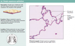
- Single layer of flattened cells with disc-shaped nuclei and sparse cytoplasm
- Functions Diffusion and filtration Provide a slick, friction-reducing lining in lymphatic and cardiovascular systems Present in the kidney glomeruli, lining of heart, blood vessels, lymphatic vessels, and serosae |
|
|
Simple Cuboidal
|
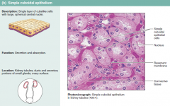
- Single layer of cube-like cells with large, spherical central nuclei
- Function in secretion and absorption - Present in kidney tubules, ducts and secretory portions of small glands, and ovary surface |
|
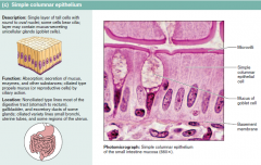
Simple Columnar
|
- Single layer of tall cells with oval nuclei; many contain cilia
- Goblet cells are often found in this layer - Function in absorption and secretion - Nonciliated type line digestive tract and gallbladder - Ciliated type line small bronchi, uterine tubes, and some regions of the uterus - Cilia help move substances through internal passageways |
|
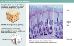
Pseudostratified Columnar
|
- Single layer of cells with different heights; some do not reach the free surface
- Nuclei are seen at different layers - Function in secretion and propulsion of mucus - Present in the male sperm-carrying ducts (nonciliated) and trachea (ciliated) |
|
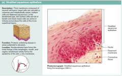
Stratified Squamous
|
- Thick membrane composed of several layers of cells
- Function in protection of underlying areas subjected to abrasion - Forms the external part of the skin’s epidermis (keratinized cells), and linings of the esophagus, mouth, and vagina (nonkeratinized cells) |
|
|
Stratified Cuboidal and Columnar
|
Stratified cuboidal
- Quite rare in the body - Found in some sweat and mammary glands - Typically two cell layers thick Stratified columnar - Limited distribution in the body - Found in the pharynx, male urethra, and lining some glandular ducts - Also occurs at transition areas between two other types of epithelia |
|
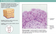
Transitional
|
- Several cell layers, basal cells are cuboidal, surface cells are dome shaped
- Stretches to permit the distension of the urinary bladder - Lines the urinary bladder, ureters, and part of the urethra |
|
|
Connective Tissue
|
- Found throughout the body; most abundant and widely distributed in primary tissues
--- Connective tissue proper --- Cartilage --- Bone --- Blood - Connective tissues have: --- Mesenchyme as their common tissue of origin --- Varying degrees of vascularity --- Nonliving extracellular matrix, consisting of ground substance and fibers - Structural Elements of Connective Tissue --- Ground substance – unstructured material that fills the space between cells --- Fibers – collagen, elastic, or reticular --- Cells – fibroblasts, chondroblasts, osteoblasts, and hematopoietic stem cells |
|
|
Fibers
|
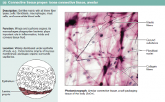
- Collagen – tough; provides high tensile strength
- Elastic – long, thin fibers that allow for stretch - Reticular – branched collagenous fibers that form delicate networks |
|
|
Special connections between cells
|
- Tight Junctions - Protein molecules in adjacent cell membranes fuse, forming an impermeable connection
--- Keeps enzymes, etc. within the proper tissue (digestive tract) - Desmosomes - Strands of protein extend and connect adjacent cell membranes; helps resist tearing --- stresses --- neck of the uterus --- Heart muscle --- skin - Gap Junctions - Cell membranes connected by hollow cylinders that allow transmission of small molecules (electrically excitable tissue) |
|
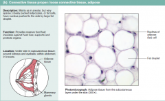
What is Adipose and what does it do?
|
- Matrix similar to areolar connective tissue with closely packed adipocytes
- Reserves food stores, insulates against heat loss, and supports and protects - Found under skin, around kidneys, within abdomen, and in breasts - Local fat deposits serve nutrient needs of highly active organs |
|
|
Connective Tissue (Function)
|
- Binding and support
- Protection - Insulation - Transportation |
|
|
What is the most abundant tissue in the body?
|
Connective
Collagen is most abundant among 3 fibers Hyaline cartilage is the most abundant cartillage |
|
|
Connective tissue Cells
|
- Fibroblasts – connective tissue proper
- Chondroblasts – cartilage - Osteoblasts – bone - Hematopoietic stem cells – blood - White blood cells, plasma cells, macrophages, and mast cells |
|
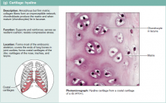
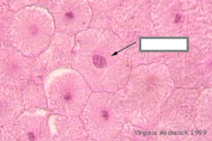
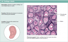
Hyaline Cartilage
|
Hyaline Cartilage
- Amorphous, firm matrix with imperceptible network of collagen fibers - Chondrocytes lie in lacunae - Supports, reinforces, cushions, and resists compression - Forms the costal cartilage - Found in embryonic skeleton, the end of long bones, nose, trachea, and larynx |
|
Elastic Cartilage
|
- Similar to hyaline cartilage but with more elastic fibers
- Maintains shape and structure while allowing flexibility - Supports external ear (pinna) and the epiglottis |
|
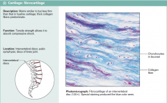
Fibrocartilage Cartilage
|
- Matrix similar to hyaline cartilage but less firm with thick collagen fibers
- Provides tensile strength and absorbs compression shock - Found in intervertebral discs, the pubic symphysis, and in discs of the knee joint |
|
|
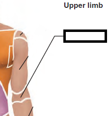
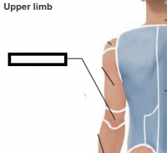
Axillary
|
|
|
|
Brachial
|
|
|
|
Antecubital
|
|
|
|
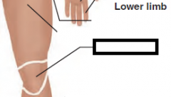
Patellar
|
|
|
|
Otic
|
|
|
|
Olecranal
|
|
|
|
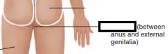
Perineal
|
|
|
|
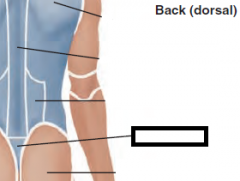
Sacral
|
|
|
|
Popliteal
|
|
|
|
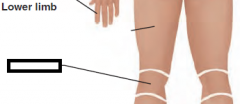
Plantar
|

|
|
|
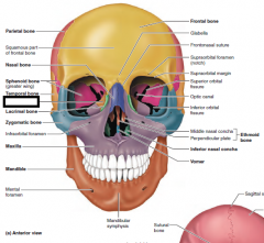
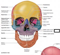
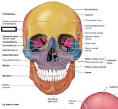
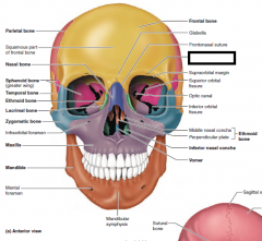
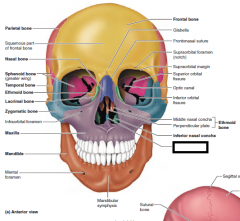
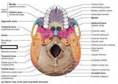
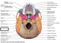
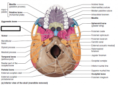
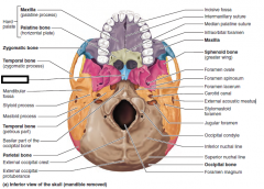
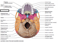
Ethmoidal Bone
|
|
|
Ethmoidal Bone(nose)
|
|

|
Inferior Nasal Concha
|
|

|
mandible
|
|

|
maxilla
|
|
|
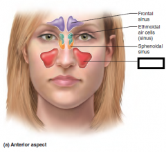
Maxillary sinus
|
|

|
Middle Nasal Concha
|
|
|
Nasal Bone
|
|

|
Optic canal
|
|
|
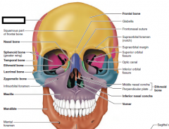
Parietal bone
|
|

|
sphenoid process
|
|

|
sphenoid sinus
|
|
|
superior orbital
|
|

|
temporal bone
|
|
|
vomer
|
|
|
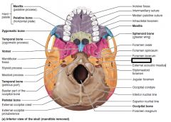
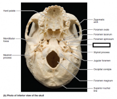
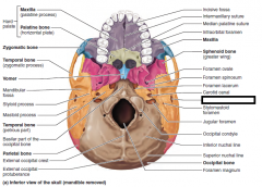
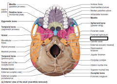
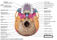
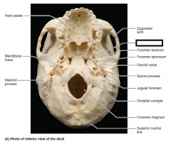
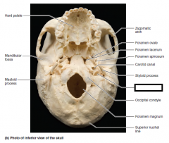
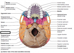
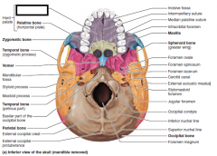
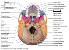
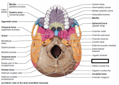
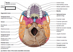
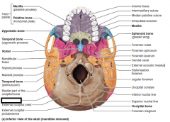
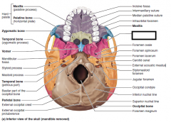
cartoid canal
|
|
|
cartoid canal
|
|
|
ext accoustic meatus
|
|
|
foramen lacerum
|
|
|
foramen magum
|
|
|
foramen ovale
|
|
|
jugular foramen
|
|
|
mastoid process
|
|
|
maxilla
|
|
|
maxilla(inferior)
|
|
|
occipital condyle
|
|
|
palentine bone
|
|
|
parietal bone
|
|
|
sphenoid bone
|
|
|
styloid process
|
|
|
temporal bone(petrous part)
|
|
|
temporal bone(zygomatic process)
|
|
|
vomer
|
|
|
zygomatic bone
|
|
|
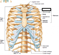
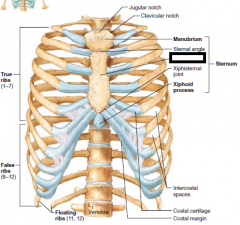
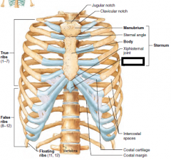
manubrium
|
|
|
sternum body
|
|
|
sternum xiphoid process
|
|
|
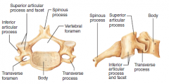
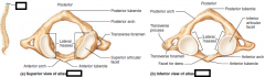
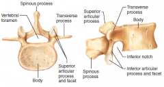
cervical
3 holes |
|
|
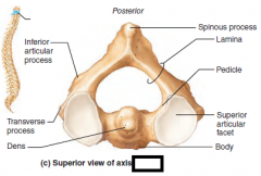
C1
|
|
|
C2
|
|
|
lumbar
|
|
|
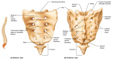
sacrum
|
|
|
thoracic
|
|
|
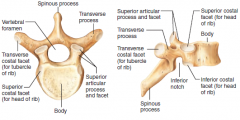
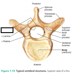
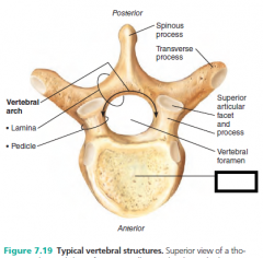
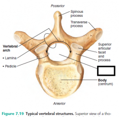
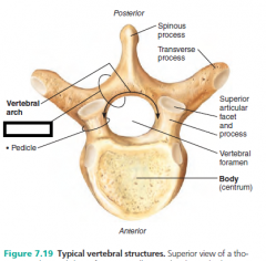
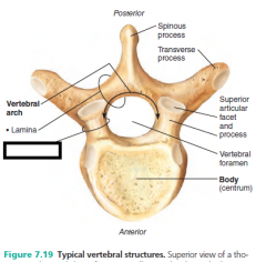
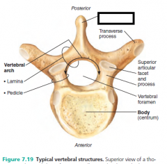
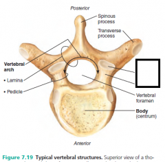
vertebral arch
|
|
|
body
|
|
|
vertebral foramen
|
|
|
lamina
|
|
|
pedicle
|
|
|
spinous process
|
|
|
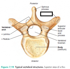
Superior articular process and facet
there's an inferior |
|
|
transverse process
|
|
|
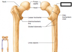
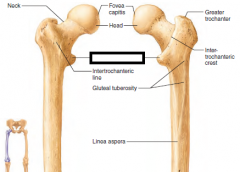
greater trochanter
|
|
|
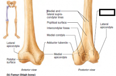
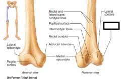
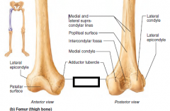
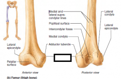
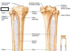
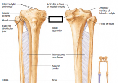
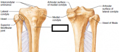
Lateral Condyle
|
|
|
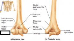
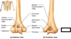
Lateral epicondyle
|
|
|
lesser trochanter
|
|
|
medial condyle
|
|
|
Medial epicondyle
|
|
|
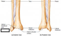
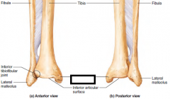
lateral malleolus
|
|
|
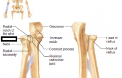
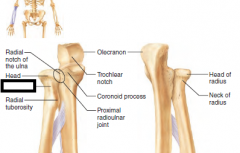
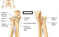
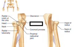
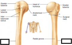
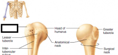
head
|
|
|
neck
|
|
|
olecranon process
|
|
|
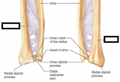
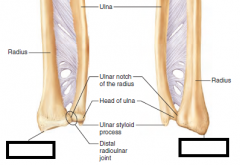
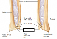
radius
|
|
|
styloid process (radius)
|
|
|
styloid process ulna
|
|
|
Trochlear notch
|
|
|
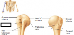
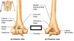
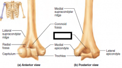
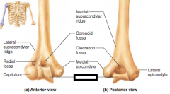
capitulum
|
|
|
deltoid tuberosity
|
|
|
greater tubercle
|
|
|
lateral epicondyle
|
|
|
lesser tubercle
|
|
|
medial epicondyle
|
|
|
olecranon fossa
|
|
|
trochlea
|
|
|
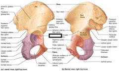
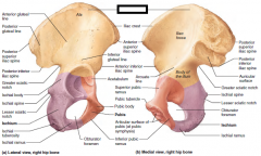
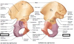
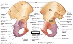
acetabulum
|
|
|
ilium
|
|
|
ischium
|
|
|
pubis
|
|
|
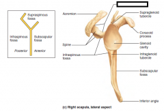
acromion
|
|
|
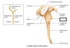
coracoid process
|
|
|
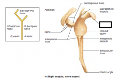
glenoid cavity
|
|
|
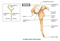
supraspinous fossa
|
|
|
lateral condyle
|
|
|
medial condyle
|
|
|
medial malleolus
|
|
|
tibial tuberosity
|
|
|
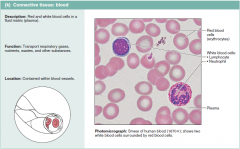
blood
|
|
|
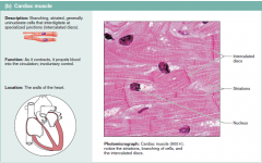
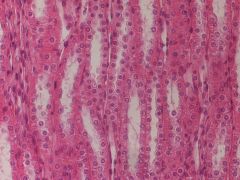
cardiac muscle
|
|
|
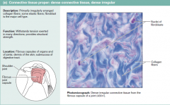
dense irregular
|
|
|
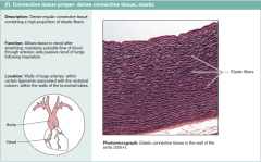
elastic
|
|
|
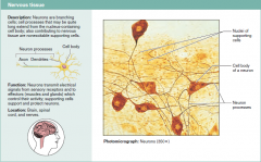
nerveous
|
|
|
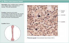
osseous
|
|
|
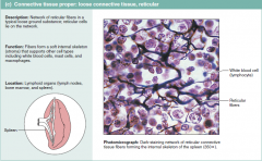
reticular
|
|
|

simple cuboidal
|
|
|
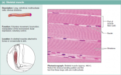
skeletal muscle
|
|
|
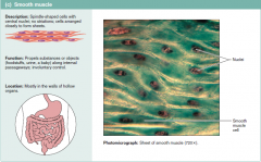
smooth muscle(pink)
|
|

|
stratified squamos
|
|

|
transitional epithelium
|