![]()
![]()
![]()
Use LEFT and RIGHT arrow keys to navigate between flashcards;
Use UP and DOWN arrow keys to flip the card;
H to show hint;
A reads text to speech;
169 Cards in this Set
- Front
- Back
|
What is the neural plate? |
In the developing embryo, the central nervous system first appears as a specialized, flattened region of cells. |
|
|
What is the neural tube? |
The neural plate invaginates and folds over to form this. |
|
|
What are the ventricles? |
The hollow space within the neural tube develops into hollow spaces within the brain called _____, which are filled with cerebrospinal fluid. |
|
|
What is the central nervous system? |
It consists of the brain and spinal cord. |
|
|
What is the peripheral nervous system? |
It consists of the rest (not the brain and spinal cord) the nerves and ganglia. |
|
|
What is the difference between gray matter and white matter? |
Gray matter is the seat of the neurosomas, dendrites, and synapses - forms a surface layer called the cortex over the cerebrum and cerebellum, and deeper masses called nuclei surrounded white matter. White matter lies deep to the cortical gray matter in most of the brain. It is composed of tracts, or bundles of axon, which here connect one part of the brain to another and to the spinal cord. |
|
|
What are sensory neurons? |
They are specialized to detect stimuli such as light, heat, pressure, and chemicals, and transmit info about them to the CNS. Beginning in almost every organ and end in the CNS. |
|
|
What are interneurons? |
They lie entirely within the CNS They receive signals from many other neurons and carry out the integrative functions of the nervous system. They process, store and retrieve information and make decisions that determine how the body responds to stimuli. |
|
|
What are motor neurons? |
They send signals predominantly to muscle and gland cells, the effectors. Lead to muscle cells, signal conduction away from the CNS. |
|
|
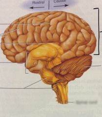
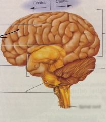
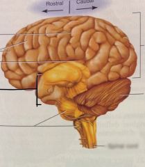
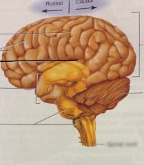
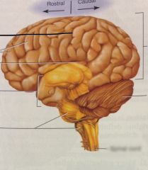
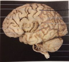
Cerebrum
|
|

|
Pons |
|

|
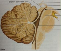
Pons
|
|

|
Cerebellum
|
|

|
Cerebellum
|
|

|
Cerebellum
|
|
|
Cerebellum
|
|
|
Medulla Oblongata
|
|
|
Medulla Oblongata
|
|
|
Medulla Oblongata
|
|
|
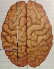
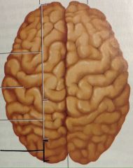
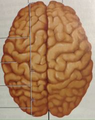
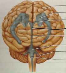
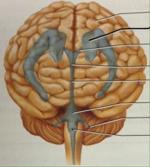
Right and Left Hemispheres
|
|
|
Gyri
|
|
|
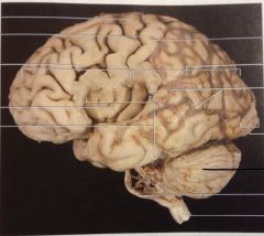
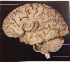
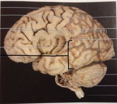
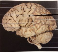
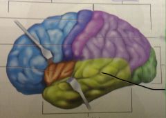
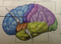
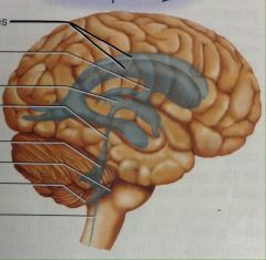
Frontal Lobe
|
|

|
Frontal Lobe
|
|

|
Frontal Lobes
|
|
|
Temporal Lobe
|
|
|
Temporal Lobe
|
|

|
Temporal Lobe
|
|
|
Parietal Lobe
|
|
|
Parietal Lobe
|
|

|
Parietal Lobe
|
|
|
Occipital Lobe
|
|

|
Occipital Lobe
|
|

|
Occipital Lobe
|
|

|
Occipital Lobe
|
|
|
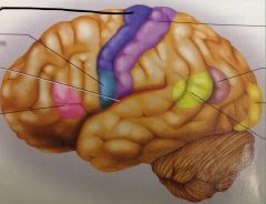
Lateral Sulcus
|
|
|
Lateral Sulcus
|
|

|
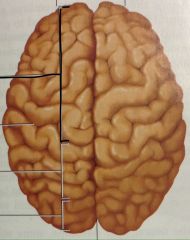
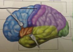
Precentral Gyrus
|
|
|
Precentral Gyrus
|
|
|
Precentral Gyrus
|
|
|
Central Sulcus
|
|

|
Central Sulcus
|
|
|
Central Sulcus
|
|
|
Central Sulcus
|
|
|
Postcentral Gyrus
|
|

|
Postcentral Gyrus
|
|

|
Postcentral Gyrus
|
|
|
Longitudinal Fissure
|
|
|
Parieto-occipital Sulcus
|
|
|
Parieto-occipital Sulcus
|
|
|
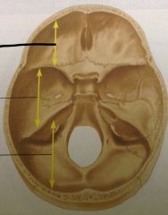
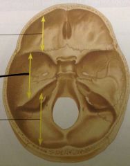
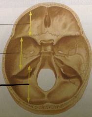
Anterior Cranial Fossa
|
|
|
Middle Cranial Fossa
|
|
|
Posterior Cranial Fossa
|
|
|
Posterior Cranial Fossa
|
|
|
Middle Cranial Fossa
|
|
|
On the cerebrum and cerebellum, the external surface you can see is a highly developed region of gray matter called the _______. |
Cortex |
|
|
Which part of the brain connects directly to the spinal cord? |
Medulla Oblongata |
|
|
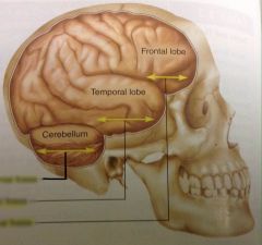
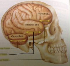
Which part of the brain rests in the anterior cranial fossa? |
Frontal Lobe |
|
|
Which part of the brain rests in the middle cranial fossa? |
Temporal Lobe |
|
|
Which part of the brain rests in the posterior cranial fossa? |
Cerebellum |
|
|
Based on the size of the sella turcica, the pituitary gland must be about the size and shape of an _______. |
Apple seed |
|
|
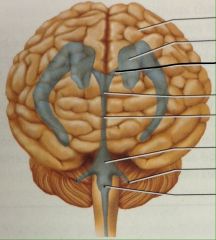
The optic nerves pass through which foramina (holes) between the cranial cavity and the orbits? |
Optic Foramen |
|
|
The optic chiasm is superior to which bone of the skull? |
Sphenoid Bone |
|
|
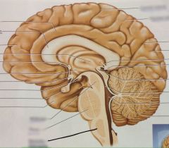
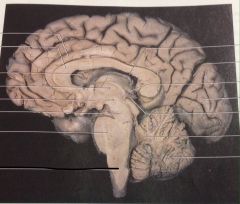
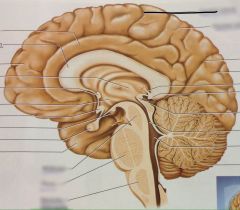
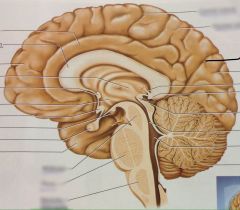
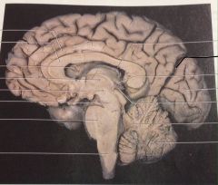
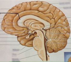
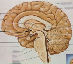
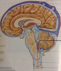
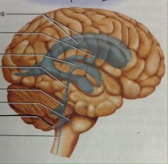
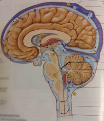
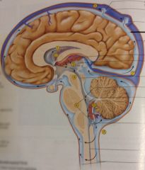
Corpus Callosum
|
|

|
Corpus Callosum
|
|

|
Corpus Callosum
|
|
|
Thalamus
|
|

|
Thalamus
|
|
|
Thalamus
|
|
|
Hypothalamus
|
|

|
Hypothalamus
|
|

|
Gyrus
|
|

|
Sulcus
|
|

|
Midbrain
|
|

|
Midbrain
|
|

|
Midbrain
|
|
|
Pons
|
|
|
Pons
|
|

|
Pons
|
|

|
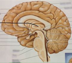
Third Ventricle
|
|
|
Third Ventricle
|
|

|
Third Ventricle
|
|

|
Third Ventricle
|
|

|
Cerebral Aqueduct
|
|

|
Cerebral Aqueduct
|
|
|
Cerebral Aqueduct
|
|

|
Cerebral Aqueduct
|
|
|
Cerebral Aqueduct
|
|
|
Fourth Ventricle
|
|

|
Fourth Ventricle
|
|
|
Fourth Ventricle
|
|

|
Fourth Ventricle
|
|
|
Which regions of the brain border the third ventricle? |
Thalamus, hypothalamus |
|
|
Which regions of the brain border the fourth ventricle? |
Pons, cerebellum |
|
|
Through which region does the cerebral aqueduct pass? |
Midbrain |
|
|
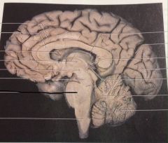
Which lobes of the cerebrum are visible in midsagittal section? |
Frontal, temporal, parietal, occipital. |
|
|
Is any cortex of the cerebrum visible in midsagittal section? |
Yes |
|
|
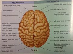
Where is the primary motor area located? |
Frontal lobe |
|
|
What is the function of the primary motor area? |
Neurons here send signals to the brain stem and spinal cord, which ultimately results in muscle contractions. |
|
|
What is the primary motor area also called? |
Precentral gyrus |
|
|
Where is the motor association area located? |
Frontal lobe |
|
|
What is the function of the motor association area? |
Where we plan our behavior, neurons compile a program for the degree and sequence of muscle contractions required for an action such as dancing, typing, or speaking. |
|
|
Where is the primary somatosensory area located? |
Parietal Lobe |
|
|
What is the function of the primary somatosensory area? |
Awareness of stimulation |
|
|
What is the primary somatosensory area also called? |
Postcentral gyrus. |
|
|
Where is the somatosensory association area located? |
Parietal Lobe |
|
|
What is the function of the somatosensory association area? |
Makes cognitive sense of stimulation |
|
|
Where is the primary visual area located? |
Occipital lobe |
|
|
What is the function of the primary visual area? |
Visual signals are received here. |
|
|
Where is the visual association area located? |
Occipital lobe |
|
|
What is the function of the visual association area? |
Spacial perception, recognize faces and other familiar objects. |
|
|
Where is the primary auditory area located? |
Temporal lobe |
|
|
What is the function of the primary auditory area? |
Auditory signals are received here. |
|
|
Where is the auditory association area located? |
Temporal Lobe |
|
|
What is the function of the auditory association area? |
Where we become capable of recognizing spoken words, a familiar piece of muscle, or a voice on the telephone. |
|
|
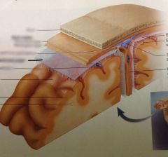
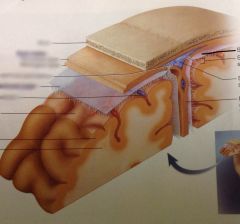
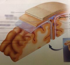
What are the meninges? |
The three concentric layers of connective tissue which surround the brain and separate it from the skull, protecting it and holding it in place during normal movements of the head. |
|
|
What is meningitis? |
The infection or inflammation of the meninges. |
|
|
What is the dura mater? |
The outermost of the meninges (literally: "tough mother"). This is a very strong membrane - you can pull on it with quite a bit for force without tearing it - which is firmly attached to the inside of the skull and contains blood vessels which nourish the bone. |
|
|
What is the falx cerebri? |
The midsagittal fold of the dura mater, it extends into the longitudinal fissure of the cerebrum and prevents lateral or side to side movement of the cerebral hemispheres within the skull. |
|
|
What is the tentorium cerebelli? |
A horizontal fold of the dura mater, it extends into the horizontal fissure which separates the occipital lobes of the cerebrum from the cerebellum, supporting the weight of the occipital lobes and limiting the vertical or up and down movement of the brain. |
|
|
What is the arachnoid mater? |
The middle layer of the meninges, much more delicate and deep to the dura mater. This bridges over sulci of the cerebral cortex, but follows the surface of the brain into larger fissures. ("spiderweb like mother") |
|
|
What is the pia mater? |
The deepest of the three layers of meninges. This is firmly attached to the nervous tissue of the brain and follows all of its convolutions into and out of sulci and fissures. ("delicate mother") |
|
|
What is the epidural space? |
It lies between the bone of the skull and the dura mater. Contains no fluid. |
|
|
What is the subdural space? |
It lies between the dura mater and the arachnoid mater. Contains no fluid. |
|
|
What is the subarachnoid space? |
It lies between the arachnoid mater and the pia mater. Contains cererospinal fluid in which the brain is floating. |
|
|
List the three layers of meninges and the three related spaces in the correct order, starting with the bone of the skull and moving toward the brain. |
Skull - epidural space - dura mater - subdural space - arachnoid mater - subarachnoid space - pia mater - brain. |
|
|
The meninges are composed of which one of the four basic tissue types? |
Connective tissue. |
|
|
Which part of the brain is superior to and rests on the tentorium cerebellum? |
Occipital lobe/cerebrum |
|
|
Which part of the brain is inferior to the tentorium cerebellum? |
Cerebellum |
|
|
Which parts of the brain are separated by the falx cerebri? |
The cerebral hemispheres |
|

|
Dura Mater
|
|
|
Arachnoid Mater
|
|

|
Pia Mater
|
|
|
Subdural Space
|
|
|
Subarachnoid Space
|
|
|
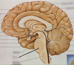
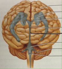
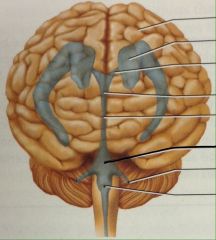
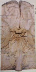
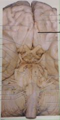
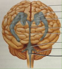
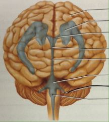
Optic Chiasm
|
|
|
Optic Chiasm
|
|
|
Optic Chiasm
|
|
|
Olfactory Tract
|
|
|
Olfactory Tract
|
|
|
Infundibulum
|
|
|
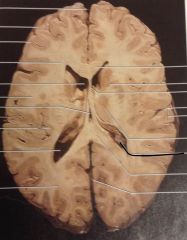
What is the choroid plexus? |
The cerebrospinal fluid is constantly being produced/secreted in the ventricles by this specialized tissue. |
|
|
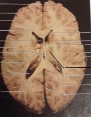
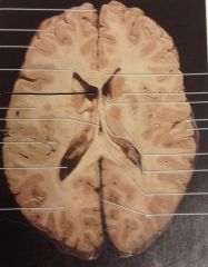
Lateral Ventricle
|
|
|
Lateral Ventricle
|
|
|
Lateral Ventricle
|
|

|
Interventricular Foramen
|
|
|
Interventricular Foramen
|
|
|
Choroid Plexus
|
|

|
Choroid Plexus
|
|
|
Median Aperture
|
|
|
Median Aperture
|
|

|
Median Aperture
|
|

|
Lateral Aperture
|
|
|
Lateral Aperture
|
|
|
Lateral Aperture
|
|

|
Arachnoid Granulations
|
|
|
Subarachnoid Space
|
|
|
Starting with the lateral ventricle, describe the flow of cerebral spinal fluid through all the ventricles and spaces until it is reabsorbed into the blood: |
1. CSF is secreted by choroid plexus in each lateral ventricle 2. CSF flows through interventricular foramina into third ventricle 3. Choroid plexus in third ventricle adds more CSF 4. CSF flows down cerebral aqueduct to fourth ventricle 5. Choroid plexus in fourth ventricle adds more CSF 6. CSF flows out two lateral apetures and one median aperture 7. CSF fills subarachnoid space and bathes external surfaces of brain and spinal cord 8. At arachnoid granulations, CSF is reabsorbed into venous blood of dural venous sinuses |
|
|
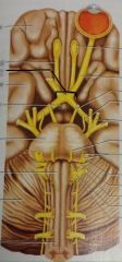
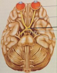
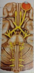
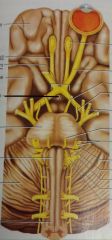
Describe Cranial nerve I |
Olfactory Nerve Cribiform foramina of ethmoid bone Sensory - Smell |
|
|
Describe cranial nerve II |
Optic Nerve Optic Foramen Sensory - Vision |
|
|
Describe cranial nerve III |
Oculomotor Nerve Superior Orbital Fissure |
|
|
Describe cranial nerve IV |
Trochlear Nerve Superior Orbital Fissure Motor - Eye movements |
|
|
Describe cranial nerve V |
Trigeminal Nerve Superior orbital fissure, foramen rotundum and infraorbital foramne, foramen ovale |
|
|
Describe cranial nerve VI |
Abducens Nerve Superior Orbital Fissure Motor - lateral eye movement |
|
|
Describe cranial nerve VII |
Facial Nerve Internal Acoustic meatus and stylomastoid foramen |
|
|
Describe cranial nerve VIII |
Vestibulocochlear Nerve Internal acoustic meatus |
|
|
Describe cranial nerve IX |
Glossopharyngeal Nerve Jugular foramen |
|
|
Describe cranial nerve X |
Vagus Nerve Jugular Foramen |
|
|
Describe cranial nerve XI |
Accessory Nerve Jugular Nerve |
|
|
Describe cranial nerve XII |
Hypoglossal Nerve Hypoglossal canal |